Traumatic Heterotrophic Ossification Of Quadriceps Femoris – A Case Report
Abstract
Background: Formation of mature lamellar bone at unusual sites like soft tissues, which normally does not exhibit properties of ossification is known as Heterotopic ossification (HO). It has a multi-factorial etiology with multiple risk factors. Trauma is one of such inciting event.
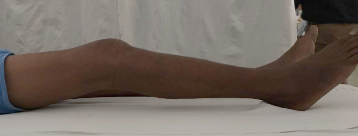
Case report- We are reporting a rare case of Heterotopic ossification of right quadriceps femoris in a 26 year old young adult, with severe knee stiffness, with no improvement following conservative treatment, which was successfully treated with surgical excision, obtaining good clinical results.
Downloads
References
Pitcock JA. Tumours and tumour like lesions of somatic tissue. In: Crenshaw AH, editor. Campbell's Operative Orthopaedics. 5th ed., Vol. 1. St. Louis: C.V. Mosby Co.; 1971. p. 1366.
Bar Oz B, Boneh A. Myositis ossificans progressiva: A 10-year follow-up on a patient treated with etidronate disodium. Acta Paediatr 1994;83(12):1332-4.
I Muni Srikanth, Amar Vishal, K Ravi Kiran (2015) Myositis Ossificans of Rectus Femoris: A Rare Case Report. by Journal of Orthpaedic Case Reports 2015 july-sep 5(3);page 92-94.
Giuseppe Rollo , Marco Pellegrino , Marco Filipponi , Gabriele Falzarano , Antonio Medici , Luigi Meccariello , Michele Bisaccia , Luigi Piscitelli , Auro Caraffa (2015). A case of the management of Heterotopic ossification as the result of acetabular fracture in a patient with traumatic brain injury. International Journal of Surgery Open 1 (2015) 30–34.
Hendifar AE, Johnson D, Arkfeld DG. Myositis ossificans: A case report. Arthritis Rheum 2005;53(5):793-5.
Myositis ossificans - DynaMed Ipswich (MA): EBSCO Publishing; 1995. Record N o . 1 1 4 6 7 1 . available from http://www.search.ebscohost.com/login.aspx?direct=true&site=dynamed&id=A N+114671.[Last updated on 2010 Jun 09; Last cited on 2010 Oct 11].
Sumiyoshi K, Tsuneyoshi M, Enjoji M. Myositis ossificans. A clinicopathologic study of 21 cases. Acta Pathol Jpn 1985;35(5):1109-22.
Cushner F D & Morwessel R M (1992) Myositis ossificans traumatica. Orthop. Rev. 21: 1319–1326.
Parikh J, Hyare H & Saifuddin A (2002) The imaging features of post-traumatic myositis ossificans, with emphasis on MRI. Clin. Radiol. 57: 1058–1066.
Clements NC Jr & Camilli A E (1993) Heterotopic ossification complicating critical illness.Chest. 104: 1526–1528.
Heinrich S D, Zembom M M & MacEwen GD (1989) Pseudomalignant myositis ossificans. Orthopedics 12: 599–602.
Ben Hamida K S, Hajri R, Kedadi H, Bouhaouala H, SalahMH, Mestiri A, Zakraoui L& Doughi M H (2004) Myositis ossificans circumscripta of the knee improved by alendronate. Joint Bone Spine. 71: 144–146.
Pape HC, Marsh S, Morley JR, Krettek C, Giannoudis PV: Current concepts in the development of heterotopic ossification; J Bone Joint Surg Br. 2004 Aug;86(6):783-7
Tsuno M M & Shu G J (1990) Myositis ossificans. J. Manipulative Physiol. Ther. 13: 340–342.
Oglvie-Harris D J, HonsCB&Fornaiser V L (1980) Pseudomalignant myositis ossificans: heterotopic new-bone formation without a history of trauma. J. Bone Joint Surg. Am. 62: 1274–1285.
Craven P L, Urist MR (1971) Osteogenesis by radioisotope labelled cell population in implants of bone matrix under the influence of ionizing radiation. Clin Orthop. 76: 231–233.
Chalmers J, Gray D H, Rush J (1975) Observation on the induction of bone in soft tissues. J Bone Joint Surg. Br. 1975: 36–45
Bosch P, Musgrave D, Ghivizzani S, Latterman C, Day C S, Huard J (2000) The efficiency of muscle-derived cell-mediated bone formation. Cell Transplant. 9: 463–470.
Thorndike A (1940) Myositis ossificans traumatica. J. Bone Joint Surg. Br. 22: 315–323.

The entire contents of the Orthopaedic Journal of Madhya Pradesh Chapter are protected under Indian and International copyrights. Orthopaedic Journal of Madhya Pradesh Chapter allow authors to retain the copyrights of their papers without restrictions, Authors grant the publisher the right of exclusive publication. The Journal then grants to all users a free, irrevocable, worldwide, perpetual right of access to, and a license to copy, use, distribute, perform and display the work publicly and to make and distribute derivative works in any digital medium for any reasonable non-commercial purpose, subject to proper attribution of authorship. The journal also grants the right to make numbers of printed copies for their personal non-commercial use under Creative Commons Attribution-Non-commercial share alike 4.0 International Public License.

 OAI - Open Archives Initiative


