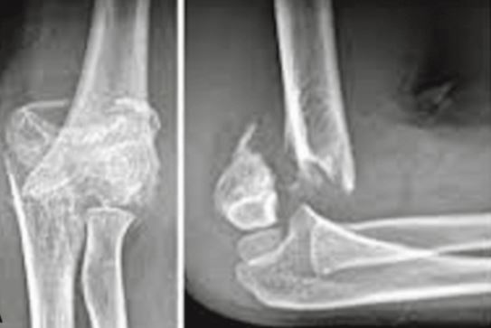
Should Postoperative X-Rays be done in Supracondylar Humerus Fractures after Pinning?
Abstract
Background: Most author advocates the need for early postoperative clinical and radiographic follow-up in operative treatment of completely displaced supracondylar fractures of the humerus in children but in central India specially rural population is there, for whom it is difficult to get weekly X-rays done. Therefore we decided to determine the necessity of postoperative x rays after k-wire fixation for the operative treatment of completely displaced supracondylar fractures of the humerus in children.
Methods: 103 patients who underwent operative management of Gartland type III fractures at our institution from November 2012 to January 2016 were reviewed. Intraoperative C-arm images were compared with postoperative radiographs to identify changes in fracture alignment and k-wire placement.
Results: A total of 103 patients (48 females, 55 males) with a mean age of 7.1 years (range, 3.1 to 12.0) were reviewed which were classified as type III. Fracture displacement or k-wire back out was seen in three patients (2.9%) at the first postoperative visit. None of these patients required further operative management. On statistical evaluation, no significant difference was seen in terms of time to first postoperative visit, days to k-wire removal, or average follow-up time. The overall complication rate was 7.76% (9/103)
Conclusions: Fracture displacement and k-wire migration observed in postoperative radiographs after k-wire fixation of supracondylar humerus fractures have little effect on clinical management or long-term complication. Radiographs can therefore be delayed until the time of k-wire removal provided sufficient intraoperative stability was obtained.
Downloads
References
McIntyre W (1996) Supracondylar fractures of the humerus. In: Letts RM (ed) Management of pediatric fractures, vol 11. Churchill Livingstone, New York. pp 167–198
Boyd DW, Aronson DD. Supracondylar fractures of the humerus: a prospective study of percutaneous k-wirening. J PediatrOrthop. 1992; 12: 189–194.
Gordon JE, Patton CM, Luhmann SJ, et al. Fracture stability after k-wirening of displaced supracondylar distal humerus fractures in children. J PediatrOrthop. 2001; 21: 313–318.
Kallio PE, Foster BK, Paterson DC. Difficult supracondylar elbow fractures in children: analysis of percutaneous k-wirening technique. J PediatrOrthop. 1992; 12: 11–15.
Ponce BA, Hedequist DJ, Zurakowski D, et al. Complications and timing of follow-up after closed reduction and percutaneous k-wirening of supracondylar humerus fractures: follow-up after percutaneous k-wirening of supracondylar humerus fractures. J PediatrOrthop. 2004; 24: 610–614.
Mehlman CT, Crawford AH, McMillon TL, et al. Operative treatment of supracondylar humerus fractures in children: the Cincinnati experience. ActaOrthop Belg. 1996; 62(suppl 1): 41–50.
Karamitopoulos MS, Dean E, Littleton AG, Kruse R. Postoperative Radiographs After Pinning of Supracondylar Humerus Fractures: Are They Necessary? J PediatrOrthop 2012; 32: 672–674.

The entire contents of the Orthopaedic Journal of Madhya Pradesh Chapter are protected under Indian and International copyrights. Orthopaedic Journal of Madhya Pradesh Chapter allow authors to retain the copyrights of their papers without restrictions, Authors grant the publisher the right of exclusive publication. The Journal then grants to all users a free, irrevocable, worldwide, perpetual right of access to, and a license to copy, use, distribute, perform and display the work publicly and to make and distribute derivative works in any digital medium for any reasonable non-commercial purpose, subject to proper attribution of authorship. The journal also grants the right to make numbers of printed copies for their personal non-commercial use under Creative Commons Attribution-Non-commercial share alike 4.0 International Public License.

 OAI - Open Archives Initiative


