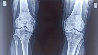
A prospective study for initial assessment of functional outcome of high tibial osteotomy in active young adults in early osteoarthritis of knee
Abstract
Introduction: Knee osteoarthritis is typically the result of wear and tear and progressive loss of articular cartilage. Common clinical symptoms include knee pain, stiffness and swelling that worsens over time. Osteoarthritis commonly affects the medial compartment of knee giving rise to varus deformity. High tibial osteotomy (HTO) is a valuable treatment modality in correcting malalignment and thereby relieving the symptoms associated with medial unicompartmental osteoarthritis.
Methodology: Twenty-eight young patients with complaints of knee pain were screened and those diagnosed as early knee osteoarthritis (grade I-III on Kellgren-Lawrence grading scale) were operated by high tibial osteotomy. Follow-up evaluation was done at 3, 6 and 9 months by Knee Society Scoring Scale and Visual Analogue Scale (VAS) for pain.
Results: The mean knee score was 53.3 pre-operatively and post-operatively the score improved gradually to the mean of 83.2 at 9 months. The visual analog scale for pain in all patients showed a significant improvement at the final follow-up.
Conclusion: High tibial medial opening wedge osteotomy is a good option in the treatment of unicompartmental osteoarthritis knee. It relieves pain and improves functional outcome. Accurate preoperative planning and good surgical technique gives better results.
Downloads
References
2. Rivero-Santana A, et al. Effectiveness of a decision aid for patients with knee osteoarthritis: a randomized controlled trial. Osteoarthritis Cartilage. 2021 Sep;29(9):1265-1274.
3. Ivarsson I, Myrnerts R, Gillquist J. High tibial osteotomy for medial osteoarthritis of the knee. A 5 to 7 and 11 year follow-up. J Bone Joint Surg Br. 1990; 72:238-244.
4. Asik M, Sen C, Kilic B, Goksan SB, Ciftci F, Taser OF. High tibial osteotomy with Puddu plate for the treatment of varus gonarthrosis. Knee Surg Sports Traumatol Arthrosc. 2006; 14:948-954. DOI: 10.1007/s00167-006- 0074-1.
5. Giuseffi, Steven A.; Replogle, William H.; Shelton, Walter R. (2015). Opening-Wedge High Tibial Osteotomy: Review of 100 Consecutive Cases. Arthroscopy: The Journal of Arthroscopic & Related Surgery, S0749806315004156. doi:10.1016/j.arthro.2015.04.097
6. Schuster P, Geßlein M, Schlumberger M, Mayer P, Mayr R, Oremek D, Frank S, Schulz-Jahrsdörfer M, Richter J. Ten-Year Results of Medial Open-Wedge High Tibial Osteotomy and Chondral Resurfacing in Severe Medial Osteoarthritis and Varus Malalignment. Am J Sports Med. 2018 May;46(6):1362-1370. doi: 10.1177/0363546518758016. Epub 2018 Mar 28. PMID: 29589953.
7. Ollivier B, Berger P, Depuydt C, Vandenneucker H. Good long-term survival and patient-reported outcomes after high tibial osteotomy for medial compartment osteoarthritis. Knee Surg Sports Traumatol Arthrosc. 2021 Nov;29(11):3569-3584. doi: 10.1007/s00167-020-06262-4. Epub 2020 Sep 9. PMID: 32909057.
8. Meding JB, Kearing EM, Ritter MA et al. Total knee arthroplasty after high tibial osteotomy. Clin orthop. 2000; (375):175-84.
9. Zhang Y, Jordan JM. Epidemiology of osteoarthritis. Clin Geriatr Med. 2010 Aug;26(3):355-69 doi: 10.1016/j.cger.2010.03.001. Erratum in: Clin Geriatr Med. 2013 May;29(2):ix. PMID: 20699159; PMCID: PMC2920533.
10. Wright RJ, Sledge CB, Poss R et al. Patient-reported outcome and survivorship after Kinemax total knee arthroplasty. J Bone Joint Surg Am. 2004; 86-A:2464-70.
11. Patond KR, Lokhande AV. Medial open wedge high tibial osteotomy in medial compartment osteoarthrosis of the knee. Natl Med J India. 1993; 6:104-8.
12. Kim, M., Ko, B. & Park, J. The proper correction of the mechanical axis in high tibial osteotomy with concomitant cartilage procedures—a retrospective comparative study. J Orthop Surg Res 14, 281 (2019).

The entire contents of the Orthopaedic Journal of Madhya Pradesh Chapter are protected under Indian and International copyrights. Orthopaedic Journal of Madhya Pradesh Chapter allow authors to retain the copyrights of their papers without restrictions, Authors grant the publisher the right of exclusive publication. The Journal then grants to all users a free, irrevocable, worldwide, perpetual right of access to, and a license to copy, use, distribute, perform and display the work publicly and to make and distribute derivative works in any digital medium for any reasonable non-commercial purpose, subject to proper attribution of authorship. The journal also grants the right to make numbers of printed copies for their personal non-commercial use under Creative Commons Attribution-Non-commercial share alike 4.0 International Public License.

 OAI - Open Archives Initiative


