Orthopaedic Journal of MP Chapter

Publisher: Madhya Pradesh Orthopaedic Association www.mpioa.com
E-ISSN:2582-7243, P-ISSN:2320-6993
2019 Volume 25 Number 1 Jan-Jun

Patterned Osteomyelitis Caused By Rubber Band

Jain S1*, Mantri D2, Rohra S3, Verma S4

1* Saurabh Jain, Assistant Professor, Department of Orthopaedics, Mahatma Gandhi Memorial Medical College and Maharaja Yashwantrao Hospital, Indore, Madhya Pradesh, India.

2 D Mantri, Department of Orthopaedics, Mahatma Gandhi Memorial Medical College and Maharaja Yashwantrao Hospital.

3 S Rohra, Department of Orthopaedics, Mahatma Gandhi Memorial Medical College and Maharaja Yashwantrao Hospital.

4 S Verma, Department of Orthopaedics, Mahatma Gandhi Memorial Medical College and Maharaja Yashwantrao Hospital.

Background: Circumferential foreign bodies left for prolonged period can migrate and cause serious constriction bands, ulcers, discharging sinus, compartment syndrome or osteomyelitis. When it involves bones of forearm, it involves typically ulna on ulna surface and radius on radial surface only, which can be seen as patterned lesion on the x-rays.

Case report: We report successful treatment by early surgical exploration, of such a case of rubber band tied on the wrist of a child and was forgotten, which migrated deep into the bones causing typical patterned osteomyelitis of both radius and ulna.

Conclusion: We report this case to create general awareness regarding complications of such foreign bodies and to consider for prompt surgical exploration for foreign body removal on visualizing the typical signature patterned lesion of the bone involving the outer surfaces of the bones only.

Keywords: Rubber band, Osteomyelitis, Daga Syndrome

Corresponding Author How to Cite this Article To Browse
Saurabh Jain, Assistant Professor, Department of Orthopaedics, Mahatma Gandhi Memorial Medical College and Maharaja Yashwantrao Hospital, Indore, Madhya Pradesh, India.
Email:
Jain S, Mantri D, Rohra S, Verma S, Patterned Osteomyelitis Caused By Rubber Band. ojmpc. 2019;25(1):38-41.
Available From
https://ojmpc.com/index.php/ojmpc/article/view/76
Manuscript Received Review Round 1 Review Round 2 Review Round 3 Accepted
2019-06-06 2019-06-12 2019-06-18 2019-06-24 2019-06-30
Conflict of Interest Funding Ethical Approval Plagiarism X-checker Note
Authors state no conflict of interest. Non Funded. The conducted research is not related to either human or animals use. 14.38 All authors have accepted responsibility for the entire content of this manuscript and approved its submission.

© 2019by Jain S, Mantri D, Rohra S, Verma Sand Published by Madhya Pradesh Orthopaedic Association. This is an Open Access article licensed under a Creative Commons Attribution 4.0 International License https://creativecommons.org/licenses/by-nc/4.0/ unported [CC BY NC 4.0].

Introduction

Unrecognized foreign bodies are focus for infection and when they are deep seated they can result in tendinitis, granuloma, deep infection and even osteomyelitis. Most common foreign bodies are wood, metal, plastic or glass [1]. Foreign bodies of circumferential nature like rubber band, animal hair, cloth, thread etc are tied on the forearm or wrist quiet commonly in India [2]. When these circumferential foreign bodies are left for prolonged period or are forgotten after applying, they can migrate and cause serious constriction bands, ulcers, discharging sinus and compartment syndrome [3].

On involvement of further deeper tissue and bones these circumferential foreign bodies can result in osteomyelitis involving typically ulna on ulnar surface and radius on radial surface only, seen as erosion of ulna and radius on medial and lateral surfaces only respectively. We report successful treatment by early surgical exploration, of such a case of rubber band tied on the wrist of a child and was forgotten, which migrated deep into the bones causing typical patterned osteomyelitis of both radius and ulna.

The purpose of reporting this case is to create general awareness regarding complications of such foreign bodies among surgeons to consider for prompt surgical exploration on visualizing the typical signature patterned lesion of the bone involving the outer surfaces of the bones only for foreign body removal.

Case report

A 4 year’s female child presented to our tertiary centre with pain, swelling and discharging sinus on an old circumferential scar at left wrist joint from 6 months. The pain was localized to the left wrist and hand, was moderate to severe and was associated with swelling at the wrist joint. She had fever on and off which was low grade.

On history, her parents denied remembering any history of trauma to left wrist. She had taken treatment by a doctor at a local clinic, regularly for past 6 months with analgesics and antibiotics, but the complaints did not improve. On inspection, there was circumferential healed scar mark at the wrist along with three discharging sinuses at the dorsolateral, dorsomedial and one on ventral aspect of the wrist on the scar itself.

There was history of continuous pus discharge from the sinus tract for the past 5 months, which was although scanty, but did not subside even after antibiotics treatment given by a local surgeon (Figure 1).

Figure 1: Pre op clinical photographs volar view (a) & dorsal view (b) showing circumferential healed scar mark at wrist along with three discharging sinus.
ojmpc_76_01.jpg

On palpation the wrist was warm and tender. The movements of the wrist and fingers were grossly restricted and there was no active extension of finger, although some flexion of fingers was present. Passive movements were very painful. Distal pulsations were palpable for both radial and ulnar arteries. Sensory supply of the hand was normal except for diffuse hyperalgesia over palm. Colour Doppler of the left upper limb showed normal vascularity. Hematological examination showed leukocytosis with WBC count of 16000/mm3, sedimentation rate was 39 mm and C reactive protein was 3.5 mg/l.

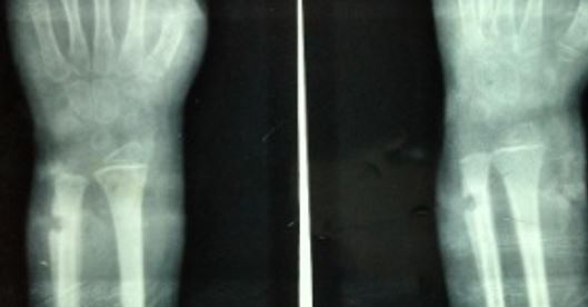
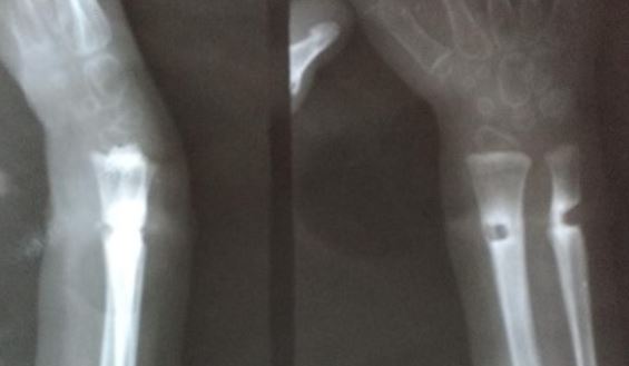
Patient was having 1 month old previous x-rays of wrist with her, which revealed a small lytic lesion of the ulna more on the ulnar side, along with the periosteal reaction but the radius seemed normal (Figure 2). Fresh x-rays were done, which showed osteomyelitis of both distal ulna and radius, with periosteal reaction and lytic lesion at both distal radius and distal ulna particularly on medial side of ulna and lateral side of radius (Figure 3).

Figure 2: 1 months old previous x-rays of wrist AP (a) & oblique (b) views showing small lytic lesion of the ulna particular on the ulnar side, along with the periosteal reaction but normal radius.
ojmpc_76_02.jpg

Figure 3: AP (a) & lateral (b) X-rays at presentation showing osteomyelitis of both distal ulna & radius, with periosteal reaction and lytic lesion on medial side of ulna and radial side of radius.
ojmpc_76_03.jpg

On visualization, of this typical signature pattern osteomyelitis causing lesion on ulnar surface of ulna and radial surface of radius, we suspected some circumferential foreign body inside and we decided to explore the wrist immediately. Under tourniquet control and under general anesthesia, we explored the wrist via circumferential incision excising the sinus tracts and the previous scar completely.


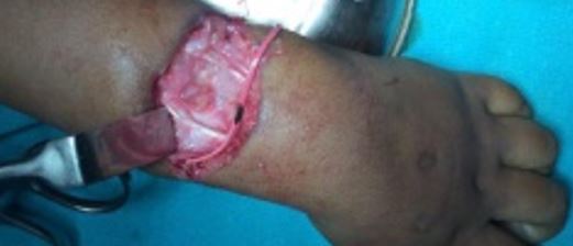
On exploration a circular rubber band was seen placed circumferential deeply buried under, which had caused the lytic lesion of the distal ulna on the ulnar side and distal radius on the radial side. This rubber band was removed and the osteolytic lesion of both radius and ulna was curetted (Figure 4).

Both the ulnar and radial vessels and the median and ulnar nerves were intact. The FDS and FDP tendons were intact but were kinked and compressed. On removing the tight rubber band, the flexor tendons got freed. The extensor tendons of fingers were found injured and were repaired end to end with Kessler’s knot.

Figure 4: Intra operative photograph of wrist showing circumferential rubber band acting as foreign body
ojmpc_76_04.jpg

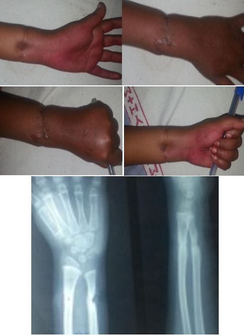
Figure 5: Clinical photographs (a to d) & X rays AP (e) & lateral (f) view at final follow up of 1 year showing full recovery and normal movements with wound & radiological healing
ojmpc_76_05.jpg

Post-operatively, showing the removed rubber band to her parents, her mother recalled putting a religious thread along with elastic rubber band around child’s wrist but didn’t remembered anything regarding removal.

Post-operatively an above elbow slab with wrist and fingers in extension was given for 3 weeks, for healing of repaired extensor tendons.Intravenous antibiotics were given for one week following which oral antibiotics were prescribed for further six weeks. At final follow up, i.e. after one year of surgery, the patient recovered fully, all wounds healed well and patient regained normal movements of finger and wrists. The radiological examination showed healing of the lesion in both radius and ulna (Figure 5).

Discussion

In India, it is common tradition to tie thread, animal hair, cloth, rubber band or ring on the wrist or limb, in order to fulfill the wishes1. This tradition is equally prevalent for both adult and child. But in a child, since these are applied by the parent, relatives for fulfilling wishes or by quacks as a malpractice, these objects may be left for prolonged period, are sometimes even forgotten after applying. When these threads, band or hairs are left for prolonged duration, these can migrate inside the skin very slowly in a child’s growing limb and can cause superficial or deep infection. Sometimes, it can migrate even deeper into the osseous region causing osteomyelitis as happened in our patient [2, 3].

When these circumferential foreign bodies, involve the underlying bones of forearm causing osteomyelitis, it involves typically ulna on ulnar surface and radius on radial surface only, which can be seen as patterned lesion on the x-rays i.e. showing erosion of ulna and radius on medial and lateral surfaces only respectively. Thus such type of osteomyelitis seen on x rays are the typical signature patterned osteomyelitis caused by the circumferential foreign body and are hallmark for the osteomyelitis caused by a circumferential foreign body. On viewing such type of signature patterned osteomyelitis on X-rays, it should be suspected of having some circular foreign body inside and it should be treated surgically promptly by exploration to remove the foreign body. In literature, few such cases have already been reported by Agrawal, Rasool, Arora etc [2-9].

All these cases have reported unrecognized foreign body in form of rubber band or thread as focus osteomyelitis of the distal radius and ulna. The x rays showed in these reports revealed the same typical signature pattern osteomyelitis i.e. lesion at the outer surfaces of the bones, but even after such patterned lesion these cases were initially treated by just antibiotics for the prolonged duration of time. This prolonged treatment just by antibiotics has caused delay and loss of precious time leading to increase in the lesion. This delay in exploration can cause serious injuries to tendon, nerves or vessels. Since our patient also presented to us late i.e. after 6 months, losing the precious time in conservative treatment by local doctor, she sustained tendon injuries, but was fortunate enough not having any vascular or neural injury.


We on viewing such typical patterned lesion on x-rays suspected it to be due to some circumferential foreign body and hence explored immediately and were successful in treating her fully without recurrence.

Hence we recommend, if such specific pattern of osteomyelitis is seen on the x rays, then we should suspect the osteomyelitis caused by foreign body of circumferential nature and should explore immediately to remove the foreign body.

Conclusion

In such specific type of lesions, the history may be inconclusive as it may be forgotten by the child and his parents and the clinical signs can be minimal. MRI and CT scan, although are useful tools in localizing foreign bodies, but are not routinely done. Thus on viewing such typical signature pattern lesion surgical debridement and removal of the foreign body which is the key to successful treatment, should be done promptly.

References

1. Greene WB. Unrecognized foreign body as a focus for delayed serratiamarce scenes osteomyelitis and septic arthritis. Two case reports. J Bone Joint Surg. Am. 1989 Jun; 71(5):754-7 [Crossref][PubMed][Google Scholar]

2. Gill SPS, Raj M, Kumar D, Singh J. Circumferential osteomyelitis of the both bone forearm bone caused by wrist band: a rare cause. Indian J orthop Surg. 2015;1(3);186-93. [Crossref][PubMed][Google Scholar]

3. Kumar KM, Shankarappa M. Rubber band syndrome of the arm. J hand Microsurg. 2013;5:2:83. [Crossref][PubMed][Google Scholar]

4. Aggarwal AN, Kini SG, Arora A, Singh AP, Gupta S, Gulati D. Rubber band syndrome - high accuracy of clinical diagnosis. J Pediatr Orthop. 2010;30(7):1-4. [Crossref][PubMed][Google Scholar]

5. Arora A, Agarwal A. Dhaga syndrome: a previously undescribed entity. J Bone Joint Surg Br. 2004;86(2):282-4. [Crossref][PubMed][Google Scholar]

6. Gaur SC, Swarup A. Progressive constriction band caused by black magic thread. J Hand Surg Br. 1991;16(3):349. [Crossref][PubMed][Google Scholar]

7. McIver MA, Gochman RF. Elastic bands on the wrist: a not so “silly” complication. Pediatr Emerg Care. 2011;27(5):428-9. [Crossref][PubMed][Google Scholar]

8. Rasool MN, Stathoulis B. Rubber band constriction of the wrist. J Hand Surg Br. 1996;21(6):806-7. [Crossref][PubMed][Google Scholar]

9. Aggarwal A, Kant KS, Verma I. The rubber band syndrome: the forgotten rubber band in the wrist. Hand Surg. 2013;18(3):407-10. [Crossref][PubMed][Google Scholar]

Disclaimer / Publisher's Note

The statements, opinions and data contained in all publications are solely those of the individual author(s) and contributor(s) and not of Journals and/or the editor(s). Journals and/or the editor(s) disclaim responsibility for any injury to people or property resulting from any ideas, methods, instructions or products referred to in the content.