Orthopaedic Journal of MP Chapter

Publisher: Madhya Pradesh Orthopaedic Association www.mpioa.com
E-ISSN:2582-7243, P-ISSN:2320-6993
2019 Volume 25 Number 1 Jan-Jun

Objective Evaluation Of Tendon Morphology By Ultrasonography In Treatment Of Recalcitrant Tennis Elbow By Autologous Platelet Rich Plasma

Jain S1*, Banodha L2, Kelkar R3, Gautam V4

1* Saurabh Jain, Assistant Professor, Department of Orthopaedics, Mahatma Gandhi Memorial Medical College and Maharaja Yashwantrao Hospital, Indore, Madhya Pradesh, India.

2 L Banodha, Department of Orthopaedics, Mahatma Gandhi Memorial Medical College and Maharaja Yashwantrao Hospital, Indore, Madhya Pradesh, India.

3 R Kelkar, Department of Orthopaedics, Mahatma Gandhi Memorial Medical College and Maharaja Yashwantrao Hospital, Indore, Madhya Pradesh, India.

4 V Gautam, Department of Orthopaedics, Mahatma Gandhi Memorial Medical College and Maharaja Yashwantrao Hospital, Indore, Madhya Pradesh, India.

Background: Traditional therapies of tennis elbow have shown inconsistent outcomes as they do not deal with poor tendon healing properties secondary to poor vascularization. Local platelet rich plasma injections, which provide locally high concentration of growth factors, have shown its efficacy in treatment of tennis elbow on a subjective basis only.

Material and methods: We tried to measure the efficacy of locally injected autologous PRP, subjectively by functional oxford elbow score and pain score as well as objectively by ultrasonographic evaluation of the morphologic changes (focal hypoechoic, odema, tendon thickness, fraying, tear, cortical erosion, calcification) in common extensor origin in 30 patients with mean age of 39.3 years of recalcitrant tennis elbow.

Results: The mean pain VAS Score improved from 7.7 before injection to 1.8 at final follow up i.e. after 6 months post injection. The Oxford elbow score improved from a mean of 19.2 prior to treatment to 41.3 after the injection at final follow up. 6 months post injection ultrasonography of the involved elbow showed decrease in focal hypoechoic, decreased edema, and improvement in thickness of the tendon and healing of the tear at the origin site.

Conclusion: This study confirms that local PRP by supplying growth factors helps to enhance the stromal and mesenchymal stem cell proliferation and increases tendon vascularity and prevents angiofibroblastic degeneration and thus improves tendon repair and healing property by releasing growth factors and increasing vascularity, which can be documented by improved tendon morphology.

Keywords: Tennis elbow, Platelet rich plasma, Ultrasonography

Corresponding Author How to Cite this Article To Browse
Saurabh Jain, Assistant Professor, Department of Orthopaedics, Mahatma Gandhi Memorial Medical College and Maharaja Yashwantrao Hospital, Indore, Madhya Pradesh, India.
Email:
Jain S, Banodha L, Kelkar R, Gautam V, Objective Evaluation Of Tendon Morphology By Ultrasonography In Treatment Of Recalcitrant Tennis Elbow By Autologous Platelet Rich Plasma. ojmpc. 2019;25(1):23-29.
Available From
https://ojmpc.com/index.php/ojmpc/article/view/73
Manuscript Received Review Round 1 Review Round 2 Review Round 3 Accepted
2019-06-06 2019-06-12 2019-06-18 2019-06-24 2019-06-30
Conflict of Interest Funding Ethical Approval Plagiarism X-checker Note
Authors state no conflict of interest. Non Funded. The conducted research is not related to either human or animals use. 13.35 All authors have accepted responsibility for the entire content of this manuscript and approved its submission.

© 2019by Jain S, Banodha L, Kelkar R, Gautam Vand Published by Madhya Pradesh Orthopaedic Association. This is an Open Access article licensed under a Creative Commons Attribution 4.0 International License https://creativecommons.org/licenses/by-nc/4.0/ unported [CC BY NC 4.0].

Introduction

Tennis elbow is treated non-operatively by rest, anti-inflammatory drugs, brace, physical therapy or by local intralesional injections of corticosteroid, dry needling or by surgical techniques [1,2]. But these therapies do not alter the common extensor tendon’s poor healing properties secondary to poor vascularization of tendon, which is the basic pathophysiology in tennis elbow [3].

Hence these traditional therapies have shown inconsistent outcomes. Recently, platelet rich plasma (PRP) an autologous biological product containing high concentrations of platelet derived growth factors has shown promising results in chronic tendinopathies, when injected locally [4-7].

Various series and RCTs have shown the efficacy of PRP in treatment of tennis elbow and the advantage of PRP over the corticosteroid injection [8-22].

But all of these studies have assessed the outcome on a subjective basis only and these series lack objective evidence of the improvement of the healing of the tendon. Ultrasonography of common extensor tendon can be used objectively to document the severity of lateral epicondylitis [23,24].

Hence, we tried to measure the efficacy of locally injected autologous PRP for treatment of recalcitrant tennis elbow, functional by Oxford elbow score as well as by ultrasonographic evaluation of the morphologic changes in common extensor origin at the lateral epicondyle before and after the injection, to document outcome objectively.

Material and method

Patients presenting with clinical signs and symptoms of lateral epicondylitis and refractory to the conventional treatment for 3 months, fulfilling the inclusion and exclusion criteria, were treated by local autologous platelet rich plasma (PRP) injection and were included in the study.

The study design was approved by the ethical committee of the institution and written consent was obtained from all the participants. All patients coming to the OPD with pain and tenderness localized to lateral epicondyle with positive Cozen test, Mills test and/or Maudslay’s test along with or without restriction of forearm rotation were diagnosed to be patients of lateral epicondylitis or tennis elbow.

These patients were initially given oral anti-inflammatory and analgesic treatment along with elbow brace and physiotherapy in form of exercises, deep tissue massage and ultrasound therapy.

Patients not responding even after 3 months of conservative treatment were labelled as the recalcitrant cases and were included in the study. Patients older than 60 years, with bilateral involvement, symptoms of carpal tunnel syndrome or cervical radiculopathy, systemic disorders (diabetes, rheumatoid arthritis, or hepatitis) or who had undergone surgery or local corticosteriod injection in the past 6 months or with local skin disease at elbow were excluded from the study.

Patients in the study were investigated with complete blood count, blood urea, serum uric acid, blood glucose levels, rheumatoid factor, liver function test and ESR. Range of motion was assessed and radiographs of the involved elbow were taken to rule out other elbow pathology.

Ultrasonography of the involved elbow was also done to evaluate tear at the common extensor origin, focal hypoechoic, oedema, cortical erosion, calcification, thickness and fraying of the common extensor tendon and probe induced tenderness and was compared with normal elbow.

Pre injection pain score (visual analogue scale) and Oxford elbow score were calculated. 10 ml of autologous blood was collected in an acid citrate dextrose vaccutainer and was passed through a two stage centrifuge (first stage at 1600 rpm for 15 minutes for separation of erythrocytes, and the next stage at 2800 rpm for 7 minutes in order to concentrate platelets)to separate the blood into three layers.

The lower most layer contain erythrocytes and leukocytes, the middle buffy layer contains most of the platelets with platelet concentration of 1,227,000 ± 250,000/μl (i.e. 4-6 times the average normal values) and the uppermost layer contains mostly plasma. The middle buffy layer of the platelet concentrate was collected in a sterile syringe.

Under all aseptic precautions, 1 ml of PRP was injected at the most tender point over the lateral epicondyle of the humerus by peppering technique. After injection, patients were rested for 30 minutes and were advised against massage or hot fomentation. Ice packs and paracetamol were advised for discomfort rather than non-steroidal anti-inflammatory drugs, as the latter may interfere with platelet function.

Patients were regularly followed at 2 weeks, 6 weeks, 3 months, and 6 months. Post injection patient’s outcome was re-assessed using the visual analogue scale (VAS) for pain and Oxford Elbow Score. Ultrasonography of the involved elbow was performed again at final outcome of 6 months by an experienced musculoskeletal sonologist to re-evaluate for tear at the common extensor origin, oedema at the common extensor origin, cortical erosion, probe-induced tenderness, and thickness of the tendon. All the gathered data was tabulated and statistical analysis done.


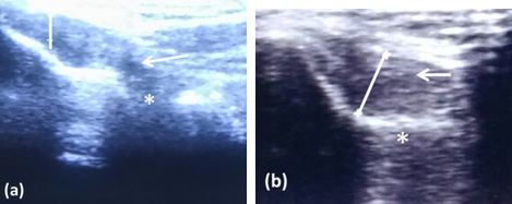
Figure 1: Longitudinal USG of patient of tennis elbow pre-injection (a) showing focal hypoechoic (white arrow), mild tear at origin(asterix), fraying & thinning of the common extensor tendon. 6 months post PRP injection (b) USG showing reduced focal hypoechoic with increased in thickness of tendon (5.6mm) and healin in the tear at the origin.
ojmpc_73_01.jpg

Result

30 patients with mean age 39.3 years (range 25 to 58) of tennis elbow were included in the study. Out of these 22 (73%) were female and 8 (27%) were male.24 (80%) of the patients of tennis elbow had complains in right elbow with right to left ratio of 4:1.Most of our patients had right dominance i.e. 23(76%) patients.

None of the cases had bilateral involvement. The mean duration of symptoms was 7.2 months (range 5 to 13 months). The mean pain VAS Score improved from 7.7 before injection to 5.4 after 2 weeks, 4.1 after 6 weeks, 3.2 at 12 weeks of injection and 1.8 at final follow up i.e. after 6 months post injection, respectively.

At the initial presentation, out of the 30 patients, 19 patients had severe pain whereas 11 patients had moderate pain before injection, which improved to only 3 patients having moderate pain and none of the patients having severe pain at elbow at final follow up after the injection. 27 patients had either no or mild pain only at the lateral elbow (table 1).

Very severe to severe tenderness at the lateral epicondyle of elbow prior to injection was seen in 28 (93%) patients, which improved to, none of the patients having severe or very severe tenderness and 28 (93%) patients having either no or only mild tenderness at the lateral elbow at final follow-up (fig 1a & b).

The functional outcome as assessed by the Oxford elbow score improved from a mean of 19.2 prior to treatment to 41.3after the injection at final follow. Pre injection elbow score was worst to severe in 28 patients, whereas, at final follow up 23 patients had elbow score as normal or mild i.e. between 40 to 48 and 6 patients had oxford elbow score between 30 to 39.

Prior to injection 6 patients had restriction in the forearm rotation less than 160 degree. At final follow-up all the patients had normal range of motion.

Prior to injection, ultrasonography of the involved elbow showed tear at the common extensor origin in 18, oedema in 14, cortical erosion in 24, calcification in 4, thinning and fraying of the common extensor tendon in 6 and probe induced tenderness in 29 patient(fig 1). Focal hypoechoic in the deep part of common extensor tendon was seen in 26 patients before injection, whereas only 6 patients showed these focal hypoechoic in deep common extensor tendon after 6 months of the injection, indicating the evidence of healing in the tendon. At final follow up only 7 patients had ultrasonographic evidence of tear at common extensor origin, oedema in 2 patients, cortical erosion in 24, 2 patients had thinning and fraying of common extensor origin, and probe induced tenderness was seen in only 6 patients (table 1).

Two patients came up with complaints of post injection pain and swelling at injection site at one week after the injection and one patient came up with fever, both of these complaints were relieved by analgesics and antibiotics medications respectively. None of the patients showed any other complication, like infection, neurovascular change or worsening of epicondylar pain.

Table 1: Results of the patients of tennis elbow after autologous PRP injection

ParameterGradingPre-injectionPost – injection
2 week6 week12 week6 month
Pain (VAS score)No (0)00128
Mild (1-2)02112019
Moderate (3-6)11261883
Severe (7-10)192000
Mean VAS score7.75.44.13.21.8
TendernessNo or mild03122528
moderate281642
Severe2218210
Very severe61000
Oxford elbow score0-19(worst)194110
20-29(severe)915821
30-39 (moderate)21016166
40-48 (mild to
normal)
0151123
MEAN OES19.228.632.439.641.3
USG of common extensorTear18---7
Oedema14---2
Thinning and fraying6---2
Probe induced tenderness29---6
Cortical erosion24---24
Calcification4---4
Focal hypo-echoic26---6
Range of motion900-110010000
1100-130052000
1300-15002428303030

Discussion

Lateral elbow epicondylar tendinosis or tennis elbow (TE) is very common condition among persons performing activities involving strong gripping and repetitive wrist extension [1,2]. Lateral epicondylitis is usually a misnomer because microscopic evaluation of tendon does not show signs of inflammation.

It has been well proven histologically that lateral epicondylitis or tennis elbow is not an acute inflammatory pathology, but instead it is failure of the normal tendon repair mechanism along with angiofibroblastic degeneration because tendons are relatively hypovascular. This hypovascularity may lead to hypoxic tendon degeneration which is main aetiology of tendinosis [3,24,25].

The traditional methods to treat tennis elbow, including rest, anti-inflammatory medications, bracing, physical therapy, ionotophoresis, extra corporal shockwave, botulinum toxin, and corticosteroid injection, do not alter the tendon’s poor healing properties secondary to poor vascularization of tendon, which is the basic pathophysiology in tennis elbow [1-3].

Hence these methods have shown inconsistent outcome. Autologous biological blood-derived product PRP releases high concentrations of platelet derived growth factors on injection which enhance tendon healing due to its effects on angiogenesis and collagen synthesis.

Various growth factors and cytokines in PRP include Platelet Derived Growth factors (PDGF-aa, PDGF-bb, PDGF-ab),Vascular Endothelial Growth Factor (VEGF),Fibroblast growth factor (FGF), Epidermal Growth Factor (EGF), Transforming Growth Factor beta (TGF-b1, TGF-b2), Insulin Like Growth Factor-1 and 2 (IGF-1, IGF-2), Interleukin – 8 (IL-8), Keratinocyte Growth Factor, Connective Tissue growth factor. The role of PRP in bone, wound and tendon healing is well established [27-29].

Recent literature is saturated with articles on the efficacy of PRP in treatment of tennis elbow. Various studies by Mishra et al, Hecthman et al, Tan et al, Palacio et al have already confirmed the efficacy of the PRP in treatment of tennis elbow [8-13].

Several studies have even compared the efficacy of PRP with corticosteroid injection or with whole blood injection like Gosen et al, Peerbooms et al, Krogh et al, Lediedzinski et al, Raeissadat et al, and Thanasas et al [13-22].

All of studies confirmed the efficacy of PRP injection and showed the advantage of PRP over the corticosteroid injection or whole blood.

But all of the studies have measured the results in terms of the pain relief as assessed on the VAS score or as assessed on the improved in functional elbow scores like Mayo, Oxford, Nirschl score etc.

But both these parameter pain as well as the functional elbow score are subjective in nature and lack to give objective evidence of healing of the tendon and are thus subject to bias. Ultrasonography of the tendon enables to visualize the tendon structures around the elbow [23,24].

Thus changes in the USG finding of the common extensor origin before the injection and after the injection can give the documented evidence of the improvement in the pathology of the tendon. Hence we tried to document the efficacy of the local autologous PRP injection in treatment of tennis elbow by pain relief as assessed by VAS score, improvement in functional elbow score as assessed by Oxford elbow score and to objectively document the improvement by seeing the changes in USG findings in the common extensor morphology so that we can get an objective evidence of the healing and improvement.

Our results subjectively as well as objectively confirmed the efficacy of the autologous PRP injection in the treatment of relcalcitrant tennis elbow as there is improvement in VAS score, Oxford elbow functional score as well as improvement seen in the sonographic appearance of the morphology of the common extensor tendon origin after the local autologous PRP injection as seen as decrease in focal hypoechoic, decreased edema, improvement in thickness of the tendon and healing of the tear at the origin site.

Thus this study confirms that local PRP by supplying growth factors helps to enhance the stromal and mesenchymal stem cell proliferation and prevents fibrous scarring of the tendon. This increase in tendon vascularity by PRP prevents angiofibroblastic degeneration due to hypovascularity of the tendon, which is the main pathophysiology in tennis elbow.

This locally injected PRP has led to improved tendon repair and healing property by releasing growth factors and increasing vascularity, which can be documented by improved tendon morphology. This study is limited by lack of randomized group, a relatively smaller sample size and short follow up period.

Conclusion

PRP is an effective mode of treatment for recalcitrant tennis elbow, as it has tendency to enhance the healing potential of the hypovascular tendon by releasing high concentration of growth factors and this improvement has been documented subjectively by improvement in elbow score and objectively by improved tendon morphology on ultrasonography.


References

1. Ahmad Z, Siddiqui N, Malik SS, Abdus-Samee M, Tytherleigh-Strong G, Rushton N. Lateral epicondylitis: a review of pathology and management. Bone Joint J. 2013 Sep;95-B(9):1158-64. [Crossref][PubMed][Google Scholar]

2. Behrens SB, Deren ME, Matson AP, Bruce B, Green A. A review of modern management of lateral epicondylitis. Phys Sports Med. 2012 May;40(2):34-40. [Crossref][PubMed][Google Scholar]

3. Smith RW, Papadopolous E, Mani R, Cawley MI. Abnormal microvascular responses in a lateral epicondylitis. Br J Rheumatol. 1994;33(12);1166-68. [Crossref][PubMed][Google Scholar]

4. Harmon K, Drezner J, Rao A. Platelet rich plasma for chronic tendinopathy. Br J Sports Med 2013;47:2. . [Crossref][PubMed][Google Scholar]

5. Foster TE, Puskas BL, Mandelbaum BR, Gerhardt MB, Rodeo SA. Platelet-rich plasma: from basic science to clinical applications. Am J Sports Med. 2009 Nov;37(11):2259-72. [Crossref][PubMed][Google Scholar]

6. Everts PA, Overdevest EP, Jakimowicz JJ, Oosterbos CJ, Schönberger JP, Knape JT, Van Zundert A. The use of autologous platelet-leukocyte gels to enhance the healing process in surgery - A review. Surg Endosc. 2007 Nov;21(11):2063-8. [Crossref][PubMed][Google Scholar]

7. Kaux JF, Crielaard JM. Platelet-rich plasma application in the management of chronic tendinopathies. Acta Orthop Belg. 2013 Feb;79(1):10-5. [Crossref][PubMed][Google Scholar]

8. Mishra A, Pavelko T. Treatment of chronic elbow tendinosis with buffered platelet-rich plasma. Am J sports Med. 2006;34(11);1774-8. [Crossref][PubMed][Google Scholar]

9. Hechtman KS, Uribe JW, Botto-van Demden A, Kiebzak GM. Platelet-rich plasma injection reduces pain in patients with recalcitrant epicondylitis. Orthop. 2011;34:92. [Crossref][PubMed][Google Scholar]

10. Tan X, Ju H, Yan W, et al. Autologous platelet lysate local injections for the treatment of refractory lateral epicondylitis. J Orthop Surg Res. 2016;11:17. [Crossref][PubMed][Google Scholar]

11. Palacio EP, Schiavetti RR, Kanematsu M, Ikeda TM, Mizobuchi RR, Galbiatti JA. Effects of platelet-rich plasma on lateral epicondylitis of the elbow: prospective randomized controlled trial. Revista Brasileira de Ortopedia. 2016;51(1):90-95. [Crossref][PubMed][Google Scholar]

12. Lhee SH, Park JY. Prospective randomized clinical study for the treatment of lateral epicondylitis: comparison among PRP (platelet-rich plasm), prolotherapy, physiotherapy and ESWT. J Shoulder Elb Surg. 2013;22(10);30-31. [Crossref][PubMed][Google Scholar]

13. Ahmad Z, Brooks R, Kang SN, Weaver H, Nunney I, Tytherleigh-Strong G, Rushton N. The effect of platelet-rich plasma on clinical outcomes in lateral epicondylitis. Arthroscopy. 2013;29:1851–1862. [Crossref][PubMed][Google Scholar]

14. Gosens T, Peerbooms JC, van Laar W, den Oudsten BL. Ongoing positive effect of platelet-rich plasma versus corticosteroid injection in lateral epicondylitis: a double-blind randomized controlled trial with 2-year follow-up. Am J Sports Med 2011;39:1200–8. . [Crossref][PubMed][Google Scholar]

15. Peerbooms JC, Sluimer J, Bruijn DJ, Gosens T. Positive effect of an autologous platelet concentrate in lateral epicondylitis in a double-blind randomized controlled trial: platelet-rich plasma versus corticosteroid injection with a 1-year follow-up. Am J Sports Med 2010;38:255–62. . [Crossref][PubMed][Google Scholar]

16. Krogh TP, Fredberg U, Stengaard-Pedersen K, Christensen R, Jensen P, Ellingsen T. Treatment of lateral epicondylitis with platelet-rich plasma, glucocorticoid, or saline: a randomized, double-blind, placebo-controlled trial. Am J Sports Med 2013;41:625–35. . [Crossref][PubMed][Google Scholar]

17. Lebiedziński, R. , Synder, M. , Buchcic, P. et al. A randomized study of autologous conditioned plasma and steroid injections in the treatment of lateral epicondylitis. Int Orthop (SICOT) (2015) 39: 2199 [Crossref][PubMed][Google Scholar]

18. Raeissadat SA, Sedighipour L, Rayegani SM, Bahrami MH, Bayat M, Rahimi R. Effect of Platelet-Rich Plasma (PRP) versus Autologous Whole Blood on Pain and Function Improvement in Tennis Elbow: A Randomized Clinical Trial. Pain Res Treat. 2014;19:52-5. [Crossref][PubMed][Google Scholar]


19. Raeissadat SA, Rayegani SM, Hassanabadi H, Rahimi R, Sedighipour L, Rostami K. Is Platelet-rich plasma superior to whole blood in the management of chronic tennis elbow: one year randomized clinical trial. BMC Sports Sci Med Rehabil. 2014, Mar 18;6:12. [Crossref][PubMed][Google Scholar]

20. Thanasas C, Papadimitriou G, Charalambidis C, Paraskevopoulos I, Papanikolaou A: Platelet-rich plasma versus autologous whole blood for the treatment of chronic lateral elbow epicondylitis. Am J Sports Med. 2011;39: 2130-2134. . [Crossref][PubMed][Google Scholar]

21. Creaney L, Wallace A, Curtis M, Connell D. Growth factor-based therapies provide additional benefit beyond physical therapy in resistant elbow tendinopathy: a prospective, single-blind, randomised trial of autologous blood injections versus platelet-rich plasma injections. Br J Sports Med. 2011;45(12):966–971. [Crossref][PubMed][Google Scholar]

22. Rabago D, Best TM, Zgierska AE, Zeisig E, Ryan M, Crane D. A systematic review of four injection therapies for lateral epicondylosis: prolotherapy, polidocanol, whole blood and platelet-rich plasma. Br J Sports Med. 2009;43(7):471–481. [Crossref][PubMed][Google Scholar]

23. Connell D, Burke F, Coombes P, McNealy S, Freeman D, Pryde D, et al. Sonographic examination of lateral epicondylitis. AJR Am J Roentgenol 2001;176:777–82. . [Crossref][PubMed][Google Scholar]

24. Levin D, Nazarian LN, Miller TT, O’Kane PL, Feld RI, Parker L, et al. Lateral epicondylitis of the elbow: US findings. Radiology 2005;237:230–4. . [Crossref][PubMed][Google Scholar]

Disclaimer / Publisher's Note

The statements, opinions and data contained in all publications are solely those of the individual author(s) and contributor(s) and not of Journals and/or the editor(s). Journals and/or the editor(s) disclaim responsibility for any injury to people or property resulting from any ideas, methods, instructions or products referred to in the content.