Orthopaedic Journal of MP Chapter

Publisher: Madhya Pradesh Orthopaedic Association www.mpioa.com
E-ISSN:2582-7243, P-ISSN:2320-6993
2019 Volume 25 Number 1 Jan-Jun

Hypovitaminosis D In Elderly Patients Presenting With Fractures

Shan Z1, Alvi Y2*, Ahmad S3, Jilani L4, Faizan M5, Asif N6

1 ZH Shan, Department of Orthopaedics, Jawaharlal Nehru Medical College and Hospital Aligarh Muslim University AMU, Aligarh, UP, India.

2* Yasir Alvi, Senior Resident, Department of Orthopaedics, Jawaharlal Nehru Medical College and Hospital Aligarh Muslim University AMU, Aligarh, UP, India.

3 S Ahmad, Department of Orthopaedics, Jawaharlal Nehru Medical College and Hospital Aligarh Muslim University AMU, Aligarh, UP, India.

4 LZ Jilani, Department of Orthopaedics, Jawaharlal Nehru Medical College and Hospital Aligarh Muslim University AMU, Aligarh, UP, India.

5 M Faizan, Department of Orthopaedics, Jawaharlal Nehru Medical College and Hospital Aligarh Muslim University AMU, Aligarh, UP, India.

6 N Asif, Department of Orthopaedics, Jawaharlal Nehru Medical College and Hospital Aligarh Muslim University AMU, Aligarh, UP, India.

Background: Vitamin D deficiency is seen in all races, age groups and ethnic backgrounds. It is estimated to affect more than one billion people worldwide. This study was done to find out vitamin D levels and its associated factors among elderly patients presenting with fractures.

Material and method: This cross-sectional study was performed at our tertiary care hospital, in patients above 45 years, presenting with fracture as a result of trivial trauma. In all patients serum 25(OH)D level was measured. A level of serum 25(OH)D < 10 ng/ml was labeled as the deficiency, between 10 to 29 ng/ml as insufficient and ≥ 30 ng/ml as sufficient.

Results: A total of 102 patients were included in our study with mean age of 60.8 ± 13.4 years, out of which 60 were males and 42 females. Mean serum 25(OH)D level in our study population was 15.82 ± 5.88 ng/ml. We found 94.1% of the patients were having a low level of serum 25(OH)D i.e. less than 30 ng/ml, with 78.4% insufficient (between 10 to 29 ng/ml) and 15.6% deficient levels (below 10 ng/ml). Higher age, female, menopause and lack of sunlight exposure were found to be significantly associated with lower vitamin D levels.

Conclusion: This study gives us important inside of a high prevalence of hypovitaminosis D in elderly patients. Detection and prompt intervention of vitamin D deficiency at an early stage can be helpful in decreasing the fracture in these elderly.

Keywords: Vitamin D deficiency, Serum 25(OH)D, Hypovitaminosis D, Ageing

Corresponding Author How to Cite this Article To Browse
Yasir Alvi, Senior Resident, Department of Orthopaedics, Jawaharlal Nehru Medical College and Hospital Aligarh Muslim University AMU, Aligarh, UP, India.
Email:
Shan Z, Alvi Y, Ahmad S, Jilani L, Faizan M, Asif N, Hypovitaminosis D In Elderly Patients Presenting With Fractures. ojmpc. 2019;25(1):17-22.
Available From
https://ojmpc.com/index.php/ojmpc/article/view/72
Manuscript Received Review Round 1 Review Round 2 Review Round 3 Accepted
2019-06-06 2019-06-12 2019-06-18 2019-06-24 2019-06-30
Conflict of Interest Funding Ethical Approval Plagiarism X-checker Note
Authors state no conflict of interest. Non Funded. The conducted research is not related to either human or animals use. 13.32 All authors have accepted responsibility for the entire content of this manuscript and approved its submission.

© 2019by Shan Z, Alvi Y, Ahmad S, Jilani L, Faizan M, Asif Nand Published by Madhya Pradesh Orthopaedic Association. This is an Open Access article licensed under a Creative Commons Attribution 4.0 International License https://creativecommons.org/licenses/by-nc/4.0/ unported [CC BY NC 4.0].

Introduction

Vitamin D is very important for the musculoskeletal health as well as overall wellbeing. Apart from rickets in children and osteomalacia in adults, vitamin D deficiency has been associated with pain and muscle weakness [1,2].

Inadequate serum vitamin D is also associated with secondary hyperparathyroidism leading to increased bone turnover and bone loss, resulting in increased risk for fractures especially in the elderly and postmenopausal women, even with minimal trauma [2-4]. Studies have documented the benefits of vitamin D supplementation in fracture reduction [5].

Sources of vitamin D in human body include diet and sunlight. Vegetarian diet lacks in fish and animal protein, which are important source of vitamin D. Further, food product available in market unlike western countries, are not fortified with vitamin D or even calcium. Few food toxins like phytates which are common in vegetarian diet make this situation more complicated by preventing the absorption of calcium from gut and further misbalancing the vitamin D-calcium homeostasis in body [6].

The study among Caucasian population have proposed a 10 minutes exposure of sunlight over, head and arms three times per week would be adequate to prevent vitamin D deficiency [4].

But same cannot be said in reference to Indian population where most of people are either wheatish or brown-black, having more melanin, preventing UV rays from sunlight to react with vitamin D precursor in skin. Studies in Indian settings have documented the need for larger contact period that too daily, for the successful reaction [7].

Due to these reasons, Vitamin D deficiency is quite prevalent in Indian subcontinent, with a prevalence of 66%–91% among various populations [8-10]. Elderly citizens are especially at higher risk of vitamin D deficiency primarily because skin and gut are now less efficient in producing and absorbing vitamin D and immobility among them decrease the sunlight exposure [4].

While the handfuls of studies are done on postmenopausal women with fractures, very few have studied the role of vitamin D among elderly. Given the importance of vitamin D in overall wellbeing as well as deficiency leading to fracture, data enlightening the prevalence of vitamin D deficiency in elderly patients may be of great value.

Therefore we conducted this study to find out the prevalence of hypovitaminosis D and related factor among elderly patients sustaining fractures with trivial injury.

Material and Methods

This cross-sectional study was conducted over a period of 12 month duration from January to December 2015 at our center after approval from Institutional Ethics and Research Advisory Committee and informed consent from each participant. Our city is a district place in north India, which receives daily sunlight for not less than 10 hours throughout the year and natives of our place have skin complexion of wheatish to dark in color. Most of our patients belong to rural populations involved in agriculture, who are exposed to long duration of sunlight, that too with minimal clothing [11].

All patients with more than 45 years age presenting with fragility fracture to hip and wrist caused by trivial trauma were included in the study. Patients with deranged kidney function or renal calculi, abnormal thyroid function, liver disease, history of esophageal disease, peptic ulcer or cancer or taking long-term therapy like estrogen replacement therapy/corticosteroids/anticonvulsants/anticoagulant/glucocorticoid were excluded from the study.

All study subjects were admitted in orthopedic ward, with detailed history followed by physical examination. A pre-designed questionnaire covering various risk factors was used to collect data from these subjects. For estimation of vitamin D levels, we measured pro-hormone 25-hydroxyvitamin D [25(OH)D], rather than the measurement of serum 1,25(OH)2D concentrations, as former is shown to related with vitamin D deficiency.

For this Vitamin D estimation, 4ml blood sample of the subject was collected in ice-cooled syringes and maintaining proper cold chain was send to endocrinology lab earliest. At endocrinology lab, quantitative determination of total serum 25(OH)D was done by direct competitive chemi-luminescence immunoassay (CLIA) methodology from serum after centrifugation at 1200 rpm for 10 minutes. For our study, Vitamin D less than < 10 ng/mL, between 10–29 ng/mL and more than 30 ng/ml was considered deficiency, insufficiency and sufficient respectively [12,13].

Statistical analysis was performed using Statistical Package for Social Sciences (SPSS) Version 20. The categorical variables were represented by percentage (%) along with 95% confidence interval and continuous variables were expressed as mean with standard deviation.

The statistical significance was tested by unpaired t-tests for independent as well as Analysis of Variance (ANOVA), wherever applicable with Bonferroni test for post-hoc assessments to identify the groups that are homogenous with respect to mean. All tests were two tailed and a p value of ≤ 0.05 was considered significant.


Results

A total of 102 patients were included in the study, out of which 60 patients (58.8%) were male while rest 42 (41.2%) females. Overall mean age was 60.8 ± 13.4 years (range 45 to 92 years), with mean age of male being 60.1 ± 13.7 years while that of female was 61.6 ± 13.1 years. Among them 54.9% were urban residents while 45.1 % were living in rural area. Majority of population were married (83.3%) and Muslim by religion (59.8%). None of the participants reported use of sunscreen ever.

34 patients presented with intertrochanteric fracture, 66 with fracture neck of femur while 2 patients presented with distal radius fracture. None of the patients had been investigated for vitamin D level or received treatment for osteoporosis in past.The mean level of serum 25(OH)D was 15.82 ± 5.88 ng/ml.

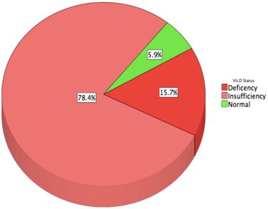
Figure 1: Patients as per to Vitamin D level
ojmpc_72_01.jpg

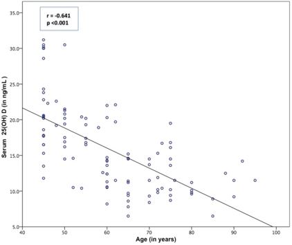
Figure 2: Age Vs Vitamin D levels
ojmpc_72_02.jpg

Out of 102 cases included in the study 6 (5.8%) patients were in sufficient range (>30 ng/mL), while rest 94.2% were classified as either deficient or insufficient level. Eight patients (78.4%) were insufficient (between 10-30 ng/mL) and 16 patients (15.6%) patient were deficient in serum 25(OH)D (< 10ng/mL) (Table 1 & Figure 1).

When compared the age in groups, the means age in deficient group (72.9±8.9 year) was higher than insufficient group (59.4±12.9 year) and normal group (45.8±2.0 year), and this was statistically significant (F = 13.352, df 2, p <0.001). On Post Hoc analysis using Bonferroni test, a significant difference was found between means of age with deficient group and insufficient group (mean difference = 13.5, p <0.001), deficient group and normal group (mean difference=27.1, p <0.001) as well as insufficient group and normal group (mean difference=13.6, p = 0.026). Thus higher age was significantly associated with vitamin D deficiency as well as insufficiency levels (Fig. 2).

Means of vitamin D level in males (17.43 ±6.39) was higher as compared to females (13.50 ± 4.14) and this was significant when we analyzed by independent t test (p<0.001). Females who had not attained menopause were having significantly higher means of vitamin D level (17.41±3.22) then those who had attained menopause (12.58±3.81, p value-0.002). When we compared religion and residence of patients with the vitamin D levels, we did not found significant relationship between means of them. Although patients who gave history of daily sunlight of 15 min or more were having significantly higher levels of vitamin D levels (16.45±6.06) in comparison to those who did not (12.60±3.55) (p value - 0.001) (Table 2).

Discussion

Vitamin D plays important role in calcium metabolism. Indians owing to cultural, religious and ethnic variation from western world are more prone to low level of vitamin D [2-4]. Most of the epidemiological researches of level of Vitamin D in fracture patients are from western countries. Hence we studied vitamin D levels and its associated factors among elderly patients presenting with fractures in 102 Indian patients and observed very high prevalence (94.1%) of hypovitaminosis D, with prevalence of insufficiency in 78.4% and deficiency in 15.6% of cases.

Table 1: Status of Vitamin D levels in study population.

Serum Vitamin D statusFrequencyPercentage95% CI
Deficiency (<10 ng/mL) 1615.79.8 – 24.1
Insufficiency(10–29 ng/mL) 8078.469.4 – 85.4
Sufficiency (30–100 ng/mL) 65.92.5 – 12.5
<20 ng/mL*7775.566.3 – 82.7
<12 ng/mL#3534.325.8 – 44.0

Table 2: Association of Vitamin D levels with various variables

VariablesFreq.Vitamin D leveltdfp value
MeanSD
SexMale6017.436.393.76699.5<0.001
Female4213.504.14
MenopauseNot attained817.413.223.311400.002
Attained3412.583.81
ReligionHindu4116.844.871.46198.50.126
Muslim6115.116.42
ResidenceRural4616.435.600.9621000.338
Urban5615.306.11
Sun exposure (<15 min/day)Inadequate1712.603.55-3.55037.60.001
Adequate8516.456.06

While there is no generalize consensus on optimal levels of serum 25(OH)D for deficiency and insufficiency, most researchers considered more than 30 ng/ml as sufficient level [6,7,12]. Lips et at has classified vitamin D deficiency at level of less than 20 ng/ml among Caucasian, but at the same time stressed that cutoff for these levels should be based on population studies and level which triggers secondary hyperparathyroidism [4,13].

Studies among Indian patients with hip fracture found prevalence of vitamin D deficiency (taken by them as <20ng/ml) at 76.7% in Delhi and 74% in South India[14,15]. If we take similar cut-off point in our study, 75.5% of patients had 25(OH)D levels less than 20ng/ml.

International studies assessing prevalence of vitamin D deficiency according to National Osteoporosis Society criteria (< 12 ng/mL), found it as 50% among US postmenopausal women with hip fracture and 78% among German vertebral fracture patients (compared to 34.3% in this study with same criteria) [16,17].

Another study done in US, among Caucasian presenting with minimal trauma fracture, documented similar 97.4% prevalence of hypovitaminosis D (<30 ng/ml) [18]. Although there is no universal consensus to what constitutes subnormal concentrations of vitamin D, most of literature shows a higher prevalence of hypovitaminosis D in fracture patients, because it is major reason of weak bones leading trivial trauma to cause fracture.

Studies have shown that when serum 25(OH)D levels fall below 30 ng/mL, calcium absorption decreases and it leads to secondary hyperparathyroidism, resulting in resorption of the calcium from bone in order to maintain serum calcium within normal range [7]. This secondary hyperparathyroidism may lead to weak and fragile bone, and further to fracture by trivial trauma. This is the same reason why studies have recognized serum 25(OH) D < 30 ng/ml be used as the cut off value for vitamin D insufficiency [4,7,19,20].

We observed the low levels of vitamin D to be significantly associated with advancing age, females, especially those who have attained menopause, and among inadequate sun exposure. Previous studies have documented elderly and postmenopausal women as risk factor for fracture [4,5] similar to our study but prior studies had conflicting results in regards to advancing age. Although higher prevalence of vitamin D deficiency among elderly and with each decade has been demonstrated by Bhat et al and Arya et al [21,22], but Marwaha et al and Narang et al in their study among health adults did not found any relation with advancing age [8,23]. Although few researches had found hypovitaminosis D irrespective of gender, majority are in notion of this being commoner among menopausal females [8-23]. Female gender, especially from north Indian population, by virtue of various customs and culture, often stay at home with minimal chances of sun exposure, and whenever they go outside, are fully or maximally covered, decreasing the chance of sunlight exposure.

Application of sunscreen, which is common in female, reduces the chances of adequate UV rays penetration in skin.

Studies have proposed minimum duration of daily 30 minutes of sun exposure is required for adequate production of vitamin D [7,15,23], although in our study this was even lesser. We observed daily exposure of 15 min of sunlight is significantly associated with higher level of serum vitamin D.

We did not observe residence and religion to have a significant effect on serum vitamin D level in our study population. Both rural and urban population as well as Hindu and Muslim, had almost similar mean of serum vitamin D level as of the mean study group, although all were in insufficient range, confirming the widely prevalent of vitamin D deficiency among various socioeconomic groups, different ethnicities, rural and urban areas as well as different professions as other have measured [7]. Therefore all older people irrespective of their ethnicity, residence or professions, need to be monitored for hypovitaminosis D and Vitamin D supplements to be given to them, which will help in resisting fracture, not only by strengthen the bone to survive trivial fall but also in averting the chances of fall by strengthening the proximal muscle [19,20].

Conclusion

With such high percentage of prevalence of vitamin D deficiency/insufficiency in elderly fracture population, we should thought toward it prevention to prevent the occurrence of fracture. Oral vitamin D supplement should be advocated as first line defense to all. Along with being fit and healthy, promoting outdoor activities would serve as a good public health measure in increasing the serum vitamin D levels.


References

1. Skaria J, Katiyar BC, Srivastava TP, Dube B. Myopathy and neuropathy associated with osteomalacia. Acta Neurol Scand. 1975;51:37–58. [Crossref][PubMed][Google Scholar]

2. Reginster JY. The high prevalence of inadequate serum vitamin D levels and implications for bone health. Curr Med Res Opin. 2005;21:579–85. [Crossref][PubMed][Google Scholar]

3. Harinarayan CV, Ramalakshmi T, Venkataprasad U. High prevalence of low dietary calcium and low vitamin D status in healthy south Indians. Asia-Pac J Clin Nutr. 2004;13:359–64. [Crossref][PubMed][Google Scholar]

4. Lips P. Vitamin D deficiency and secondary hyperparathyroidism in the elderly: consequences for bone loss and fractures and therapeutic implications. Endocr Rev. 2001;22:477–501. [Crossref][PubMed][Google Scholar]

5. Bischoff-Ferrari HA, Willett WC, Wong JB, Stuck AE, Staehelin HB, Orav EJ, et al. Prevention of nonvertebral fractures with oral vitamin D and dose dependency. Arch Int Med. 2009;169:551. [Crossref][PubMed][Google Scholar]

6. Ritu G, Gupta A. Vitamin D deficiency in India: prevalence, causalities and interventions. Nutr. 2014;6:729–75. [Crossref][PubMed][Google Scholar]

7. Harinarayan CV, Joshi SR. Vitamin D status in India - its implications and remedial measures. J Assoc Physicians India. 2009;57:40–8. [Crossref][PubMed][Google Scholar]

8. Marwaha RK, Tandon N, Garg MK, Kanwar R, Narang A, Sastry A, et al. Vitamin D status in healthy Indians aged 50 years and above. J Assoc Physician India. 2011;59:706–9. [Crossref][PubMed][Google Scholar]

9. Harinarayan CV, Sachan A, Reddy PA, Satish KM, Prasad U V, Srivani P. Vitamin D status and bone mineral density in women of reproductive and postmenopausal age groups: a cross-sectional study from south India. J Assoc Physicians India. 2011;59:698–704. [Crossref][PubMed][Google Scholar]

10. Sachan A, Gupta R, Das V, Agarwal A, Awasthi PK, Bhatia V. High prevalence of vitamin D deficiency among pregnant women and their newborns in northern India. Am J Clin Nutr. 2005;81:1060–4. [Crossref][PubMed][Google Scholar]

11. Economist T. Comparing Indian states and territories with countries: An Indian summary. The Economist 2011. http://www. economist.com/content/indian-summary (accessed March 8, 2018) [Crossref][PubMed][Google Scholar]

12. Looned K, Banerjee A, Landge J, Pandit D. Intergenerational decline in Vitamin D status: A cross-sectional study among medical students and their teachers. Int J Nutr Pharmacol Neurol Dis. 2017;7:12. [Crossref][PubMed][Google Scholar]

13. Thacher TD, Clarke BL. Vitamin D insufficiency. Mayo Clin Proc. 2011;86:50–60. [Crossref][PubMed][Google Scholar]

14. Dhanwal DK, Sahoo S, Gautam VK, Saha R. Hip fracture patients in India have vitamin D deficiency and secondary hyperparathyroidism. Osteoporos Int. 2013;24:553–7. [Crossref][PubMed][Google Scholar]

15. Paul TV, Selvan SA, Asha HS, Thomas N, Venkatesh K, Oommen AT, et al. Hypovitaminosis D and other risk factors of femoral neck fracture in South Indian postmenopausal women: a pilot study. J Clin Diagn Res 2015;9:19-22. . [Crossref][PubMed][Google Scholar]

16. LeBoff MS, Kohlmeier L, Hurwitz S, Franklin J, Wright J, Glowacki J. Occult vitamin D deficiency in postmenopausal US women with acute hip fracture. JAMA. 1999;281:1505–11. [Crossref][PubMed][Google Scholar]

17. Maier GS, Seeger JB, Horas K, Roth KE, Kurth AA, Maus U. The prevalence of vitamin D deficiency in patients with vertebral fragility fractures. Bone Joint J. 2015;97–B:89–93. [Crossref][PubMed][Google Scholar]

18. Simonelli C, Weiss TW, Morancey J, Swanson L, Chen YT. Prevalence of vitamin D inadequacy in a minimal trauma fracture population. Curr Med Res Opin. 2005;21:1069–74. [Crossref][PubMed][Google Scholar]

19. Bischoff HA, Stähelin HB, Dick W, Akos R, Knecht M, Salis C, et al. Effects of Vitamin D and calcium supplementation on falls: a randomized controlled trial. J Bone Mineral Res 2003;18:343–51. . [Crossref][PubMed][Google Scholar]

20. Trivedi DP, Doll R, Khaw KT. Effect of four monthly oral vitamin D3 (cholecalciferol) supplementation on fractures and mortality in men and women living in the community: randomised double blind controlled trial. BMJ (Clin Res Ed) 2003;326:469. . [Crossref][PubMed][Google Scholar]

21. Bhat KA, Kakaji M, Awasthi A, Shukla M, Dubey M, Srivastava R, et al. High prevalence of osteoporosis and morphometric vertebral fractures in indian males aged 60 years and above: should age for screening be lowered? J Clin Densitom. 2018;21(4):517-23. . [Crossref][PubMed][Google Scholar]

22. Arya V, Bhambri R, Godbole MM, Mithal A. Vitamin D status and its relationship with bone mineral density in healthy Asian Indians. Osteoporos Int. 2004. 15:56–61 [Crossref][PubMed][Google Scholar]


23. Narang APS, Batra S, Sabharwal S, Ahuja SC. 1, 25-Dihydroxycholecalciferol (1,25-(OH)(2) D(3)) levels in osteoporosis. Ind J Clin Biochem 2004. 19:111–3. [Crossref][PubMed][Google Scholar]

Disclaimer / Publisher's Note

The statements, opinions and data contained in all publications are solely those of the individual author(s) and contributor(s) and not of Journals and/or the editor(s). Journals and/or the editor(s) disclaim responsibility for any injury to people or property resulting from any ideas, methods, instructions or products referred to in the content.