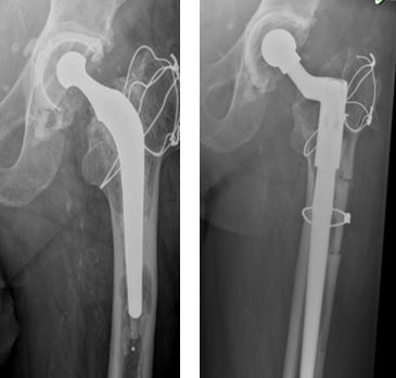
8 patients (62 %) had revision because of aseptic loosening, 2 due to infection and 3 patients (23%) had fracture as the indication for revision surgery. Almost all patients (12 patients) of cortical window for cemented stem extraction had cemented femoral component except one who needed cortical window for removal of uncemented implant. 12 patients out of 13 had revision of both femoral and acetabular component and 1 patient had revision of only the femoral stem.
As per the operative notes, 11 femoral stems (85%) were extracted very easily after making a cortical window while 2 femoral stem implant removal was done with difficulty. In 9 patients, window was made to remove either cement or cement restrictor, whereas in 3 cases window was made to remove the broken part of femoral stem.
Revision THA was done with “Reclaim” (Depuy) prosthesis in 10 patients, whereas Wagner type uncemented long modular revision prosthesis (Depuy), “Reef” prosthesis which is distally interlocked modular revision femoral stem (Depuy) and cemented “C Stem which is triple taper polished femoral stem (Depuy) was used in one case each.
In almost all the cases (12 cases) size of cortical window was 2.5 cm x 5 cm, whereas in one case the size was slightly bigger window i.e. 7.5 x 2.5 cm. In 10 cases only metal cable wires were used to fix back the cortical cover of the window. In 3 cases additional plates were also used to increase the stability of a pre-existing fracture as these cases of peri-prosthetic fractures. None of the cases needed bone grafting except in one case with longer cortical window, in which reaming material obtained were used as bone graft around cortical window.
Full radiological healing of cortical window was seen in 9 cases in less than 3 months whereas 2 cases took upto 18 months to heal. There was insufficient follow up available for 2 cases to comment on healing. In one 1 case of subsidence was found on initial x rays which was stable as seen in subsequent follow ups.
Initially at 6 weeks, 2 patients were pain free and 11 patients had mild pain whereas one had moderate pain. No patients reported severe pain after Revision Total Hip Replacement. At 6 months 7 (54%) patients were pain free and 5 (38 %) of patients had mild pain. All patients had good mobility with able to do all daily routine work comfortably.
Discussion
Although Total hip arthroplasty is provides excellent short and long term outcomes, but like all procedures, it is not free of complications [3-5]. Complications arising out of the primary total hip arthroplasty may demand a revision surgery with removal or exchange of previous components. Reasons which need revision total hip arthroplaty are usually aseptic loosening, infection, peri-prosthetic fracture, recurrent dislocation and mal-positioning of components [6-9]. The primary and crucial step in revision surgery is to remove the previously implanted components without much iatrogenic tissue trauma.
Thus removal of old implants is a challenging arduous task for surgeon which is extremely demanding, time consuming and can potentially cause more damage to host bone [10]. Various techniques and instrumentation for approaching the femoral part of component have been mentioned in literature, all having their own set of complications like invasive, non-union or migration of osteotomies and delayed weight bearing [11-18]. A conventional trochanteric osteotomy, which is too proximal, has limited value in removal of well-fixed femoral implant and cement distally. It also has associated complications of non-union, proximal migration of trochanter and trendelenburg gait disturbance [11,12].
Complications reported with a sliding trochanteric osteotomy were non-union and minor fractures [13,14]. An extended trochanteric osteotomy gives better exposure femoral implant, cement mantle and cement plug removal [15,16]. However, Scott King et al reported 18 % intraoperative fracture with trochanteric tip fracture and trochanteric migration rate of 18 % was also noticed [17].
Antal et al recommended use of retrograde genocephalic removal in selected cases of broken femoral stem but this may lead to fracture while impacting and infected cases can lead to a spread to the knee joint [18]. Ultrasonic devices are costly and not available in all hospitals.
Cortical window method is a novel method used for femoral stem extraction [19-23]. Some of the surgeons have used cortical window creation for extraction of stem, but they created window proximally on anterio-medial aspect, but we created a cortical window distally, 1cm below the tip of the prosthesis on lateral aspect, with exact site estimation done by x-rays and CT scans.
We used an oscillating saw by joining the corners of pre-drilled holes at corners of window, to create a controlled defect. Window, thus created in femoral canal allowed access to the femoral implant, cement and cement restrictor. After the procedure, the cortical window fragment is replaced back and is secured in place with circlage metal cables.
Cemented femoral stems require revision more than uncemented stems. In our series, 12 were (92%) cemented stem and only one was uncemented stem. During assessment it was noted that in cortical window was required to remove the femoral stem in only 15% cases, which was difficult. In rest of the cases, stem was easily removed even without making the cortical window, but the cortical window technique was still required to remove cement restrictor and cement and for canal preparation.
In two cases, access to acetabulum was difficult to establish because of femoral implant, hence in these cases cortical window can be performed first to aid removal of stem, to increase acetabular exposure.
In all reported series majority of the revision THA are done for aspetic loosening like in our series, which takes years to occur after the primary surgery [6,8]. Hence the higher mean age and delay after the primary surgery is understandable.


