Mina D K et al. : Management of Soft Tissue Complications Associated with Leg Bone Injuries by Local Muscle Pedicle Flap

Management of Soft Tissue Complications Associated with Leg Bone Injuries by Local Muscle Pedicle Flap


Mina D K, Gupta S, Tiwari A K, Meena R, Nama K G


Investigation performed at Department of Orthopaedics, Govt. Medical College, Kota, Rajasthan, India


Abstract

Background: Subcutaneous nature of tibia and poor blood supply to leg bone area causes increased complications and poor wound healing which can be managed by applying principles of plastic surgery
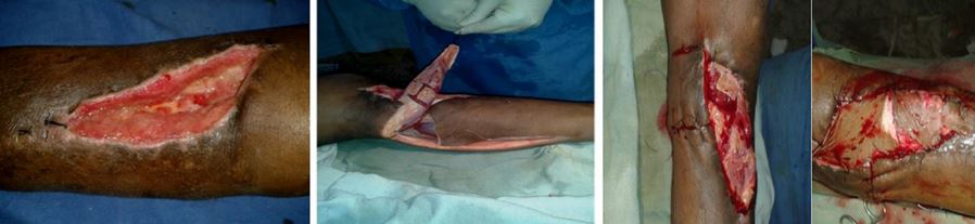
Aims & objectives: To evaluate the use of gastrocnemius, soleus and sural flap by orthoapedic surgeon  in management of complications of open leg bone fractures i.e. exposed implants, soft tissue defects.

Material & methods: We prospectively studied 20patients with soft tissue complications in patients having both bone leg fracture. Out of which12 patients were managed by gastrocnemius flap,5 patients by soleus flap & 3 patients were treated by sural flap according to the location of the wound. Patients with age between 18-60 years with no or minimal infection, size of wound <50cm2 for middle leg and <9×12cm for lower leg were included in study. The mean follow up was of 6 months.

Results: Only two sural flap and one soleus flap had marginal necrosis. Hypoaesthesia over lateral border of foot was noted in two patients of sural flap which was not troublesome. Not any patient had developed significant functional loss. All flaps resulted in a good wound coverage with early healing.

Conclusions: Local muscle flap is efficient and easy method of treating wound defects over leg and allows an orthopaedician to manage the compound fractures comprehensively.

Keyword : compound fractures; gastrocnemius flap; Muscle pedicle flap; neurocutaneous flap.


Address for Correspondence:Dr. Dinesh Kumar Mina . Govt. Medical College, Kota, Rajsthan

Email:dinesh18feb@gmail.com


How to site this article:Mina D K, Gupta S, Tiwari A K, Meena R, Nama K GManagement of Soft Tissue Complications Associated with Leg Bone Injuries by Local Muscle Pedicle Flap. OrthopJMPC 2016;22(2):31-36.


Introduction

High velocity trauma leads to open leg bone injuries and their operative complications i.e. exposed bone & implants and infection remain a challenge for orthopaedic surgeons from many years. The subcutaneous location of tibia, poor blood flow to the region are attributable factors for these complications. [1,2].  From management point of view these


complications of soft tissue defects are managed by plastic surgeons, and the centers where these facilities are not available these patients are referred to the higher centers.

The goal in management of the open wounds is to provide stable soft tissue coverage. Soft tissue coverage with muscle can seal the joint and bone from ongoing contamination and help to revascularize underlying structures. Muscle flaps are first introduced by Ger R.[3]. For leg bone important muscle flaps are medial and lateral head gastrocnemius flap for proximal tibia, patellar tendon & patella [4] ,soleus flap for closure of exposed middle third of leg bone [5], and reverse artery sural flap for exposed part of lower 1/3rd tibia [6].

The consistent vascular anatomy and superficial location of gastrocnemius muscle flap have made it a workhorse for coverage of defects around knee joint and in patellar region.

Soleus is a suitable muscle to repair soft tissue defects in the middle third of the leg due to its unique anatomic features.Its main advantage is its binary blood flow supply which allows us to cut it into two longitudinal parts [5] to be used as hemisoleus flap.

For the wounds over distal third of leg, free flap is the preferred choice but it has several major disadvantages [7] like it is a difficult long duration surgery and needs microsurgical instruments & expertise.

Reverse sural artery flaps for injuries of distal third of tibia provide good coverage both form functional and aesthetic point of view. It can be used in traumatic legs with major arteries defect [8].It provides durable skin coverage with ideal contouring with minimal postoperative discomfort and can be performed quickly.

By learning and using these techniques and procedures orthopaedic surgeons can also manage these complications successfully. Advantage of these local procedures are increased vascularity of the area and thus decreased infection [9], which promote early fracture healing [10] and it results in decrease morbidity of the patients, decrease referral and burden on health care facilities. With this background, we conducted a study with the aim to evaluate the outcome of local muscle pedicle flap surgeries done by an orthopaedician for management of compound leg bone fractures.

Material and Methods

We have prospectively studied 20 patients between the period of July 2013 and August 2015  admitted in orthopaedic department having Gustillo type 3 fracture leg bone or  exposed implant in leg area.The male female ratio was 3:2. with an average age of 37 years. We had 12 patients with soft tissue defects or exposed implant in proximal leg/patellar region, 5 patients in middle leg and 3 patients in distal third leg / ankle. We treated them with gastrocnemius flap, soleus/reverse soleus flap and reverse sural artery flap for soft tissue defects in proximal, middle and lower third of leg/ankle respectively. All the patients of fracture leg bone were primarily operated with either external fixator or internal fixation methods. The mean interval between the flap surgery and pervious bony fixation surgery was 4.5 weeks.This delay in surgery was due to presence of  infection, which was controlled by appropriate antibiotics and repeated debridements. The mean follow up was of 6 months after definitive surgery.

The inclusion criteria were-

A. Patients with age <60 years.

B. Patients having either exposed implant or bone in leg area, with no or minimal infection

C. Size of wound <50cm2 for soleus and <9×12cm for sural flap.

The exclusion criteria were-

A. Patients >60 years.

B. Presence of  active/florid  infection.

C. Presence of peripheral vascular disease and insufficient peroneal,posterior   tibial, sural arteries and lesser saphenous vein.

D. Compound leg bone fractures with damaged/crushed  gastrocnemius or soleus muscle

E. History of recent deep vein thrombosis in the involved extremity.

Pre-operative planning-

After determining the size of recipient site defect,patients have been assessed for presence of infection,if it was present thorough debridement were done and a clean granulated wound bed had been achieved.A vascular examination for patency of popliteal,peroneal, posterior tibial artery and lesser saphenous vein was done.Status of donor site muscle(either traumatized or presence of hematoma),and the function of remaining muscles of lower leg(to determine if loss of gastrosoleus due to flap could result in significant loss of planter flexion) had also been assessed.

Surgical techniques-

Gastrocnemius , Soleus and Reverse Sural Artery flaps were done in the patients with defects in proximal middle and distal third leg respectively as per the standard operating technique.

Post Operative management-

To prevent pedicle compression and vascular congestion limb elevation and immobilization was done. The drain was removed once the drainage was <30ml/day. The dressing was changed after 5days and then compression bandages/elastic wraps were advised. Follow up was done (ranges from 1.5 to 9 months) to look for donor site morbidity (scar, contracture or functional limitation),condition of flap and associated complications. Mobilisation was started after 5 days. Range of motion for joints were assessed.The final results were assessed according to healing of wound and stability of the flap coverage.

Results

We operated 20 patients out of which 12 with gastrocnemius flap,5 patients with soleus and 3 with reverse sural artery flap.Results for gastrocnemius flap were excellent. Thegastrocnemius flap for proximal leg & patellar region were healed completely. Results for soleus flap were good. Two soleus flap develop partial necrosis and one reverse sural flap develop partial necrosis with infection.Some showed venous congestion which was managed by limb elevation. One of the soleus flap had ugly scar at donor site.The final outcome was unaffected in all of the cases.None of the patients with gastrocnemius& soleus flap had planter flexion weakness at final follow-up.Hypoesthesia over lateral border of foot was noted in all patients of sural flap but it was not troublesome.At the final follow-up range of motion at knee and ankle joints was marginally compromised. One of the patient had donor site scar contracture around knee joint.Patients with reverse sural flap had non-cosmetic bulbous shaped flap but it was acceptable to them and had no difficulty in wearing shoes.


Figure 1 : Intraoperative pictures of Gastrocnemius flap.


Figure 2 : Intraoperative pictures of Soleus flap.


Discussion

Comparing the vascularity and results of fasciocutaneous flaps with the muscle flaps there is more advanced healing of fractures and wounds by muscle flaps compared to fasciocutaneous flap despite the high vascularity density of the latter. [11]

Cross leg flaps may be an option to cover the soft tissue defects but patient compliance is poor and they may lead to joint stiffness and pressure sores.So we used ipsilateral pedicle flap.

Gastrocnemius flap is most commonly used for knee and upper tibial defects. We used medial head in most of the cases because of its long arc of rotation. The damage to common peroneal nerve may occur during elevation of lateral head, but it is preventable by carefuldissection. The territory of medial head extends to within 5cm of medial malleolus and for lateral head the distal limit is within 10 cm of lateral malleolus [12].

The soleus is a type 2 flap according to Mathes and Nahai [13]. It can be used in various ways i.e. proximally or distally based, hemisoleus flap or composite flap which are based on the vascular supply of the muscle [14] The hemisoleus flap is a better option for wound defects and exposed bone over middle leg [15].  Some studies [16]show high failure rates associated with soleus flap.The reason for this is because the distal end of soleus is muscular, not tendinous and it is friable  and vulnerable to damage to its blood supply. By dissecting and freeing the first


perforator to increase its arc of rotation, the distal reach of this muscle can be enhanced [16]  The elevation of soleus flap requires more time and microsurgical techniques.Despite these disadvantages soleus flap is the workhorse for middle third of leg.

Reverse sural artery flap was first described by Masquelet et al [17] as neurocutaneous island flap in 1992. It has numerous advantages, it is a one stage operation, which does not require microsurgical techniques. Elevation of the flap is easy and quick. The donor site has minimal morbidity as it can be closed primarily when small flap is raised and skin grafted when large flap is raised [18]. The vascular supply to the arterial network of the sural area is constant and reliable, and have been studied in details and there is no need to sacrifice any major artery and or sensory nerve [19].As the chances of survival of sural and soleus flaps dependsupon location and presence of patent perforators, so the preoperative colour Doppler evaluation of perforators may help in pre op planning.but it is not essential or recommended routinely.One of the few disadvantages of sural flap is hypoesthesia at lateral border of foot due to damage to sural nerve. Damage to Sural nerve may also occur during gastrosoleus flap,but with careful dissection it can be avoided.In our study, none of the patients of gastrosoleus flap had this complication.Injury to sural nerve may lead to painful neuroma formation which may be prevented by deep burial of nerve ends.All these flaps take minimum of 2 weeks for being taken up, requiring immobilization, but it may lead to stiffness of knee or ankle joint.To prevent this we started mobilization of the joints after one week,depending on the fracture union.

The cosmetic results of flaps remain a problem. In gastrosoleus flap long ugly posterior scar may form which may result in contracture formation.Sural artery flap usually gives a bulbous appearance, but these do not affect the final functional outcome.There occurs minimal functional limitation of donor site muscle after flap surgeries.Normal walking is unaffected, however functionally more demanding activities like walking uphill and running are affected [20].The major limitation of this study is small sample size. Considering the complications Neale et al [21] have reviewed the complications of muscle flap transposition for traumatic leg defects and concluded that the causes of complications were mainly technical errors, inadequate debridement, use of diseased and traumatized muscle and unrealistic objectives.      

Conclusion

The complex open injuries of leg can be successfully managed by proper evaluation, adequate debridement, use of appropriate muscle flap resulting in early good wound healing, creating environment for faster fracture healing with good functional outcome.


References

1.                Canale ST, Beaty JH.Campbell’s Operative  Orthopaedics 12th ed. Missouri: Mosby; 2012. ; pp. 1405-6

2.                Bucholz RW, Heckman JD, Court­Brown CM. Rockwood and Green's Fractures in Adults 7th ed. Philadelphia: Lippincott Williams & Wilkins; 2009 ; pp. 376–7.

3.                Ger R.  The technique of muscle transposition in the operative treat¬ment   of traumatic and ulcerative lesion of leg.  J Trauma 1971; II 6:  502-510

4.                Colen L, Mathes S. Lower extremity traumatic injury. In: Jurkiewicz MJ, Krizek T, Mathes S, Ariyan S, editors. Plastic Surgery. St. Louis: CV Mosby; 1990. p. 935-81

5.                Wright JK, Watkins RP. Use of the soleus muscle flap to cover part of the distal tibia. Plast Reconstr Surg 1981;68:957-8

6.                Rios-Luna, Villanueva-Martinez, Fahandezh-Saddi, Villanueva López, Del Cerro-Gutierrez. Versatility of the sural fasciocutaneous flap in coverage defect of the lower limb. Injury 2007;38:824-31

7.                Heller L, Levin LS. Lower extremity microvascular reconstruction. Plast Reconstr Surg. 2002;108(4):1029–41.

8.                Alam MK, Shaheen MS, Hossain S,  Anam S, Rahman S. Sural Island   Flap – A Good Option For Coverage Of The Exposed Heel (Tendoachillis). J Dhaka Med. Coll. 2010;19(1):19-24.

9.                Arnold PG, Yugueros P, Hanssen AD. Muscle flaps in  osteomyelitis  of the lower extremity: a 20 year account. PlastReconstr Surg. 1999;104(1):107­10

10.                Davis KM. et al, Muscle-bone interactions during fracture healing. J Musculoskelet Neuronal Interact 2015;15(1):1-9

11.                Harry Lorraine E,  Miael F,  Ewa M. Comparison of the Vascularity of   Fasciocutaneous Tissue and Muscle for Coverage of Open Tibial      Fractures. Plast  Recons Surg: 2009;124:1211­19

12.                Sanders R, O’neill T. The Gastronmius Myocutaneus  Used as A Cover for the Exposed Knee Prosthesis. JBJS: 1981;63-B:383-86.  Http://Www.Boneandjoint.Org.Uk/Content/Jbjsbr/63-B/3/383.Full.Pdf

13.                Mathes SJ, Nahai F. Clinical applications for muscle and musculocutaneous flaps.  New York;  CV Mosby Company Publishers. 1982 : 16-26, 514-524

14.                G.G. Hallock. Getting the most from the soleus muscle. Am Plast Surg, 36 (1996), pp. 139–146

15.                Ata-ul-Haq, Tarar MN, Malik FS, Khalid K, Riaz A, Mehrose MY, Khan H. Hemisoleus muscle flap, a better option for coverage of open fractures involving middle third of tibia. J Ayub Med Coll Abbottabad. 2009 Oct-Dec;21(4):154-8

16.                Ademola SA, Adebayo WO, Lawani O. Reconstruction of soft tissue defects in the distal third of leg,ankle, and foot: A meta-analysis. Nigerian J Plast Surg 2015;11:1-7

17.                Masquelet AC, Romana MC, Wolf G. Skin island flaps supplied by the vascular axis of the sensitive superficial nerves: anatomic study and clinical experience in the leg. Plast Reconstr Surg. 1992;89:1115–21


18.                Pirwani MA, Samo S, Soomro YH. Distally Based Sural Artery Flap: A workhorse to cover the soft tissue defects of lower 1/3 Tibia and Foot. Pak J Med Sci. 2007;23:103-7

19.                Hasegawa M, Torii S, Katoh H, Esaki S. The distally based superficial sural artery flap. Plast Reconstr Surg. 1994 Apr;93(5):1012-20

20.                Kramers-de Quervain IA, Läuffer JM, Käch K, Trentz O, Stüssi E.      Functional donor-site morbidity during level and uphill gait after a gastrocnemius or soleus muscle-flap procedure. J Bone Joint Surg Am  2001 Feb;83-A(2):239-46

21.                Neale HW,  Stern PJ,  Kreitein JG et al. Complications of muscle flap transpositions for traumatic defects of leg. Plastic Reconstr Surg 1983; 72: 512-515.Schemitsch EH, Bhandari M. Fractures of the diaphyseal humerus. Skeletal trauma, 3rd edn. WB Saunders, Toronto. 2001:1481-511.

22.