Orthopaedic Journal of MP Chapter

Publisher: Madhya Pradesh Orthopaedic Association www.mpioa.com
E-ISSN:2582-7243, P-ISSN:2320-6993
2023 Volume 29 Number 2 Jul-Dec

Core decompression and non-vascularized iliac crest graft in avascular necrosis of hip

Khare A1*

1* Ajay Khare, Director, Global Hospital, Ujjain, Madhya Pradesh, India.

Background: Avascular necrosis of femoral head occurs due to impaired blood supply of the head of femur. Core decompression with non-vascularized iliac crest graft has been the treatment of choice in cases of early AVN.

Material and Method: This prospective, interventional study was conducted in Global hospital, Ujjain from jan-2022 to march -2023. We studied 20 hips having AVN of the femoral head treated by core decompression and autologous cancellous bone grafting with a graft taken from the iliac crest. Total number of 20 patients with AVN of the femoral head (up to grade 2b of Ficat and Arlet classification) were treated by use of non-vascularized bone graft.

Results: The mean HHS was 69.45 preoperatively and 83.55 at six months postoperatively (p ≤ 0.0003). Out of 20 hips, 14 had excellent (HHS >90) to good (HHS 80-90) outcomes, while six had fair (HHS 70- 80) to poor (HHS >70) outcomes. The mean VAS score was 6.3 preoperatively and 3.8 at six months postoperatively (p ≤ 0.0001). On radiographic and clinical evaluation, the patients showed significant improvement on the six-month follow-up.

Conclusion: Our study suggests that core decompression with autologous cancellous bone grafting might not reverse AVN progression, but can delay the progression of AVN, avoid collapse, reduce pain, and provide improvement in functional outcomes at least in the short term.

Keywords: Core decompression, iliac crest graft, avascular necrosis of hip

Corresponding Author How to Cite this Article To Browse
Ajay Khare, Director, , Global Hospital, Ujjain, Madhya Pradesh, India.
Email:
Khare A, Core decompression and non-vascularized iliac crest graft in avascular necrosis of hip. ojmpc. 2023;29 (2):50-55.
Available From
https://ojmpc.com/index.php/ojmpc/article/view/178
Manuscript Received Review Round 1 Review Round 2 Review Round 3 Accepted
2023-12-07 2023-12-13 2023-12-19 2023-12-25 2023-12-31
Conflict of Interest Funding Ethical Approval Plagiarism X-checker Note
Authors state no conflict of interest. Non Funded. The conducted research is not related to either human or animals use. 12.34 All authors have accepted responsibility for the entire content of this manuscript and approved its submission.

© 2023by Khare Aand Published by Madhya Pradesh Orthopaedic Association. This is an Open Access article licensed under a Creative Commons Attribution 4.0 International License https://creativecommons.org/licenses/by-nc/4.0/ unported [CC BY NC 4.0].

Introduction

Avascular necrosis of femoral head occurs due to impaired blood supply to the head of femur. It can be caused by fractures, dislocations, chronic steroid use, chronic alcohol use, coagulopathy, congenital causes and many other factors. But most common cause is Idiopathic. (1-4) The use of a non-vascularized bone graft is more attractive in treating the early stages of avascular necrosis of femoral head than vascularized graft because, it is significantly less technically demanding. (5,6)

Non-vascularized autologous bone grafting has several theoretical advantages. this method leads to the decompression of the avascular lesion and the elimination of the necrotic bone, breaking the cycle of ischemia and intraosseous hypertension in addition to providing growth factors from the graft [7].

The regenerative properties due to the presence of osteoblasts in the trabecular bone and the ability to procure a large amount of graft make the iliac crest the gold standard site of cancellous bone graft harvesting. [8,9]

core decompression can be considered an effective treatment modality in the early stages (up to stage 2b) of AVN of the femoral head [10]. Therefore, this study was conceptualized to study the effect of core decompression along with cancellous bone grafting on cases of AVN of the femoral head.

Material and method

This prospective, interventional study was conducted in Global hospital, Ujjain from jan-2022 to march-2023. We studied 20 hips having early AVN of the femoral head treated by core decompression and autologous cancellous bone grafting with a graft taken from the iliac crest.

AVN of femoral head (up to grade 2b of Ficat and Arlet classification) who met the inclusion and exclusion criteria are included in this study.

Inclusion criteria was patients with avascular necrocis of the femoral head up to stage 2b (Ficat and Arlet), patients aged between 18 and 60 years and patients giving consent for the surgical procedure and participation in the study.

Exclusion criteria was patients with previous infection of the hip Joint, neuromuscular disorder of the hip joint and severe osteoporosis

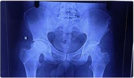
AVN stage was confirmed preoperatively using an X-ray of the pelvis with both hip joints and an MRI of both hips. A preoperative workup was done. After taking written and informed consent, patients were taken for the operation theatre.

Patients were placed on a traction table in a supine position. Sterile painting and draping were done. Subsequently, a 2-3 cm mid- lateral longitudinal incision was made over the subtrochanteric region.

The tensor fascia lata was split in the direction of its fibers, and the vastus lateralis muscle was elevated. Under C-arm guidance, a 3.2 mm threaded guide pin was inserted through the lateral cortex into the affected part of the femoral head, with the entry point between the lesser trochanter and greater trochanter.

The guide pin was directed toward the centre of the necrotic area of the femoral head. The guide pin was over-reamed with an 8 mm reamer. The cancellous bone graft was taken from the outer table of the iliac crest of the patient.

The gap created with the cancellous bone graft was packed. Both the operative sites were washed thoroughly with normal saline and layer-wise closure was done with vicryl and ethilon. Finally, sterile dressing was done.

178_1.jpg
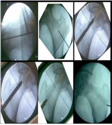
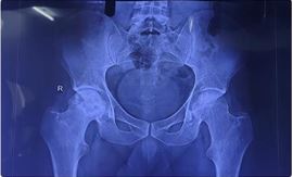
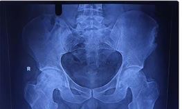
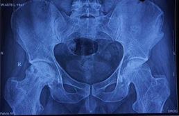
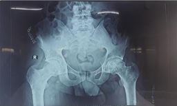
Figure 1: Core decompression of femoral head & bone grafting

Post operative management-Intravenous (IV) antibiotics were given till the third postoperative day, followed by oral antibiotics until suture removal. Dressing was done on the first, third, and fifth postoperative days. Physiotherapy started on day one which included knee mobilization, static quadriceps exercises, ankle pump, and toe movement. After one-week, partial weight bearing was started.

Follow-up was done at 1.5 months, three months, and, finally, six months. Patients were assessed using the Harris Hip Score (HHS) and Visual Analog Scale (VAS) scores at follow-up.

Results

In our study, the majority (50%) of patients were in the 20-30-year age group, making it the most common age group, followed by the 31- 40-year age group (40%).


There was a male predominance, with 17 out of 20 patients being male. All patients became symptomatic within the last year (range = 2-12 months). Of 20 patients, 11 presented to us within three months of developing symptoms (Table 1). Out of 20 patients, 10 were able to sit cross-legged, squat, and climb stairs without any significant problems. Four patients were not able to sit cross-legged or squat, while five patients were not able to climb stairs.

Life style modification was advised to all patients. Five patients who were alcoholics were advised to stop taking alcohol. Seven patients had a history of steroid use due to COVID-19 infection. No specific aetiology was identified in eight patients

Table 1: Age distribution, sex distribution and duration of pain (in months)

CharacteristicsParametersNumber of patients
Age distribution (in years)20-30
31-40
41-50
51-60
10
8
1
1
Sex distributionMale
Female
17
3
Duration of pain (in months)0-3 months
4-6 months
7-9 months
10-12 months
11
6
2
1

Table 2: Risk factor/ Etiology

Risk factors/EtiologyNumber of patients
Idiopathic8
Steroid7
Alcohol 5

Table 3: The mean HHS was 69.45 preoperatively and 83.55 at six months postoperatively (p ≤ 0.0003)

DegreePreoperativeAt 6 months
≤7094
70-80 72
81-9045
>9009

Table 4: Visual Analog Scale score preoperatively and at six months postoperatively. The mean VAS score was 6.3 preoperatively and 3.8 at six months postoperatively (p ≤ 0.0001)

Vas scorePreoperativeAt 6 months postoperatively
0-3 012
4-6144
7-964

Table 5: Complications

Complication Number of patients
Secondary arthritis6
Pain at the graft donor site (postoperatively) 17
Pain at the donor site (at 1.5months)5
Pain at the donor site (at 3months)1
Pain at the donor site (at 6months)0

Overall, 30% of patients developed secondary arthritis after six months and progressed to Ficat and Arlet stage 3/4. In total, 17 patients had pain at the donor site immediately after surgery.

After three months, only one patient had pain at the donor site which was managed with analgesics. After six months, no patients had pain at the donor site. Apart from this, no significant donor site morbidity was seen.

Figures 2-6 are the radiographic images of one of the study patients showing preoperative, postoperative, and follow-up X- rays. On radiographic evaluation, the patient operated on for AVN of the femoral head with core decompression and cancellous bone grafting showed significant improvement on the six-month follow up.

178_2.jpg
Figure 2: Preoperative X-ray of the patient showing avascular necrosis of the femoral head.

178_3.jpg
Figure 3: Immediate postoperative X-ray

178_4.jpg
Figure 4: X-ray at 1.5 months of follow-up.


178_5.jpg
Figure 5: X-ray at three months of follow-up.

178_6.jpg
Figure 6: X-ray at the final follow-up.

Discussion

The blood supply of the femoral head is so tenuous that even a slight vascular injury can predispose to AVN. Hence, AVN is commonly seen in the femoral head. Studies have shown that core decompression can arrest or even reverse the process of AVN and can avoid femoral head collapse and its sequelae. Core decompression can preserve the femoral head at an early stage of the pathology [4,5].

Steinberg et al. reported that 92% of patients who underwent non-operative management had femoral head collapse at the end of two years. They found that patients with early- stage AVN who had undergone core decompression and cancellous bone graft had about a 70% success rate [11]. Core biopsies were performed in the early sixties in a small number of patients with AVN of the femoral head to examine the pathological changes. The increased intraosseous pressure was relieved by core decompression, thereby providing pain relief to the patient.

According to Steinberg, treating early AVN of the femoral head with core decompression and cancellous bone grafting is a safe and efficient method [12]. We studied 20 hips having AVN of the femoral head treated by core decompression and autologous cancellous bone grafting with a graft taken from the iliac crest. In a study, Babhulkar [13 ] treated 32 patients with AVN of the femoral head by core decompression and iliac crest grafting and reported pain relief in all patients. Similar results were found in our studies as well.

At the final follow-up, 16 patients had pain relief. Core decompression with vascularized fibular graft has been the treatment of choice in cases of early AVN. Vascularized fibular grafting not only provides subchondral support but also vascularization at the site. However, the disadvantage is morbidity at the donor site and the need for microvascular anastomosis [14].

Therefore, a cancellous graft from the iliac crest has been considered as an alternative. Osteogenesis and osteoinduction are two of the key characteristics of bone grafts, and they are frequently attributed to cancellous grafts rather than cortical grafts. This makes it advantageous for revascularization as well. Moreover, it avoids donor site morbidity associated with fibular graft. A densely filled graft may provide structural stability [15,16].

Therefore, we preferred core decompression with autologous cancellous bone grafting with a graft taken from the iliac crest. In our study, all patients were kept non- weight bearing for 1.5 months, and after that partial weight bearing for 1.5 months. This was done because the femoral head would need enough time for regeneration after core decompression. Therefore, if weight bearing is started before sufficient bone growth, it could result in a collapse of the femoral head due to stress [13].

In our study, 90% of the patients were in the 20-40-year age group, and the remaining 10% of patients were in the 41-60-year age group. Our study showed a male predominance as 17 (85%) patients were male. Similar male predominance was also reported by Babhulkar [13] where 81.25% of patients were males. Activities of daily living were assessed by cross-legged sitting, squatting, and stair climbing. Overall, 50% of patients faced difficulty in these activities, suggesting that activities of daily living are affected even in the early stages of AVN.

The most common stage according to Ficat and Arlet classification at which the patients presented was stage 2B (45%), followed by stage 2A (40%) and stage 1 (15%). No difference was noted in the preoperative and postoperative MRI stages in 11 patients over six months. Three patients reverted from stage 2B to 2A on MRI grading. Six patients showed progression to later stages on MRI grading.

The mean HHS was 69.45 preoperatively and 83.55 at six months postoperatively. Out of 20 hips, 14 had excellent (HHS >90) to good (HHS 80-90) outcomes, while six had fair (HHS 70- 80) to poor (HHS >70) outcomes. The mean VAS score was 6.3 preoperatively and 3.8 at six months postoperatively. Out of 20 patients, 16 had excellent to good outcomes and four had fair to poor outcomes in terms of pain relief.

Although no significant improvement was seen in the MRI findings, as interpreted by HHS results, excellent functional outcomes were found in 66% of grade 1 patients, 50% of grade 2A patients, and 33% of grade 2B patients. Poor outcomes were found in 33% of grade 1 patients, 12.5% of grade 2A patients, and 22% of grade 2B patients.


Conclusion

Our study suggests that core decompression with autologous cancellous bone grafting might not reverse AVN progression but can delay the progression of AVN, avoid collapse, reduce pain, and provide improvement in functional outcomes at least in the short term. The study had a relatively shorter follow-up period of six months. Probably, a longer follow-up is needed for evaluating long-term functional outcomes. A shorter follow-up period was because of time constraints. Another limitation was the small sample size as overestimation of treatment effect is more likely in a small sample compared to a large sample.

Patient compliance was also a limitation of our study. The limitation of the surgical method was that it could only be done in the early stage of the disease (Ficat and Arlet stages I and II) and did not have any significant outcomes, if done in the advanced stages of the disease (Ficat and Arlet stages III and IV). Other surgical treatments such as muscle pedicle bone grafting and vascular grafting were not done due to time, technical, and financial constraints. Core decompression with cancellous bone grafting is a promising procedure in stages 1 and 2 as it reduces the symptoms in the majority of cases and provides improvement in functional outcomes.

References

1. Mont MA, Jones LC, Hungerford DS: Nontraumatic osteonecrosis of the femoral head: ten years later. J Bone Joint Surg Am. 2006; 88:1107-29. . [Crossref][PubMed][Google Scholar]

2. Sharma D, Maulik Jhaveri D, Urang Patel D, Jha A, Shah P, Golwala P: Pain relief with core decompression and autologous bone graft in osteonecrosis of femoral head in grade 2. Int J Orthop Sci. 2019, 5:369-72. 10. 22271/ortho.2019.v5.i4g.1699 06, 88:1117- [Crossref][PubMed][Google Scholar]

3. Hua KC, Yang XG, Feng JT, Wang F, Yang L, Zhang H, Hu YC. The efficacy and safety of core decompression for the treatment of femoral head necrosis. A systematic review and meta analysis, journal ortho surg research. Sept 2019 14. 1.306 [Crossref][PubMed][Google Scholar]

4. Lavernia CJ, Sierra RJ: Core decompression in atraumatic osteonecrosis of the hip. J Arthroplasty. 2000, 15:171-8. 10. 1016/s0883-5403(00)90132-3 [ Crossref][PubMed][Google Scholar]

5. Urbaniak JR, Coogan PG, Gunneson EB, Nunley JA: Treatment of osteonecrosis of the femoral head with free vascularized fibular grafting. A long-term follow-up study of onehundred and three hips. J Bone Joint Surg Am. 1995, 77:681-94. 10.2106/00004623-199505000-00004 [Crossref][PubMed][Google Scholar]

6. Mont MA, Hungerford DS. Non-traumatic avascular necrosis of the femoral head. J Bone Joint Surg Am. 1995; 77:459. [Crossref ][PubMed][Google Scholar]

7. Lavernia CJ, Sierra RJ, Grieco FR. Osteonecrosis of the femoral head. J Am Acad Orthop Surg. 1999;7(4):250-61. [Crossref][PubMed][Google Scholar]

8. Steinberg ME. Diagnostic imaging and role of stage and lesion size in determining outcome in osteonecrosis of the femoral head. Tech Orthop. 2001;16:6-15. [Crossref][PubMed][Google Scholar]

9. Harkess JW. Arthroplasty of hip. ,Campbells Operative Orthpeadics, Edited by Crenshaw AH, 8th edition, Vol. 1: CV Mosby Company, St. Louis, Washington DC, Torto, 1982 [Crossref ][PubMed][Google Scholar]

10. Eftekhar NS. Total hip replacement using principles of lowfriction arthroplasty: The hip surgery of the musculoskeletal system, Edited by CM Evarts, Vol. 3: Churchill Livingston, 1983. . [Crossref][ PubMed][Google Scholar]

11. Steinberg ME, Larcom PG, Strafford B, Hosick WB, Corces A, Bands RE, Hartman KE: Core decompression with bone grafting for osteonecrosis of the femoral head. Clin Orthop Relat Res. 2001, 71-8. 10. 1097/00003086- 200105000-00009 [Crossref][PubMed][Google Scholar]

12. Steinberg ME: Core decompression of the femoral head for avascular necrosis: indications and results. Can J Surg. 1995, 38 Suppl 1:S18-24. . [Crossref][PubMed][Google Scholar]

13. Babhulkar S: Osteonecrosis of femoral head: treatment by core decompression and vascular pedicle grafting. Indian J Orthop. 2009, 43:27- 35. 10. 4103/0019-5413.45320 [Crossref][PubMed][Google Scholar]

14. Yoo MC, Kim KI, Hahn CS, Parvizi J: Long-term followup of vascularized fibular grafting for femoral head necrosis. Clin Orthop Relat Res. 2008, 466:1133-40. 10. 1007/s11999-008-0204- 9 [Crossref][PubMed][Google Scholar]

15. Sen RK: Management of avascular necrosis of femoral head at pre- collapse stage. Indian J Orthop. 2009, 43:6-16. 10. 4103/0019-5413.45318 [Crossref][PubMed][Google Scholar]

16. Sanap A, Rabari YB, Teja V, Shah S. Management of avascular necrosis of famoral head by core decompression. International journal of research in orthopaedics. vol. 4, no. 3. May- June 2018 [Crossref][PubMed][Google Scholar]

Disclaimer / Publisher's Note

The statements, opinions and data contained in all publications are solely those of the individual author(s) and contributor(s) and not of Journals and/or the editor(s). Journals and/or the editor(s) disclaim responsibility for any injury to people or property resulting from any ideas, methods, instructions or products referred to in the content.