Orthopaedic Journal of MP Chapter

Publisher: Madhya Pradesh Orthopaedic Association www.mpioa.com
E-ISSN:2582-7243, P-ISSN:2320-6993
2023 Volume 29 Number 2 Jul-Dec

Supracutaneous LCP as a definite fixation method in compound metaphyseal and intraarticular fractures of tibia

Bajoria RS1*

1* R S Bajoria, Professor, Department of Orthopedics, Gajra Raja Medical College, Gwalior, Mp, India.

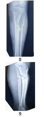
Introduction: Open fractures of distal and proximal tibia are common injuries encountered by orthopedic surgeons. Metaphyseal compound fractures are not suitable for ORIF with greater chances of infection, soft tissue complications and risk of plate exposure. With external fixator, treatment & fracture healing period usually increased & complication rates are quite high, so we managed these fractures by supracutenous LCP as external fixator; and results evaluated. We find out it a better alternative method to conventional external fixator. We evaluated the feasibility of supracutenous LCP as definite fixation methods in open fractures.

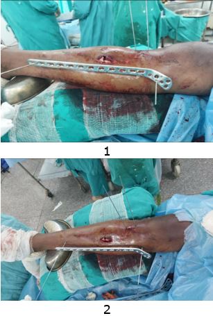
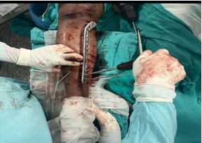
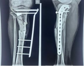
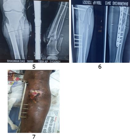
Material and methods: This study conducted from April 2020 to October 2023. Total 30 cases were included in this study. Open GA II & IIIA of tibia metaphyseal fractures in 18 cases and intra articular tibia fractures in 12 cases included in this study. Debridement & LCP plates fixation was done in external fixator manner. Proximal & distal tibia LCP fixed with long screws. Most fixations were done within 1 - 5 days of trauma. Patients were followed up till bony union. Minimum follow up was 6 months.

Results: 8 cases united in 4 months, 8 cases united in 6 months, 9 cases united in 6-9 months and 5 cases went in non-union. Average duration of union was 5.6 months. Functional outcomes evaluated by AKSS score for proximal fractures & by AOFAS for distal fractures. 15 cases have shown excellent results; with union, 11 cases have shown good results; soft tissues reconstruction required, 4 cases had bad results; due to infection. Overall, 26 cases were had good to excellent results.

Conclusion: We observed that external fixation in open fractures usually results in infection, delayed or non-union, requirement of further surgeries. Supracutaneous LCP as primary definite fixation can give excellent results in terms of stable & rigid fixation, early fracture union, less chances of second surgery, cost effective and better patient compliance & better functional outcomes.

Keywords: Supracutaneous LCP, definite fixation, compound fractures of tibia

Corresponding Author How to Cite this Article To Browse
R S Bajoria, Professor, Department of Orthopedics, Gajra Raja Medical College, Gwalior, Mp, India.
Email:
Bajoria RS, Supracutaneous LCP as a definite fixation method in compound metaphyseal and intraarticular fractures of tibia. ojmpc. 2023;29(2):44-47.
Available From
https://ojmpc.com/index.php/ojmpc/article/view/177
Manuscript Received Review Round 1 Review Round 2 Review Round 3 Accepted
2023-12-07 2023-12-13 2023-12-19 2023-12-25 2023-12-31
Conflict of Interest Funding Ethical Approval Plagiarism X-checker Note
Authors state no conflict of interest. Non Funded. The conducted research is not related to either human or animals use. 14.32 All authors have accepted responsibility for the entire content of this manuscript and approved its submission.

© 2023by Bajoria RSand Published by Madhya Pradesh Orthopaedic Association. This is an Open Access article licensed under a Creative Commons Attribution 4.0 International License https://creativecommons.org/licenses/by-nc/4.0/ unported [CC BY NC 4.0].

Introduction

Primary plating as an external fixator of compound fracture is a good method of management. There is a high rate of union with a low complication rate when using external locked plating for open fractures of distal femur and proximal tibia[1].

LCP can be used as a definitive external fixator for compound tibial fractures as it gives good results and low complication rates, with satisfactory stability. It also has the advantage of facilitating wound healing.

It is also cosmetically acceptable and non-cumbersome while mobilization as it does not strike the opposite leg. It allows easy assessment of fracture healing on x rays due to non-overlapping of the implant[2].

The consistent good outcome using this “Supracutaneous technique” support our opinion of using Locking compression plate as external fixator in distal tibial fractures which are very well tolerated by patients and address the challenging problems of compound wound healing, non-union and osteomyelitis[3,4].

Material and Method

This study is conducted from April 2020 to October 2023. Total 30 cases were operated. Open GA II & IIIA metaphyseal fractures in 18 cases and intra articular fractures in 12 cases were included in this study. Debridement, & LCP plates fixation was done in external fixator manner. Proximal and distal tibia LCP fixed with long screws. Most fixations were done within 1 - 5 days of trauma.

Patients were followed up till bony union. Minimum follow up was 6 months. In intraarticular fractures fixed with preliminary k-wire fixation and 2-3 cc screws are used for articular reduction. 1-2 k wires used for metaphyseal fractures. Long LCP is measured & both ends fixed with k wires. Proximal & distal ends of plate stabilized by 4 screws on both sides. Maintained a 1- 2 cm space on surface between skin & plate for better soft tissue care.

Locking compression plate (LCP) applied as an external fixator achieves indirect reduction & gives early union & maximum function. Its fixed-on surface of skin so is called as Supracutaneus plating (Giovannini et al. 2016).

This can be an important tool in armamentarium of an orthopaedic surgeon especially in metaphysical fractures with open injury Giovannini5 et al. or closed injuries with precarious soft tissue Kerkhoffs [6] et al.

For compound metaphysial fracture, debridement is done and closed reduction & temporary fixation is done by percutaneous K wires. Long LCP plating is selected & fixed in bridging fashion.

In intraarticular fracture, condylar reduction is done and fixation is achieved by sub articular cancellous screw fixation (1-3 screws). Followed by supracutenous LCP fixation.

Secondary soft tissue surgery, if required is done by plastic surgeon in form of skin grafting or free or vascular flap. Secondary bony procedure, if required, is done in form of bone marrow injection or bone grafting. In some cases, secondary surgery in form of external fixator or intramedullary nailing is done. Follow up of every patient is done at weekly for 1 month, bimonthly for 3 months and monthly till 6-8 months or until union is achieved at fracture site.


Figure 1 and 2: Intra operative picture


Figure 3: Plate application


Figure 4: Post operative X-ray


Figure 5: Pre op x ray, Figure-6 Post op xray Figure 7- Follow up


Figure 8 and 9: Follow up xray at 6 months

Table 1: Union time

Number of casesUnion time
84 months
86 months
96-9months
5Non-union

Results: 8 cases united in 4 months, 8 cases united in 6 months, 9 cases united in 6-9 months and 5 cases went in non-union. Average duration of union was 5.6 months. Functional outcomes evaluated by AKSS score for proximal fractures & by AOFAS for distal fractures. 15 cases shown excellent results; with union, 11 cases shown good results; soft tissues reconstruction required, 4 cases had bad results; due to infection. Overall, 26 cases were had good to excellent results.

Discussion

In an observational study by Panda [1] et al of eight patients with compound injuries to distal femur and proximal tibia or both during a period of one year. Total 8 fractures were treated by external fixation i.e precontoured anatomical locking plate application. Out of 8 cases 7 had full range of motion (87.5%) and 1 had terminal lag of movements (12.5%). The final outcome of the study based on knee society score was, 4 excellent (50%), 3 good (37.5%) and 1 fair (12.5%).

Bansal [2] et al prospectively evaluated 30 cases of fresh compound tibial fractures. The mean fracture healing time was 14 week (range, 12-18 weeks) for proximal tibia, 24 weeks (range, 18-26 weeks) for tibial diaphysis / multi segmental fractures, 18 weeks (range, 14-20 weeks) for distal tibia. Once biplaner radiographic cortical bridging was observed, full weight bearing for 1 month before implant removal was advised. According to the Johner and Wruhs7 criteria results were excellent in 75%, good in 18% and poor in 7% cases.

In a study by Venkatesh [3] et al total of five (05) patients underwent “supracutaneous plating” of the tibia using a metaphyseal locking compression plate. In all five patients the plate was kept in place until there was complete consolidation both clinically and radiologically. At the latest follow-up (average 15 months). All patients were fully weight bearing with a fully healed tibia. In the study by Rajshekharan [4] et al, four patients with Grade II compound fractures of the tibia included. The average age of the patients was 52 years (range 45-60). The average time of presentation was 36 hours (range 10 h - 3 days). The plate was in situ for average 20 weeks. In all patients, plate was removed after radiological and clinical union. No screw tract infection found.
Results of our study are comparable to other studies.

Conclusion

We observed that external fixation in open fractures usually results in infection, delayed or non-union, requirement of further surgeries. Supracutaneous LCP as primary definite fixation can give excellent results in terms of stable & rigid fixation, early fracture union, less chances of second surgery, cost effective and better patient compliance & better functional outcomes.


References

1. Panda Shakti S. Panda Damodar and Suri Nikhil. Supracutenous plating. Use of locking. [Crossref][PubMed][Google Scholar]

2. compression plate as external fixator for intra-articular compound fractures. International Journal of Medical Research & Health Sciences. 2016, 5. 8:62-67. [Crossref][PubMed][Google Scholar]

3. Akhil Bansal, Sourabh Alawa, Deependra Sonkar, Arvind Karoria, Anshul Khare and Sanjiv Gaur. Supracutaneous locking compression plate as an external fixator in compound metaphyseal and diaphyseal fractures of tibia. International journal of orthopedics science 2020. 6 (3);633-636. [Crossref][PubMed][Google Scholar]

4. S. K. Venkatesh Gupta, Shyam Prasad Parimala. Open journal of orthopaedics 2013. 106-109 [Crossref][PubMed][Google Scholar]

5. Supracutenous. locking compression plate for grade 1 & 2. compound fractures of distal tibia. A case series. [Crossref][PubMed][Google Scholar]

6. Rajasekharn. Jai kumar SC. locking compression plate for grade. 2 compound fractures of. [Crossref][PubMed][Google Scholar]

7. tibia –a case series. International journal of science study 2016. original article. . [Crossref][PubMed][Google Scholar]

8. Francesca Giovannini 1, Luigi de Palma 2, Andrea Panfighi 2, Mario Marinelli 2. Intramedullary nailing versus external fixation in Gustilo type III open tibial shaft fractures. a meta-analysis of randomised controlled trials. Strategies Trauma Limb Reconstruction, 2016 Apr. 11(1).1-4. doi: 10.1007/s11751-016-0245-7. Epub 2016 Feb 26 [Crossref][PubMed][Google Scholar]

9. Rademakers, Kerkhoffs, Sierevelt, Raaymakers, Marti. Operative Treatment of 109 Tibial Plateau Fractures: Five- to 27-Year Follow-up Results. Journal of Orthopaedic Trauma 21(1). p 5-10, January 2007. [Crossref][PubMed][Google Scholar]

10. Johner R, Wruhs O. Classification of tibial shaft fractures and correlation with results after rigid internal fixation. Clinical orthopaedics and related research. 1983; (178):7 - 25. [Crossref][PubMed][Google Scholar]

Disclaimer / Publisher's Note

The statements, opinions and data contained in all publications are solely those of the individual author(s) and contributor(s) and not of Journals and/or the editor(s). Journals and/or the editor(s) disclaim responsibility for any injury to people or property resulting from any ideas, methods, instructions or products referred to in the content.