Orthopaedic Journal of MP Chapter

Publisher: Madhya Pradesh Orthopaedic Association www.mpioa.com
E-ISSN:2582-7243, P-ISSN:2320-6993
2022 Volume 28 Number 2 Jul-Dec

A case report on nerve sheath tumour of median nerve at distal end radius damaging distal radius ulna joint

Girdhar S1, Raheja P2, Bajoria R3*

1 S Girdhar, Department of Orthopedics and Trauma Centre, JA Group of Hospitals, Gwalior, Mp, India.

2 P Raheja, Department of Orthopedics and Trauma Centre, JA Group of Hospitals, Gwalior, Mp, India.

3* RS Bajoria, Professor, Department of Orthopedics and Trauma Centre, JA Group of Hospitals, Gwalior, Mp, India.

PNSTs (Peripheral Nerve sheath tumors) are common tumors of hand present as solitary swelling along the course of nerve. However, Multiple swellings may be present along the course of nerve in patients of neurofibromatosis. The most common benign PNSTs are neurofibroma and schwannoma, which account for approximately 10% to 12% of all benign soft tissue neoplasms and may occur in upper and lower extremities. PNSTs generally presents with painless swelling.

In this paper, we present a 67-year-old female with swelling on her right wrist from last 6 months which was increasing gradually over the time for which she took treatment at various hospitals and was investigated. Patient was investigated radiographically and excisional biopsy was done and diagnosis was confirmed. On final follow up for last 6 months patient did not have any recurrence of swelling with complete movements at wrist joint.

Keywords: nerve sheath tumour, median nerve, distal radius ulna joint

Corresponding Author How to Cite this Article To Browse
RS Bajoria, Professor, Department of Orthopedics and Trauma Centre, JA Group of Hospitals, Gwalior, Mp, India.
Email:
Girdhar S, Raheja P, Bajoria R, A case report on nerve sheath tumour of median nerve at distal end radius damaging distal radius ulna joint. ojmpc. 2022;28(2):88-90.
Available From
https://ojmpc.com/index.php/ojmpc/article/view/165
Manuscript Received Review Round 1 Review Round 2 Review Round 3 Accepted
2022-12-03 2022-12-10 2022-12-17 2022-12-24 2022-12-31
Conflict of Interest Funding Ethical Approval Plagiarism X-checker Note
Authors state no conflict of interest. Non Funded. The conducted research is not related to either human or animals use. 14.32 All authors have accepted responsibility for the entire content of this manuscript and approved its submission.

© 2022by Girdhar S, Raheja P, Bajoria Rand Published by Madhya Pradesh Orthopaedic Association. This is an Open Access article licensed under a Creative Commons Attribution 4.0 International License https://creativecommons.org/licenses/by-nc/4.0/ unported [CC BY NC 4.0].

Introduction

Soft tissue tumours of bone are infrequent in Orthopaedics practice. Therefore peripheral nerve sheath tumours are uncommon and mostly mistaken for Ganglion of the wrist and have diagnostic and treatment challenges.1 PNSTs (Peripheral Nerve sheath tumors) are tumors of hand present as solitary swelling along the course of nerve with incidence of 5% of all tumors of upper extremity.2 More common in females and have known to show association with Neurofibromatosis type 1.3 They usually grow slowly and appear as painless swellings for several years before diagnosed.4 Bony involvement is extremely rare, especially in the upper extremities5. Although the diagnosis is established with imaging in most of the cases, the gold standard for diagnosis still remains histopathological examination6

Case Report

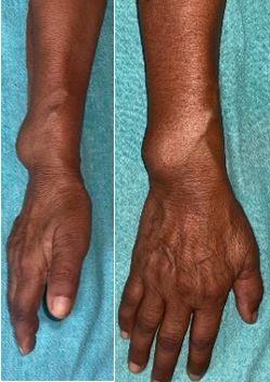
A 67-year-old female presented to our OPD with swelling on her right wrist from last 6 months which was increasing gradually, over time for which took treatment at various hospitals. Regarding her family history, there were no reports of related systematic or neoplasm diseases. On clinical examination, swelling was firm, non-tender and no signs of inflammation were present and percussion over mass produced a Tinel-like sensation along median nerve. Neither motor weakness nor muscular atrophy was observed. Other laboratory findings were normal. For final excision, a longitudinal incision which was centred over sweeling was made, without releasing transverse ligament of carpal tunnel. The lesion was revealed in an eccentric position along median nerve. A marginal swelling excision with preservation of median nerve was done after careful surgical manipulations. After complete excision of mass, clear damage to distal ulna and radius was seen. The defect of bone and soft tissue was postoperatively protected with a forearm splint for six weeks and final diagnosis of PNST was made on basis of Histological report. On final follow up for last 6 months patient did not have any recurrence of swelling with complete movements at wrist joint

Discussion and Conclusion

Pathology behind Peripheral Nerve Sheath tumours of hand is not clear, most of these tumors are misdiagnosed as ganglion cyst.[1] Histopathological examination is the most important for diagnosis. Kubiena et al.[7] showed that these lesions arise from the cells of the nerve sheath and engulf some nerve fascicles. However, bone destruction due to a benign NF is not so common. This presentation of bone damage is commonly seen in schwannomas, neuromas, and malignant tumors.[8] The diagnosis of Peripheral nerve sheath tumor always remains a challenge and multiple differential diagnosis even after using MRI remains challenging and following tumors can be considered: begin solitary neoplasms such as lipomas, fibromas, xanthomas, ganglion tumors, mucous cysts, glomus tumors, giant cell tumors of the tendon sheath, vascular tumors, as well as post-traumatic neuromas, in addition to low-grade malignant neoplasms.[9] A challenging step in the treatment is the complete tumor excision and the simultaneous preservation of nerve function. On the other hand, wide local dissection is difficult in patients who presents with large masses which infiltrate the surrounding soft tissue and bone.[6]

ojmpc_165_01.jpg
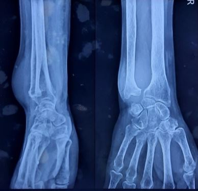
Figure 1 and 2: Swelling present over the wrist MRI and FNAC was done at some private institution reporting it as Ganglion cyst. On radiographic imaging, X-Ray showed the characteristic pattern of a large soft tissue mass damaging distal radio ulnar joint was noted.

ojmpc_165_03.jpg
Figure 3 and 4: Xray suggestive of mass in between radius ulna destroying radius, Ulna and DRUJ anatomy

ojmpc_165_05.jpg
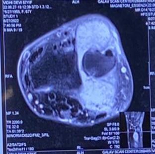
Figure 5:


ojmpc_165_06.jpg
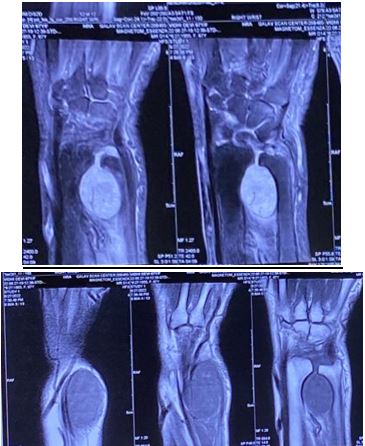
Figure 6 and 7: Well-defined PD FS hyperintense/T1 hypointense lesion noted in the distal forearm with widening of distal radioulnar joint. The lesion is seen the dorsal and volar aspect of distal forearm traversing in between distal radius and ulna. Th lesion is displacing the triangular fibrocartilaginous complex caudally.

ojmpc_165_08.jpg
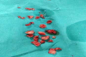
Figure 8: Intra operative Swelling Excision

ojmpc_165_09.jpg
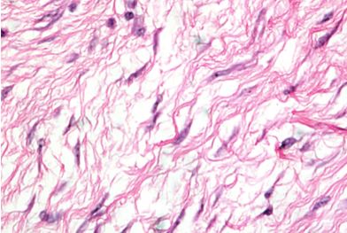
Figure 9: Histopathological slide showing tumour cells with elongated nuclei in collagen rich matrix

References

1. D. S. LouisandF. M. Hankin,“Benignnervetumorsoftheupper extremity,” Bulletin of the New York Academy of Medicine, vol. 61, no. 7, pp. 611–620, 1985 [Crossref][PubMed][Google Scholar]

2. World Health Organization. Lyon, France: International Agency for Research on Cancer; 2020. Soft Tissue and Bone Tumours. WHO Classification of Tumours, 5th edition. [Crossref][PubMed][Google Scholar]

3. The surgical management of symptomatic peripheral nerve sheath tumors. Levi AD, Ross AL, Cuartas E, Qadir R, Temple HT. Neurosurgery. 2010;66:833–840. [Crossref][PubMed][Google Scholar]

4. O. Ozdemir, C. Kurt, E. Coskunol, I. Calli, and M. H. Ozsoy, “Schwannomas of the hand and wrist: long-term results and review of the literature,” Journal of Orthopaedic Surgery, vol. 13, no. 3, pp. 267–272, 2005 [Crossref][PubMed][Google Scholar]

5. Surgical management of neuromas of the hand and wrist. Regal S, Tang P. J Am Acad Orthop Surg. 2019;27:356–363. [Crossref][PubMed][Google Scholar]

6. The surgical management of symptomatic benign peripheral nerve sheath tumors of the neck and extremities: an experience of 442 cases. Desai KI. Neurosurgery. 2017;81:568–580. [Crossref][PubMed][Google Scholar]

7. Peripheral neural sheath tumors (PNST)--what a radiologist should know. Kubiena H, Entner T, Schmidt M, Frey M. Eur J Radiol. 2013;82:51–55. [Crossref][PubMed][Google Scholar]

8. Schwannoma: a rare benign tumor of soft tissues. Mankin HJ, Mankin KP. Musculoskelet Surg. 2014;98:95–99. [Crossref][PubMed][Google Scholar]

9. Benign nerve tumours of the hand (excluding wrist) Gosk J, Gutkowska O, Urban M, Martynkiewicz J, Bąk M, Ziółkowski P. Arch Orthop Trauma Surg. 2015;135:1763–1769. . [Crossref][PubMed][Google Scholar]

Disclaimer / Publisher's Note

The statements, opinions and data contained in all publications are solely those of the individual author(s) and contributor(s) and not of Journals and/or the editor(s). Journals and/or the editor(s) disclaim responsibility for any injury to people or property resulting from any ideas, methods, instructions or products referred to in the content.