Orthopaedic Journal of MP Chapter

Publisher: Madhya Pradesh Orthopaedic Association www.mpioa.com
E-ISSN:2582-7243, P-ISSN:2320-6993
2022 Volume 28 Number 2 Jul-Dec

A rare solitary intraosseous calcaneal lipoma: case report

Kaunteya1, Bajoria R2*, Sahil3

1 Kaunteya, Department of Orthopedics, Gajra Raja Medical College, Gwalior, Mp, India.

2* RS Bajoria, Professor, Department of Orthopedics, Gajra Raja Medical College, Gwalior, Mp, India.

3 Sahil, Department of Orthopedics, Gajra Raja Medical College, Gwalior, Mp, India.

Intraosseous lipoma, a rare primary benign tumor of the skeleton, is most found in the calcaneus. It may contain homogenous fat, but it may also contain necrosis, calcification, or ossification. It usually does not show contrast enhancement, but there is an interface enhancement between the outer fat plain and the inner fluid part. Differential diagnoses for calcaneal lipoma include aneurysmal bone cyst, fibrous dysplasia, enchondroma, and intraosseous ganglia and UBC. Herein, we present an unusual 47-year-old female case of calcaneal intraosseous lipoma. Nonoperative treatment may be a viable option for many patients with small or asymptomatic lesions. Interventions include intralesional steroid injection, open curettage and bone grafting, decompression and percutaneous injection of bone marrow or graft substitutes. Attention is directed to the use of injectable steroid as an alternative means to open surgical intervention. Certain potential problems exist, despite the various surgical approaches utilized, including infection, postoperative fracture, recurrence, immobilization, and prolonged hospitalization.

Keywords: Intraosseous lipoma, calcaneus, intralesional steroid injection

Corresponding Author How to Cite this Article To Browse
RS Bajoria, Professor, Department of Orthopedics, Gajra Raja Medical College, Gwalior, Mp, India.
Email:
Kaunteya, Bajoria R, Sahil, A rare solitary intraosseous calcaneal lipoma: case report. ojmpc. 2022;28(2):83-85.
Available From
https://ojmpc.com/index.php/ojmpc/article/view/163
Manuscript Received Review Round 1 Review Round 2 Review Round 3 Accepted
2022-12-03 2022-12-10 2022-12-17 2022-12-24 2022-12-31
Conflict of Interest Funding Ethical Approval Plagiarism X-checker Note
Authors state no conflict of interest. Non Funded. The conducted research is not related to either human or animals use. 13.32 All authors have accepted responsibility for the entire content of this manuscript and approved its submission.

© 2022by Kaunteya, Bajoria R, Sahiland Published by Madhya Pradesh Orthopaedic Association. This is an Open Access article licensed under a Creative Commons Attribution 4.0 International License https://creativecommons.org/licenses/by-nc/4.0/ unported [CC BY NC 4.0].

Lipoma of bone is a benign neoplasm of adipocytes that typically arises within the medullary cavity of bone. Although there is a large amount of fatty tissue in bone marrow, intraosseous lipoma occurs very rarely, with a 0.1-2.1% prevalence [1-3]. However, this probably is not the actual incidence number because the lesions are frequently asymptomatic. Calcaneus bone is reported to be the most frequent site of intraosseous lipoma [4]. Herein, we present a case of a rare calcaneal intraosseous lipoma.

CASE PRESENTATION

A 47-year-old female patient presented with pain of approx. 6 months duration in the left heel (Figure1). The patient complained of pain on palpation of the medial aspect of the heel and hind foot. The pain increased on weight bearing and shoe contact and subsided with rest. There is no history of focal trauma, and the patient denied any pre-existing inflammatory conditions such as RA, or other metabolic disorders. The patient had been previously treated with NSAIDS with partial relief, but the pain continued.

ojmpc_163_01.jpg
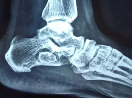
Figure 1:
Left foot lateral x-ray radiograph showing a well-defined, osteolytic lesion in the anterior portion of the body of the calcaneus, with a centralized radiodensity.

ojmpc_163_02.jpg
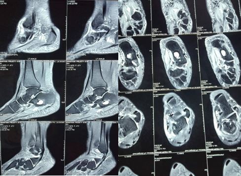
Figure 2:
MRI study of the Left ankle; small calcaneal lipoma (measuring ~ 1.7*1.2*1.0 cm).

Her medical history was unremarkable, and the physical examination was only significant for tenderness and edema localized to the medial aspect of the right heel. The range of motion of the ankle was normal;. Laboratory findings, including a complete blood count with differential cell count, erythrocyte sedimentation rate, C-reactive protein, and a biochemical profile that included serum alkaline phosphatase, calcium and phosphate levels were all in the normal range.

Initially a Xray of left foot advised, showing a well-defined, osteolytic lesion in the anterior portion of body of calcaneus, with a centralized radiodensity. A Non-contrast MRI shows small calcaneal lipoma (measuring ~ 1.7*1.2*1.0 cm) (Fig 2). According to Milgram's classification, lesion was graded as a stage 3 calcaneal lipoma because of necrosis, centrally cystic transformed areas and sclerosis in wall. Under aseptic precaution intralesional steroid injection (methyl prednisolone) applied and patient follow up done for 6 months. The result shows good functional outcome. Patient relieved from pain.

DISCUSSION

Intraosseous lipomas, which occur in both males and females, are present in fourth and fifth decades, with a prevalence of 0.1-2.1%. However, real prevalence is thought to be higher than this estimate because most cases are asymptomatic [2,3]. Therefore, most cases have been detected incidentally. Pain is major complication in symptomatic patients, and they present with pathological fractures.

Although intraosseous lipoma can occur anywhere in the skeletal system, 71% of these lipomas are in the lower extremities, and usually appears in the calcaneus (32%), in which the most frequently found tumor is also the intraosseous lipoma. The upper extremities, skull, mandible, and spine are involved with decreasing frequencies. Intraosseous lipomas located in long bones usually affect the metaphysis [5-8]. Basically, simple bone cysts called intraosseous ganglia are present in intraosseous lipomas. On X-rays, intraosseous lipomas appear as benign, osteolytic lesions with well-defined limits, and it is difficult to identify them only based on X-ray findings. Computerized tomography (CT), especially MRI, is useful in differential diagnosis. Radiographic diagnosis of a lipoma may not be straightforward and so there may be differential diagnosis that may include simple bone cyst, nonossifying fibroma, aneurysmal bone cyst, fibrous dysplasia, bone infarct, giant cell tumor, chondroid tumor or fungal infections. The appearance of these lesions on radiographs, CT scans and MRIs can vary as result of their degree of involution and necrosis. It is important to make a correct diagnosis based on multi-method imaging studies, especially MRI.

In classifications performed by Milgram et al. [4], intraosseous lipomas are divided into three stages based on fat necrosis: Stage 1A- sharply limited lesion with homogenous fat content; Stage 2- Dominantly fatty lesions with central necrosis, calcification, or ossification, and Stage 3- A heterogeneous, fat- containing lesion involving multiple necrotic areas, cystic transformations, sclerosis, or ossification in wall. Our case was staged as 3 according to this classification.


Because calcaneal lipomas do not affect bone stability, conservative treatment can be applied. The fracture can be treated with curettage and bone graft in cases of doubt. There is usually no recurrence after surgery [5]. Non-operative treatment may be a viable option for many patients with small or asymptomatic lesions. Interventions include intralesional steroid injection, open curettage and bone grafting, decompression and percutaneous injection of bone marrow or graft substitutes.

Certain potential problems exist, despite the various surgical approaches utilized, including infection, postoperative fracture, recurrence, immobilization, and prolonged hospitalization.

CONCLUSION

Proper diagnosis and appropriate therapeutic management are necessary not only prevent the recurrence but also to exclude other cystic lesions of calcaneum. Intraosseous lipomas can be diagnosed easily with their fatty component, especially via MRI. Steroid injection remains a reliable method for treating calcaneal lipoma owing to its low invasiveness.Steroid injection therapy for calcaneal lipoma yielded a good functional result. Patient got relieved from pain and procedure is economical with less complications. As it is an outdoor procedure, so hospital stay is avoided. Therefore, this procedure could be a choice for treatment of calcaneal lipoma/SBCs patient. To furthermore strengthen this conclusion, Further study needs to be conducted with more sample size.

Informed consent

Written informed consent was obtained from the patient for the publication of this case report.

Conflict of interest

The authors declared that there are no potential conflicts of interest with respect to the research, authorship, and/or publication of this article.

References

1. Campanacci M, Enneking WF. Bone and soft tissue tumors: clinical features, imaging, pathology and treatment. 2nd ed. Berlin: Springer, 1999, pp. 683-9 [Crossref][PubMed][Google Scholar]

2. Myhre-Jensen OA. Consecutive 7-year series of 1331 benign soft tissue tumors. Clinicopathologic data. Comparison with sarcomas. Acta Orthop Scand 1981;52:287-93 [Crossref][PubMed][Google Scholar]

3. Unni KK, Inwards CY. Dahlin's bone tumors: general aspect and data on 10165 cases. 6th ed. Philadelphia: Lippincott Williams & Wilkins, 2006, pp. 298-300 [Crossref][PubMed][Google Scholar]

4. Milgram JW. Intraosseous lipomas. Clin Orthop Relat Res 1988;231:277-302. . [Crossref][PubMed][Google Scholar]

5. Campbell RS, Grainger AJ, Mangham DC, Beggs I, Teh J, Davies AM. Intraosseous lipoma: report of 35 new cases and a review of the literature. Skeletal Radiol 2003;32:209-22. . [Crossref][PubMed][Google Scholar]

6. Blacksin MF, Ende N, Benvenia J. Magnetic resonance imaging of intraoeeous lipomas: a radiologic-pathologic correlation. Skletel Radiol. 1995; 24:37-41. [Crossref][PubMed][Google Scholar]

7. Bagatur AE, Yalcinkaya M, Dogan A, Gur S, Mumcuoglu E, Albayrak M. Surgery is not always necessary in intraosseous lipoma. Orthopedics 2010;33:306. . [Crossref][PubMed][Google Scholar]

8. Hassani M, Gharehdaghi M, Khooei AR, Ghodsi E, Nazarzadeh H. Bilateral intraosseous tumor of the calcaneus with imaging-pathologic discordance. a case report and literatures review. Arch Bone Jt Surg 2014;2:238-42. [Crossref][PubMed][Google Scholar]

Disclaimer / Publisher's Note

The statements, opinions and data contained in all publications are solely those of the individual author(s) and contributor(s) and not of Journals and/or the editor(s). Journals and/or the editor(s) disclaim responsibility for any injury to people or property resulting from any ideas, methods, instructions or products referred to in the content.