Orthopaedic Journal of MP Chapter

Publisher: Madhya Pradesh Orthopaedic Association www.mpioa.com
E-ISSN:2582-7243, P-ISSN:2320-6993
2022 Volume 28 Number 2 Jul-Dec

A Prospective Analysis of Functional Outcome of Surgical Stabilization of Distal End Radius fractures with Plate Osteosynthesis.

Barua VK1*, Sirsikar A2, Naik S3, Sharma T4

1* Vinay Kumar Barua, Department of Orthopaedics, Netaji Subhash Chandra Bose Medical College, Jabalpur, M P, India.

2 A Sirsikar, Department of Orthopaedics, Netaji Subhash Chandra Bose Medical College.

3 S Naik, Department of Orthopaedics, Netaji Subhash Chandra Bose Medical College.

4 T Sharma, Department of Orthopaedics, Netaji Subhash Chandra Bose Medical College.

Introduction: Distal radius fractures (DRF) are the most common fractures of the upper extremities and due to population explosion, with an ageing society & enormous increase of high-speed motor vehicle accidents, the number of distal radial fractures can be expected to increase in the coming decades. Plate osteosynthesis has become the standard treatment for comminuted intra articular DRF. Main aim of this study was to analyse the functional outcome in patients stabilized by plate osteosynthesis with minimum follow up of 6 months.

Material and Methods: A prospective study was done on 46 patients with distal radius fractures who were operated with plate osteosynthesis, type of plate was decided as per fracture pattern and followed up at 1, 3, and 6 months and outcomes measured using Modified Mayo score, Grip strength tested by Dynamometer.

Observations: Volar plating was done in 34 patients; dual plating was done in 4 patients dorsal column plating was done in 8 patients. Average time of radiological union of fracture was 10 weeks, average time of clinical union of fractures was 8 weeks, average time to return to normal activity was 3 weeks, average time to return to professional activity was 4 weeks.

Results: According to Modified MAYO score 18 patients had excellent results, 10 had good results, 8 had fair and 4 had poor results. Grip strength was >80% compared to normal side in 29 patients, >60% in 8 patients and, <60% compared to normal side in 3 patients measured with a Dynamometer at 6 months.

Conclusion: Fractures of distal end radius managed with plate osteosynthesis is a good treatment modality with excellent results provided the surgeon has a sound knowledge of literature and a good surgical hand.

Keywords: distal end radius, plate osteosynthesis

Corresponding Author How to Cite this Article To Browse
Vinay Kumar Barua, , Department of Orthopaedics, Netaji Subhash Chandra Bose Medical College, Jabalpur, M P, India.
Email:
Barua VK, Sirsikar A, Naik S, Sharma T, A Prospective Analysis of Functional Outcome of Surgical Stabilization of Distal End Radius fractures with Plate Osteosynthesis.. ojmpc. 2022;28(2):51-59.
Available From
https://ojmpc.com/index.php/ojmpc/article/view/157
Manuscript Received Review Round 1 Review Round 2 Review Round 3 Accepted
2022-12-03 2022-12-10 2022-12-17 2022-12-24 2022-12-31
Conflict of Interest Funding Ethical Approval Plagiarism X-checker Note
Authors state no conflict of interest. Non Funded. The conducted research is not related to either human or animals use. 15.65 All authors have accepted responsibility for the entire content of this manuscript and approved its submission.

© 2022by Barua VK, Sirsikar A, Naik S, Sharma Tand Published by Madhya Pradesh Orthopaedic Association. This is an Open Access article licensed under a Creative Commons Attribution 4.0 International License https://creativecommons.org/licenses/by-nc/4.0/ unported [CC BY NC 4.0].

Introduction

Distal radius fractures (DRF) are the most common type of fracture of the upper extremities and incidence is expected to rise due to a growing elderly population1. Especially women have a 15% higher life time risk of DRF, than men of similar age. In addition, DRF in the elderly are often associated with poor bone quality and osteoporosis2-4.

Historically, DRF were conservatively treated by closed reduction and immobilization or K-wires. Following the introduction of angular stable locking plates and the excellent results using internal fixation, a treatment shift occurred away from K-wires or external fixator to plate osteosynthesis. Thus, displaced DRF (Barton fractures) can be stabilized from plate osteosynthesis. In addition, stabilization by plate osteosynthesis provides enough stability to enable early active wrist rehabilitation without immobilization. Multiple studies showed a significantly improved functional outcome compared to immobilization and an early mobilization post-surgery has no increased risk of secondary loss of reduction and complications8-10.

As incidence of DRF rises and the number of patients treated by plate osteosynthesis increases, literature remains interested in the optimal treatment method, clinical outcomes and complication rates10,11. Complication rates after palmarly stabilized DRF are reported up to 39% and complication rates after dorsally stabilized DRF are reported upto 28%(17) whereas other studies documenting outcome after DRF showed good functional and radiological results5,8,12-15.

Main aim of this study was to evaluate the functional outcome in cases of fracture distal end radius treated by open reduction and internal fixation using different plates decided according to fracture pattern and to study the complications related to the use of these plates.

Materials and methods

Institutional review board and Institutional Ethical approval was obtained for this prospective follow-up study. All patients treated with plate osteosynthesis from 01 January 2021 to 31 June 2022 that met inclusion/excision criteria, were included in this study and invited in writing and by telephone to attend the follow-up investigation. Three invitation letters were sent to each patient. Failure to reply after the third invitations was classified as a non-responder. Written informed consent was obtained from all participating patients. They were treated exclusively at our hospital, NSCB Medical College and Hospital, Jabalpur.

Indications for surgery included a displaced DRF with a dorsal tilt of more than 15 degrees, an intra-articular step of more than 1mm, a radial shortening of more than 2mm or an incongruency in the distal radioulnar joint in the standard radiographs.

Inclusion criteria included patients (aged between 18 years and 55 years) with unstable, comminuted or intra articular and extra articular fractures of distal end radius.

Exclusion criteria included patients aged below 18 years, patients medically unfit for surgery, pathological fractures, compound fractures, patients who are not willing for surgery. and trauma cases > 4 weeks.

From 2021 to 2022, a total of 46 patients were stabilized by plate osteosynthesis. Of these, 34 patients were stabilised with volar plating 8 patients with dorsal plating and 4 with dual plating. Out of 46 patients 40 were followed up for 24 weeks and 6 patients were lost, 4 at 8th week and 2 at 12th week. Therefore, the final analysis totalled 40 patients.

All procedures were performed using either general or regional anesthesia in a supine position, with fluoroscopic assistance and a pneumatic arm tourniquet of 250mmHg. A standard Modified Henry’s approach between the flexor carpi radialis tendon and radial artery was chosen for volar plating. The flexor carpi radialis tendon was retracted ulnarly and the forearm fascia was dissected. The pronator quadratus was incised radially and elevated of the radius.

For dorsal approach about 8 cm midline incision taken (halfway between radial and ulnar styloid) which can extend proximally or distally as needed subcutaneous fat incised in line with skin incision to expose extensor retinaculum, extensor retinaculum incised over the extensor digitorum communis and extensor indicis proprius(fourth compartment) tendons are mobilised radially and ulnarly to expose the underlying radius and joint capsule, the joint capsule is incised longitudinally on the dorsal radius and carpus dissection is continued below the capsule (dorsal radiocarpal ligament) toward the radial and ulnar sides of the radius to expose the entire distal radius in dorsal approach.

The fracture was reduced under image intensification and, when necessary, temporarily fixed with K-wires. The plate was placed and initially fixed with a bicortical screw through the gliding hole. After ensuring exact positioning of the plate under image intensifier, the remaining plate holes were filled with angular stable screws. Care was taken that the screws at the articular surface were placed subchondrally to prevent dorsal protrusion. Screw length was taken 2mm shorter to prevent protrusion.


In 34 patients volar plating was done in 8 patients dorsal plating was done and in 4 dual plating was done. Routine antibiotics and anti-inflammatory drugs were given. Check x-ray were taken on 3rd postoperative day after Check Dress 1. Sterile dressings were done on 3rd and 5th postoperative day. Sutures were removed on 10th to 15th post operative day and patient were discharged with below elbow pop slab. Patients were assessed clinically and radiographically at 8 weeks, 12 weeks, and 24 weeks to assess the fracture union and the progress of patients recovery were documented.

All patients started handtherapy of the free joints (shoulder, elbow, fingers) for both upper extremities on the first postoperative day. After slab removal the wrist was then included in physiotherapy programme.

Outcome evaluation each of the patients, who returned for the follow-up investigation, underwent a standard X-ray of the wrist in two planes (anteroposterior and lateral view.

Range of motion (ROM) was measured in palmar flexion, dorsi flexion, supination, pronation, radial- and ulnar deviation at the follow-up investigation. Demographic data included age, gender, injured hand, mode of injury and interval between surgery and follow-up. In addition functional outcome analysed with Modified Mayo score and Demerit Point System of Gartland and Werley, grip strength by dynamometer were analyzed.

All the intraoperative and postoperative complications that were documented in the surgical write ups were recorded and each return evaluation was analysed for complication. Complex regional pain syndrome (CRPS) was diagnosed clinically based on the Veldman’s criteria (16,17). Frequency and causes of complication were analysed.

The primary (pre-reduction), immediate postoperative as well as final radiographs were checked for alignment and intra-articular step-of. The fractures were classified according to the Frykmann classification. An acceptable reduction was defined as 10 degrees of dorsal tilt, 15 degrees in radial inclination, 2mm ulnar variance and 2mm of articular incongruity (12,18).

In the anteroposterior radiographs, radial inclination and radial length and in the lateral radiographs, the palmar tilt was measured (19) Fracture healing was defined as bony bridging of the radial, ulnar, and dorsal cortical aspects of the distal part of the radius (12) . The lateral X-ray verified the plate position and was subsequently classified according to Soong etal. in Grade 0, I and II. (20)

The statistical analysis was performed by SPSS 23.0 (Statistical Package For Social Sciences). This was a prospective study. Descriptive statistics were performed to all study variables.

Continuous variables are described as mean and standard deviation. Categorical variables were described as frequency and percentage and were described with graphs, bar charts and pie charts.To compare scaled parameters Paired t-test was used. Chi-square was used for testing categorical data. If p value <0.05 data was considered significant at 5% level of significance and if p value ≤0.01 was considered significant at level of significance.

Results

A total of 46 patients (28 males, 18 females) returned for the follow-up investigation with a mean ± SD age of 34.71 ± 6.80years (range 18-55years) and follow-up of 6 months. Detailed demographic data is presented below.

In our study the average radial inclination preoperatively was 7.76±5.8 degrees, the average postoperative radial inclination was 18.2±3.3degrees. The average radial inclination achieved was 10.44 degrees study. Preoperative mean radial length 3.66±1.79 mm was observed preoperatively with an immediate postoperative radial length of 9.08±1.65 mm, we achieved a mean correction of 6.15±2.66 mm during the surgical procedure.

The preoperative mean volar tilt was -17.1±7.82 degrees and mean postoperative volar tilt was 6.95±4.54 degrees the total correction achieved was 19.63±7.56 degrees the higher degree of correction achieved was due to the fact that the dorsal tilt was expressed in negative value and hence the correction achieved was greater than the normal range (0-11degrees). (Table 1)

Table 1: Detailed functional outcome measuring range of motion.

MovementsNMinMaxMean ± SD
Week- 8 Pronation46518065.61 ± 8.60
Week- 12 Pronation405810082.30 ± 10.70
Week- 24 Pronation4068135111.35 ± 18.40
Week 8 Supination46153223.35 ± 4.61
Week 12 Supination40244234.98 ± 5.40
Week 24 Supination40284136.5 ± 2.9
Week 8 Palmar Flexion46154124.76 ± 6.34
Week 12 Palmar Flexion40244937.90 ± 5.7
Week 24 Palmar Flexion40297054.55 ± 8.80
Week 8 Dorsi Flexion46163925.04 ± 5.70
Week 12 Dorsi Flexion40264939.83 ± 5.54
Week 24 Dorsi Flexion40296955.33 ± 8.37
Week 8 Radial Deviation464107.04 ± 1.67
Week 12 Radial Deviation4061210.63 ± 1.46
Week 24 Radial Deviation4091814.65 ± 2.30
Week 8 Ulnar Deviation4691814.02 ± 2.22
Week 12 Ulnar Deviation40122520.40 ± 2.75
Week 24 Ulnar Deviation40143025.78 ± 3.93

The mean range of motion achieved in our study was as follows palmar flexion of 54.55 ± 8.80 degrees, dorsiflexion of 55.33 ± 8.37 degrees, radial deviation of 14.65 ± 2.30 degrees, ulnar deviation of 25.78 ± 3.93degrees, supination of 55.55 ± 57.99degrees, pronation of 111.35 ± 18.40degrees. these results were taken at 6 months postoperatively and were compared with the normal side, they required 18.2±16 physiotherapy sessions to attain range of motion described at 6 months. (Table 1)

ojmpc_157_01.jpg
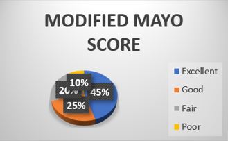
Figure 1: Figure 1. Figure showing Using the MODIFIED MAYO score, we had 18 (45%) excellent results, 10 (25%) good results, 8 (20%) fair results and 4 (10%) poor results with a mean ± SD Modified Mayo score of 85.6 ± 10.21.

ojmpc_157_02.jpg
Figure 2:According to Demerit point system of Gartland and Werley we had 22 (55%) excellent results, 10 (25%) good results, 5 (12.5%) fair results, 3 (7.5%) poor results with a mean±SD of Demerit Point System Of Gartland and Werley of 7.23 ± 5.39.

Using the MODIFIED MAYO score, we had 18 (45%) excellent results, 10 (25%) good results, 8 (20%) fair results and 4 (10%) poor results with a mean ± SD Modified Mayo score of 85.6 ± 10.21.

Table 2: Functional outcome using scoring systems.

ScoreNMinMaxMean ± SD
Modified Mayo Score40599785.6 ± 10.21
Demerit point system of Gartland and Werley401197.23 ± 5.39

A total of 8 complications (17.3%) occurred in 46 patients. Most common complications included CRPS in 4 patients (8.7%). Complications are summarized in Table 3 below.

Table 3: Percentage of complications.

CRPS48.7
Median Nerve Entrapment under Surgical Scar12.2
Dorsal Tendon Attrition12.2
Superficial Wound Infection24.3
No Complication3882.6
Total46100.0

Intra articular fractures showed the highest complication rate of 7/34 (20.5%), whereas 1/12 (8.4%) were found in extraarticular fractures. Median Nerve Entrapment under Surgical Scar was found in 1 patient which was confirmed by ultrasonography and was managed with excision of surgical scar followed by median nerve neurolysis and carpal tunnel release it showed a complete regression. All patients with CRPS were treated conservatively with splinting, hand therapy, dimethylsulphoxide (DMSO) ointment, non-steroidal anti-inflammatory drugs and vitamin E. Superficial wound infection was observed in two patients, which was treated conservatively with antibiotics and splinting.

Dorsal tendon attrition was found in one patient of dorsal plating who was managed with hardware removal after radiological union. No significant differences could be found in incidence of complications and plate type (p=0.22), age (p=0.47], gender (p=0.50), or post-operative immobilization [cast/thermoplastic splint (p=0.31)].

Discussion

Distal radius fractures are one of the most common fractures in the upper extremities and the incidence is expected to continue rising due to the growing elderly population(22). Since the introduction of plate osteosynthesis in the early 2000s plus the initial reports of low complication rates and good functional outcomes, plate osteosynthesis has gained popularity in treating DRF(5.46.47).

Palmar locking plate fixation enables a stabilization of dorsally displaced fractures without the increased risk of tendon irritation compared to dorsal stabilization(5,6,23). Fixation of DRF provides enough stability to allow an early rehabilitation with active wrist mobilization. Thereby, better functional outcomes can be achieved in the early rehabilitation phase without the increased risk of a loss of reduction or further complications(10,24). Therefore, functional outcome and reported complications after operatively treated DRF remain current in the literature including a comparison of the various available treatment options(7,25).


Today the optimal treatment options for DRF are under debate, but a recent Network Meta-analysis concluded, that plate fixation offers the best results in terms of early functional outcome and reduction of fracture healing complications. Patients in this study, with a mean age of 34.7years and a minimum follow-up of six months showed a good functional and in mean an “acceptable” radiological outcome. The Modified Mayo Score averaged 86 points and Demerit Point system of Gartland and Werley 7 points.

Each patient’s MODIFIED MAYO score were taken at 8 weeks, 3 months and 6 months interval along with range of motion. Phadnis J et al in 2011 to report the functional outcome of a large number of patients at a significant follow up time after fixation of their distal radius with a volar locking pate reported 74% of the patients with good or excellent DASH and MODIFIED MAYO score. Statistical analysis showed that no specific variable including gender, age, fracture type, post-operative immobilisation or surgeon grade significantly affected outcome. Complication occurred in 27 patients (15%) and in 11 patients was major (6%) study demonstrated good to excellent results in the majority of patients after volar locking plate fixation of the distal radius, with complication rates comparable to other non-operative and operative treatment modalities and recommended this mode of fixation for distal radius fractures requiring operative intervention. Rozental et al. showed mostly good and excellent functional outcomes in 45 patients at 17 months mean follow up. Like our study these both showed good to excellent functional outcome using the MODIFIED MAYO score. Rohit Arora et al. used modified Green and Obrein score and reported 31 excellent, 54 good, 23 fair and 6 poor results. Minegishi H et al in 2011 to evaluate the functional and radiological results of treating unstable distal radius fractures with the volar locking plated among 15 patients reported 5 patients with excellent outcome, 7 with good outcome, and 3 with fair outcome according to Cooney’s clinical scoring chart.

K .Egol et al showed a mean Modified MAYO of 78.2±7.7 in external fixator group and 87±4.9 in volar plating group at 12months, but their functional MODIFIED MAYO score at 6 months was 72.6±23.8 in external fixator group ,89.0±21.7 in plating group at 3 months interval their DASH scores was 71.4±21.1 in external fixator group and 89.5±2.1 in plating they did not compare MODIFIED MAYO scores at 8 weeks interval. Adani R et al evaluated MODIFIED MAYO scores sequentially weekly upto 12 weeks and at final examination they reported 94.6±6.3 in the conventional group and 96.2±6.8 in the MIPPO group at 12 weeks.

Jirangkul P et al recorded a modified MAYO score 87±7 at 6weeks in ORIF group and 73±28 in CRPP group at 6 weeks they reported a MODIFIED MAYO score 89±13 in ORIF group and 74±23 in CRPP group at 12 weeks with significant P value of 0.01 they recorded another MODIFIED MAYO score at 1 year in which the ORIF group fared with 4±8 score and CRPP fared 9±18 with no significant P value. Ballal A et al reviewed 20 patients, five patients had excellent modified Mayo wrist score, 9 had good scores, 4 had satisfactory and two patients had poor results. Seven patients had a RUSS score less than five points and four patients had RUSS score of five points, four patients had six points, two patients had seven points and three patients had eight points. One patient was noted to have dorsal collapse of the fracture during the final review. But, no evident of cosmetic deformity or any diminution in functional outcome of wrist was noted. In present study MODIFIED MAYO score at 6months follow up is 85.6 ± 10.21. Sohael M. Khan et al recorded a DEMERIT POINT SYSTEM OF GARTLAND AND WERLEY score of 3.75 in ORIF group and 7.55 in CRPP group at 36 weeks with significant P value of <0.05. in their study 70% patients had excellent results, 20% patients had good results and 10% had fair results. In our study 22 (55%) patients had excellent results, 10 (25%) had good results, 5(12.5%) patients had fair results and 3(7.5%) patients had poor results. Mean Demerit Point system of Gartland and Werley at the end of 6 months follow up was 7.23 ± 5.39.

Complications after plate osteosynthesis for DRF are well reported in the literature. First reports from Orbay etal. suggested a complication rate of 3%(26), respectively 4%(27) but later studies reported complications up to 60% (5,7,21,25). In a recent systemic review Alter etal.(25) analyzed complications, they reported a complication rate of 15% in 3.911 operatively treated DRF with palmar locking plate. Jie Wei et al in 2013 ( ) did a meta analysis and found that dorsal fixation offers a lower risk of neuropathy and carpal tunnel syndrome than the volar approach, but a higher risk of tendon irritation. Patients with a distal radius fracture can expect similar outcomes after volar or dorsal surgery. Complication rate of 17.4% in this study is comparable to previously published studies and the low complication rate reflects the familiarity with the implant and large numbers of treated DRF by plate osteosynthesis (average 262 DRF per year). The most common complications in this study included CRPS (8.6%) and superficial wound infection (4.3%). No significant impact on the complication rate could be found for age, gender or type of post-operative immobilization.

Occurrence of a complication showed no significant or clinical important influence on the final functional outcome.


Intra-articular screws are also frequently reported in the literature between 0.5 and 1.3%(5,28) and not only caused by malpositioning, but also due to loss of reduction or secondary fracture dislocation. Even the use of angular stable screws does not preclude secondary displacement(5). Intra-articular screw penetration can result in a destruction of the radiocarpal joint, causing malunion, osteoarthritis and clinical failure. CRPS is closely associated to fractures of the distal radius with an incidence between 1 and 6%(5), but is also commonly seen in injuries to the upper extremities (most commonly in DRF)(5,25,29,30). However, it remains a clinical diagnosis and the pathomechanism is still not fully researched. This may, however, be related to an over excretion of cytokinins, mitochondrial dysfunction in the affected upper extremity, as well a genetic predisposition does exist(31,32). We agree with Esenwein etal. that CRPS is a complication, that cannot be influenced by the surgeon(5). Some of the limitations should be addressed before interpreting this study. The study included a total of 46 operatively treated DRF in the study period as due to COVID-19 the number of accidents and number of patients visiting the hospitals decreased significantly. Of these 6(13%) couldn’t be followed-up for several reasons. Thus, clinical results and complication rate could be biased. On the one hand, one might assume that patients who do not return have no complications and are asymptomatic, indicating that the complication rate is overestimated and the clinical results are better than reported. Alternatively, patients with complications or problems could simply have gone to another hospital. In addition, there is no unique definition in the literature for an “acceptable” postoperative radiological result and a wide range for cut-of values does exist. It would be desirable, that further studies focus on specific cut-of values to determine which radiological parameters would affect range of motion. At the follow-up radiographs were only taken of the injured wrist and not from the contralateral wrist. Therefore, no comparison with the uninjured wrist was possible. This might explain the discrepancy in the results in ulnar variance, showing a significant correlation to grip strength and range of motion, but no differences between an unacceptable and acceptable ulnar variance. Further studies should consider this issue and investigate

the impact of radiological differences between the injured and healthy wrist. Another limitation is that the study was not focused on one particular outcome parameter (e.g., Modified Mayo Score at the last follow-up), resulting in multiple testing. P-value had to be corrected and therefore, the study might be underpowered in some subgroup analyses. At the final follow-up examination, the X-rays of only 2/46, respectively, 3/46 patients showed an unacceptable palmar tilt or radial inclination.

Due to the small sample size a comparison between an acceptable and unacceptable radiological result depending on functional outcome was not possible.The minimum follow-up interval of this study was six months. Therefore, not all complications that typically occur later, for example, tendon rupture, are covered in this study.

Conclusion

Due to aging society, & enormous increase of high-speed motor vehicle accidents, the number of distal radial fractures can be expected to increase in the coming decades. In this study, forty six cases of distal radius fractures who were treated with open reduction and internal fixation with plate osteosynthesis were followed up and functional outcomes were analysed and discussed. From this sample study, we conclude that plate osteosynthesis provides successful results for the treatment of both extra articular and intra articular unstable fractures of distal radius. This method allows restoration of the anatomy, stable internal fixation, a decreased period of im-mobilisation and early return of wrist function. This method, which is effective in anatomic realignment, allows early joint motion, owing to its fixation strength. In the subjects of our study, a successful anatomic alignment was acquired, regardless of the direction of fracture angulation. The patients who were young adults in majority, went back to their daily activities with 90% recovery. Close placement to joint interface and screwing capability in different orders are biomechanical superiorities of a locking plate. The pre-contoured anatomical LCP not only provide restoration of radial length but also helps in stabilizing angulation. They maintain intra-articular congruity thus reducing radio carpal arthritis and decrease in grip strength. They also provide quicker recovery and better functional range of movement and provide better fixation in a osteoporotic bone. In our study excellent to good results suggests that stabilizing the fracture fragments with locking plate is an effective method to maintain the reduction till union and prevent collapse of the fracture fragments, even when the distal radius fracture is grossly comminuted intra-articular / unstable and or the bone is osteoporotic. It is a simple and reproducible procedure that improves recovery from this common injury. The technique emphasis that ORIF with plating has excellent functional outcome with minimal complications thus proving that it is the prime modality of

treatment for distal radius fractures. The procedure is applicable for AO types A, B and C fractures of the distal radius, in young patients with a good bone stock as well as in elderly osteoporotic patients. In conclusion, we looked at Functional results of locking compression plates and found an improved range of movement and radiological outcome at eight, twelve and twenty-four weeks follow up.


Thus, this study demonstrates that with the execution of good surgical techniques, including proper plate position, proper insertion of screws and avoidance of past pointing, and proper patient selection, a satisfactory functional and radiological outcome can be obtained for a great majority of patients with most of the distal radius fracture’s (incl. Complex intra-articular) by using a locking plate fixation.

References

1. MacIntyre NJ, Dewan N (2016). Epidemiology of distal radius fractures and factors predicting risk and prognosis. J Hand Ther 29:136–145. . [Crossref][PubMed][Google Scholar]

2. Figl M, Weninger P, Liska M et al (2009). Volar fixed-angle plate osteosynthesis of unstable distal radius fractures. 12 months results. Arch Orthop Trauma Surg. 129:661–669 [Crossref][PubMed][Google Scholar]

3. Figl M, Weninger P, Jurkowitsch J et al (2010). Unstable distal radius fractures in the elderly patient-volar fixed-angle plate osteosynthesis prevents secondary loss of reduction. J Trauma Inj Infect Crit Care. 68:992–998. [Crossref][PubMed][Google Scholar]

4. Gologan RE, Koeck M, Suda AJ, Obertacke U (2019). %3e 10-year outcome of dislocated radial fractures with concomitant intracarpal lesions as proven by MRI and CT. Arch Orthop Trauma Surg. 139:877–881. [Crossref][PubMed][Google Scholar]

5. Esenwein P, Sonderegger J, Gruenert J et al (2013). Complications following palmar plate fixation of distal radius fractures. a review of 665 cases. Arch Orthop Trauma Surg. 133:1155–1162 [Crossref][PubMed][Google Scholar]

6. Le ZS, Kan SL, Su LX, Wang B (2015). Meta-analysis for dorsally displaced distal radius fracture fixation. volar locking plate versus percutaneous Kirschner wires. J Orthop Surg Res. [Crossref][PubMed][Google Scholar]

7. Quadlbauer S, Pezzei C, Jurkowitsch J et al (2018). Early complications and radiological outcome after distal radius fractures stabilized by volar angular stable locking plate. Arch Orthop Trauma Surg. 138:1773–1782. [Crossref][PubMed][Google Scholar]

8. Quadlbauer S, Pezzei C, Jurkowitsch J et al (2016). Early rehabilitation of distal radius fractures stabilized by volar locking plate. a prospective randomized pilot study. J Wrist Surg 06:102–112. [Crossref][PubMed][Google Scholar]

9. Lozano-Calderón SA, Souer S, Mudgal C et al (2008). Wrist mobilization following volar plate fxation of fractures of the distal part of the radius. J Bone Jt Surg Ser A. 90:1297–1304. [Crossref][PubMed][Google Scholar]

10. Osada D, Kamei S, Masuzaki K et al (2008). Prospective study of distal radius fractures treated with a volar locking plate system. J Hand Surg Am. 33:691–700. [Crossref][PubMed][Google Scholar]

11. Lameijer CM, ten Duis HJ, van Dusseldorp I et al (2017). Prevalence of posttraumatic arthritis and the association with outcome measures following distal radius fractures in non-osteoporotic patients. a systematic review. Arch Orthop Trauma Surg 137:1499–1513. [Crossref][PubMed][Google Scholar]

12. Arora R, Lutz M, Deml C et al (2011). A Prospective randomized trial comparing nonoperative treatment with volar locking plate fxation for displaced and unstable distal radial fractures in patients sixty-fve years of age and older. J Bone Jt Surg Am. 93:2146–2153. [Crossref][PubMed][Google Scholar]

13. Arora R, Lutz M, Hennerbichler A et al (2007). Complications following internal fxation of unstable distal radius fracture witha palmar locking-plate. J Orthop Trauma. 21:316–322. [Crossref][PubMed][Google Scholar]

14. Haug LCP, Deml C, Blauth M, Arora R (2011). Dorsal screw penetration following implant removal after volar locked plating of distal radius fracture. Arch Orthop Trauma Surg. 131:1279–1282. [Crossref][PubMed][Google Scholar]

15. Erhart S, Toth S, Kaiser P et al (2018). Comparison of volarly and dorsally displaced distal radius fracture treated by volar locking plate fixation. Arch Orthop Trauma Surg. 138:879–885. [Crossref][PubMed][Google Scholar]

16. Veldman PH, Reynen HM, Arntz IE, Goris RJ (1993). Signs and symptoms of refex sympathetic dystrophy. prospective study of 829 patients. Lancet (London, England). 342:1012–1016 [Crossref][PubMed][Google Scholar]

17. Crijns TJ, van der Gronde BATD, Ring D, Leung N (2018). Complex regional pain syndrome after distal radius fracture is uncommon and is often associated with fibromyalgia. Clin Orthop Relat Res. 476:744–750. [Crossref][PubMed][Google Scholar]

18. Ng CY, McQueen MM (2011). What are the radiological predictors of functional outcome following fractures of the distal radius. J Bone Joint Surg Br. 93(B):145–150. [Crossref][PubMed][Google Scholar]

19. Schmitt R, Pommersberger K (2014). Karpale Funktion und Morphometerie. In: Schmitt R, Lanz U (eds) Bildgebende Diagnostik der Hand, 3rd edn. Thieme, Germany. pp 184–197 [Crossref][PubMed][Google Scholar]

20. Soong M, Earp BE, Bishop G et al (2011). Volar locking plate implant prominence and flexor tendon rupture. J Bone Jt Surg Ser A. 93:328–335. [Crossref][PubMed][Google Scholar]


21. Schlickum L, Quadlbauer S, Pezzei C et al (2018). Three-dimensional kinematics of. the fexor pollicis longus tendon in relation to the position of the FPL plate and distal radius width. Arch Orthop Trauma Surg. [Crossref][PubMed][Google Scholar]

22. Pillukat T, Fuhrmann R, Windolf J, van Schoonhoven J (2016). Die palmare winkelstabile Plattenosteosynthese bei Extensions frakturen des distalen Radius. Oper Orthop Traumatol. 28:47–64. [Crossref][PubMed][Google Scholar]

23. Zhang B, Chang H, Yu K et al (2017). Intramedullary nail versus volar locking plate fxation for the treatment of extra-articular or simple intra-articular distal radius fractures. systematic review and meta-analysis. Int Orthop 41:2161–2169. [Crossref][PubMed][Google Scholar]

24. Quadlbauer S, Pezzei C, Jurkowitsch J et al (2020). Rehabilitation after distal radius fractures. is there a need for immobilization and. physiotherapy Arch Orthop Trauma Surg. [Crossref][PubMed][Google Scholar]

25. Alter TH, Sandrowski K, Gallant G et al (2019). Complications of volar plating of distal radius fractures. a systematic review. J Wrist Surg. 08:255–262 [Crossref][PubMed][Google Scholar]

26. Orbay JL, Fernandez DL (2002). Volar fixation for dorsally displaced fractures of the distal radius. a preliminary report. J Hand Surg Am. 27:205–215 [Crossref][PubMed][Google Scholar]

27. Orbay JL, Fernandez DL (2004). Volar fixed-angle plate fixation for unstable distal radius fractures in the elderly patient. J Hand Surg Am. 29:96–102. [Crossref][PubMed][Google Scholar]

28. Soong M, Van Leerdam R, Guitton TG et al (2011). Fracture of the distal radius: risk factors for complications after locked volar plate fixation. J Hand Surg Am. 36:3–9. [Crossref][PubMed][Google Scholar]

29. Quadlbauer S, Leixnering M, Rosenauer R et al (2020). Palmar radioscapholunate arthrodesis with distal scaphoidectomy. Oper Orthop Traumatol. . [Crossref][PubMed][Google Scholar]

30. Roh YH, Lee BK, Noh JH et al (2014). Factors associated with complex regional pain syndrome type I in patients with surgically treated distal radius fracture. Arch Orthop Trauma Surg. 134:1775–1781. [Crossref][PubMed][Google Scholar]

31. Üçeyler N, Eberle T, Rolke R et al (2007). Differential expression patterns of cytokines in complex regional pain syndrome. Pain 132:195–205. . [Crossref][PubMed][Google Scholar]

32. Tanl ECT, Janssenl AJM, Roestenbergl P et al (2011). Mitochondrial dysfunction in muscle tissue of complex regional pain syndrome type I patients. Eur J Pain. 15:708–715. [Crossref][PubMed][Google Scholar]

Disclaimer / Publisher's Note

The statements, opinions and data contained in all publications are solely those of the individual author(s) and contributor(s) and not of Journals and/or the editor(s). Journals and/or the editor(s) disclaim responsibility for any injury to people or property resulting from any ideas, methods, instructions or products referred to in the content.