Orthopaedic Journal of MP Chapter

Publisher: Madhya Pradesh Orthopaedic Association www.mpioa.com
E-ISSN:2582-7243, P-ISSN:2320-6993
2022 Volume 28 Number 1 Jan-Jun

Functional and radiological outcome of parallel plate technique in distal humerus fractures: a prospective study

Singh V1*, Bhinde S2, Patidar A3, Agrawal A4, Sharma S5

1* Vivek Singh, Professor, Department of Orthopaedics, R D Gardi Medical College and CRG Hospital and Associated Charitable Hospital, Ujjain, Mp, India.

2 S Bhinde, Department of Orthopaedics, R D Gardi Medical College and CRG Hospital and Associated Charitable Hospital, Ujjain, Mp, India.

3 A Patidar, Department of Orthopaedics, R D Gardi Medical College and CRG Hospital and Associated Charitable Hospital, Ujjain, Mp, India.

4 A Agrawal, Department of Orthopaedics, R D Gardi Medical College and CRG Hospital and Associated Charitable Hospital, Ujjain, Mp, India.

5 SK Sharma, Department of Orthopaedics, R D Gardi Medical College and CRG Hospital and Associated Charitable Hospital, Ujjain, Mp, India.

Background: Distal humerus fractures account for 2-6% of all fractures and 30% of all elbow fractures.These are difficult to treat because of their complex anatomy, metaphyseal comminution of fracture, subchondral bone and articular involvement with small fragments.

Aim: To study the results of intercondylar fracture distal end humerus treated with open reduction internal fixation with parallel plating technique in terms of final range of motion of elbow, union time and rate of complications.

Material and Methods: A total of 25 patients with distal humerus intercondylar fractures treated with parallel plating technique were included in this prospective randomised study. At each follow up patients were evaluated clinically and radiologically for union and outcomes were measured in terms of MAYO Elbow Performance Score(MEPS) consisting of pain intensity, range of motion, stability and function.

Results: Eight (32%) patients got excellent outcome, fourteen(56%) got good outcome and 3(12%) got poor outcomes and complications observed were infection, non union, hardware prominence and elbow stiffness.

Conclusion: Open reduction internal fixation(ORIF) with parallel plating technique can be a successful technique for fixation of distal humerus intercondylar fractures when its principles are strictly adhered to.

Keywords: Distal humerus fractures, parallel plating, MEPS score

Corresponding Author How to Cite this Article To Browse
Vivek Singh, Professor, Department of Orthopaedics, R D Gardi Medical College and CRG Hospital and Associated Charitable Hospital, Ujjain, Mp, India.
Email:
Singh V, Bhinde S, Patidar A, Agrawal A, Sharma S, Functional and radiological outcome of parallel plate technique in distal humerus fractures: a prospective study. ojmpc. 2022;28(1):12-17.
Available From
https://ojmpc.com/index.php/ojmpc/article/view/153
Manuscript Received Review Round 1 Review Round 2 Review Round 3 Accepted
2022-06-06 2022-06-12 2022-06-18 2022-06-24 2022-06-30
Conflict of Interest Funding Ethical Approval Plagiarism X-checker Note
Authors state no conflict of interest. Non Funded. The conducted research is not related to either human or animals use. 11.56 All authors have accepted responsibility for the entire content of this manuscript and approved its submission.

© 2022by Singh V, Bhinde S, Patidar A, Agrawal A, Sharma Sand Published by Madhya Pradesh Orthopaedic Association. This is an Open Access article licensed under a Creative Commons Attribution 4.0 International License https://creativecommons.org/licenses/by-nc/4.0/ unported [CC BY NC 4.0].

Introduction

Distal humerus intraarticular fractures are relatively uncommon and comprise approximately 2 to 6% of all the fractures and 1/3rd of all humeral fractures (1).

In this modern society with a growing elderly population and extremely active young population, incidences of distal humerus fractures have increased and have a bimodal age distribution. High energy injuries like Road Traffic Accidents (RTA) and side swipe injuries are common in younger patients, while low energy injuries like a simple fall or trivial trauma are more prevalent in elderly patients with osteoporosis.

These are the fractures which occur within a square of lower end humerus, whose base is the distance between epicondyles, on an antero posterior radiograph. The treatment of these fractures is complex due to their proximity to vital structures, metaphyseal comminution in the fracture, complex anatomic injury patterns with articular involvement of small fragments, limited subchondral bone and therefore limited space for instrumentation (2).

Poor outcomes like contracture secondary to prolonged immobilisation thought to be necessary to protect the fixation, on union, high failure rate is also noted with old internal fixation techniques. The chances of functional impairment and deformity are very high following conservative treatment of distal humerus fractures, and hence they are deemed unacceptable for modern practice and they are operated upon to meet the AO principles. The recent trend for managing these fractures is by open reduction and internal fixation and stable osteosynthesis, with early rehabilitation as immobilization may lead to stiffness of the elbow joint (3).

The goal of restoring a painless and functional elbow, in a fractured distal humerus, requires anatomical re-construction and stable fixation. The distal humerus consists of an articular block, connected to the shaft with two pillars (medial and lateral pillar). The main principle of managing these fractures is re-construction of the articular block and stable internal fixation of this re-constructed block with the shaft by plating on both pillars.(4) Without this dual plate arrangement, stability of fixation can be inadequate and this has been proven beyond doubt .(5)

Many studies have validated the superiority of double plating technique which consists of two major configurations, the orthogonal/ perpendicular/ 90-90degree plating and the parallel plating.(6)

The orthogonal plating is performed by placing one plate medially and one posterolaterally and the parallel plate by one medially and the other laterally.(7)

In order to obtain a more stable initial fixation, parallel (180˚) plating has been introduced with the concept that the screws will interdigitate with the distal fracture fragments and restore the ‘tie beam arch’ of the distal humerus.

Biomechanical comparison studies concluded that parallel system under physiological loads provided significantly higher stability in terms of stiffness, as well as a tendency for higher stiffness under torsion (8).

Materials and methods

25 cases of distal humerus fractures admitted and operated with bicolumnar parallel plate technique between August 2019 to July 2021 for the period of 2 years were included in our study. All the patients of distal humerus fractures with >18 years of age with medical fitness for surgery with closed fractures, Grade 1 and Grade 2 compound injuries and AO type A2, A3 and C injuries were included in the study. Patients medically unfit for surgery, those not willing for surgery, patients with neurovascular injuries and Grade 3 compound open fractures were not included in the study.

All the necessary pre-operative work up was done in the form of radiological and hematological investigations. Well written informed consent was taken from all the patients enrolled in the study. Prior Ethics Committee approval was obtained. MAYO Elbow Performance Score (MEPS) was calculated.

A detailed history regarding name, age, sex, date of injury, mechanism of injury, residential address, occupational status and associated injuries were recorded. Patients general condition, vitals were noted. Patients affected limb were x rayed in both true antero-posterior and true lateral views in slight traction after removing slab if applied previously.

The patients were submitted to a battery of routine investigations such as CBC, electrolytes, urea creatinine ratio, viral markers and Covid 19 –RTPCR test required for pre-anesthetic checkup. Associated medical comorbidities were dealt with if present. Our aim was to achieve eight technical principles based on two goals:(1) Maximizing fixation in the distal fragments. (2)

Ensuring that all fixation in the distal segment contributes to stability at the supracondylar level. Once the fracture was exposed the articular reduction of the fracture fragment was carried out by anatomical reduction of the distal articular fragments with temporary K-wires and bone clamp in a way that the wires do not interfere in plate placement. Plate placement and provisional fixation with Pre -bended 3.5mm plates were placed in the medial and lateral ridges in a way that both end up at different levels at the humeral shaft. Both the plates were fixed with at least 3 screws in the shaft.


A (first proximal) screw was placed in each plate but not fully tightened to allow movement of the plate for later compression. Distal fragment was fixed with temporary K wires. Long screws were applied in the medial and lateral distal fragments for articular fixation. Supra condylar compression was achieved by backing out the proximal screw on one side and application of a large bone clamp distally on that side and proximally on the opposite cortex to eccentrically load the supracondylar region.

A second proximal screw was inserted through the plate in compression mode, and then the backed-out screw was retightened. This step was repeated for other column also. Diaphyseal screws were to be applied to achieve residual compression through under contoured plates. Temporary K wires in the distal fragments were removed and replaced with screws and thus, final fixation was achieved. After final fixation of the fracture fragments and osteotomized olecranon was fixed with a Tension Band Wire (TBW).

A suction drain was attached and closure was done in layers.Post operatively, patients were placed in a well padded above elbow extension splint and limb was kept elevated for first 3 post op days. Active range of motion and finger movements were started from day 1. Intravenous antibiotics were given for 5 days; Oral antibiotics were given for 6 days. Drain removal after 48 hours. Suture removal done on 13th post op day. Elbow range of motion exercises were performed from day 3 onwards as patient tolerated. Generally, active-assisted and active range of motion exercises were encouraged (flexion, pronation, and supination) of elbow. Patients were allowed to return to their normal routine activities after 6 months.

Follow up at 2nd, 6th, 12th, and 24th week. Patients were evaluated clinically and radiologically for union at each follow up, and the  outcomes were measured in terms of Mayo elbow performance score (MEPS).

Results

Maximum numbers of patient in this study were of middle-aged group and the mean age was 45.24 years. Incidence was more in females (56%) as compared to males (44%). Incidence was slightly more on right side (52%) as compared to left side (48%). Most common mode of trauma was Motor Vehicle Accident (44%) followed by fall from height (36%). Associated injuries were encountered in 7 cases (28%). Fracture distal end radius was the most common associated injury encountered (8%).

Mean trauma and surgery time interval was 6.64 days. Mean duration of surgery was 111 minutes. Mean blood loss was 49.2 ml.

ojmpc_153_01.jpg
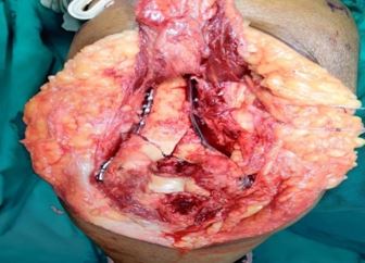
Fig.1: articular reduction of fragments with provisional K-wires.

ojmpc_153_02.jpg
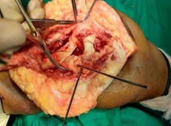
Fig.2:-Plates placed in medial and lateral columns of distal humerus.

ojmpc_153_03.jpg
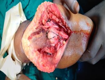
Fig.3: Final construct with TBW of the osteomised olecranon.

The most common approach used was olecranon osteotomy (76%), followed by triceps reflecting anconeus pedicle-TRAP approach (16%) and para-tricipital approach (8%).


ojmpc_153_04.jpg
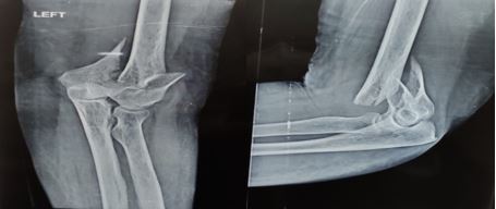
Fig.4: Pre-operative radiograph

ojmpc_153_05.jpg
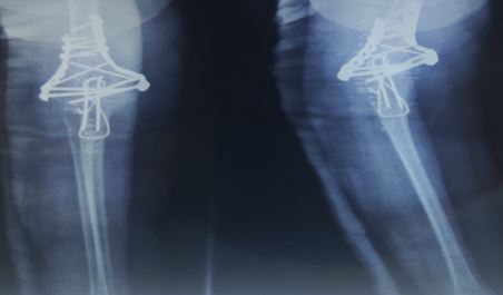
Fig.5: Immediate post-operative radiograph

ojmpc_153_06.jpg
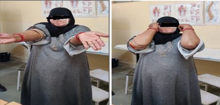
Fig.6: 24weekspost operative clinical

The range of movements that is flexion, extension, supination, pronation was good to excellent in majority of cases. Average time of union was about 13.6 weeks. 14 patients had good, 8 patients had excellent and 3 patients had poor MEPS scores at final follow up. 76% of patients had nil complications. 12% complained of elbow stiffness. 8% complained of superficial wound infection. 4% complained of hardware prominence.

Discussion

A well-functioning elbow is a prerequisite for performing day to day activities of daily living. Intercondylar humerus fractures directly affect the mobility and stability of the elbow joint and hinder the functional capacity of the elbow, thus affecting the person economically and socially.

Ulnohumeral joint and radiohumeral joint relationship should be perfect to achieve a better functional outcome. Open reduction and internal fixation with parallel plate technique has provided a good functional outcome for distal humerus fractures management. The mean age of our study group was 45.24 years. It was seen from our study that mean age for distal humerus fractures is on the comparative decline as more and more younger age group patients are involved now, especially in a developing country like India.

This can be attributed to the rise of Motor Vehicle Accidents amongst the young. The age range for our study was 19-71 years. Sanchez Sotello (2) had a mean age range 16-91 years, Sanders et al (12) had a mean age range of 12-85 years, whereas, Gofton et al (13) had an age range of 16-80 years. More females were affected in our study group as compared to males and the male female ratio (M: F) of our group was 1:1.2, which was discordant with other study groups. Majority of patients suffering from distal humerus fractures arrived at our centres as they were part of motor vehicle injuries (44%).

This was followed by fall from height (36%). The results of our study were not concordant with study of Sanchez Sotello (2) where the major mode of injury was fall from height (56%). Our results suggested that motor vehicle accidents are on the rise as more and more young population are travelling in roads now a days and are parts of rash driving.

Fracture configuration according to AO type was significantly associated with functional outcomes in our study group. Majority of our patients suffered from AO type C1 injury (40%) followed by AO type C2 injury (28%).

Group C had poor MEPS scores than Group A patients which suggested direct association between fracture configuration and functional results. Since Group C fractures are more common than Group A fractures, it complements the fact that high velocity motor vehicle injuries are more prevalent these days.

In our study group, average time of union was 13.28 weeks, with a range between 10 weeks and 18 weeks, which was in concordance with the other study groups. This is the radiological outcome of our study. Time of union had a significantly positive relation with the AO classification of fracture with a p value of 0.021.


According to the MEPS grading criteria, we obtained excellent results in 32%, good results in 56% and poor results in 12% of our cases, with a mean MEPS score of 82.2. It was in concordance with other study groups. This suggested parallel plating technique for distal humerus fractures provided good to excellent functional outcomes in the majority of cases. Complications rates of 24% was observed in our study which was divided into-elbow stiffness in 12%, superficial wound infection in 8% and hardware prominence in 4%. Patients who developed superficial wound infection were treated conservatively with antibiotics.

Elbow stiffness was resolved with aggressive physiotherapy.Sanchez Sotello et al(2) encountered complication rates of 43% which was divided into- heterotopic ossification in 16%, ulnar neuropathy in 6%, superficial infection in 6%, posttraumatic arthritis in 6% non union , deep infections and osteonecrosis in 3%, which was not in concordance with our study results.

The rates of infection, elbow stiffness and heterotopic ossifications were higher in type B and C fractures as compared to type A fractures as seen in a study conducted by Robinson et al. (14)

Table 1:Comparing our study with other study groups

StudiesMean age in yearsComplication RatesAverage time of fracture union (weeks)MAYO Elbow Performance Score(MEPS)Number of patientsM:F ratio
Sanchez Sotello et al(2)5843%1285321.4:1
Atalar et al(9)4748%Not specified86.1±12.6371.6:1
Athwal et al(10)5253%12.884212:1
Dinesh S et al(11)3941%13.482242:1
Present study45.2424%13.2882.2251.2:1

Conclusion-Distal humerus fractures continue to be a complex fracture for surgeons to treat. Open reduction and internal fixation is the treatment of choice for distal humerus fractures. Anatomical reduction, stable fixation and early elbow mobilisation are the prerequisites for better functional outcome.

Parallel plating technique provides greater stability in osteoporotic bones and highly complex comminuted fractures as compared to other traditional plates used for the same. Parallel plating may be the preferred technique utilised for very distal fracture patterns since more stability can be obtained by placing additional screws in the distal fragment. Therefore, parallel plating can be a successful technique for internal fixation of distal humerus when its principles are strictly adhered to.

References

1. Kulkarni, V. , Mahesh, U. , Jumani, M. S. , Shivalingaiah, K., YS, H., & Qureshi, A. (2017). To study the outcome of intercondylar fractures of distal humerus using dual plating and its functional outcome. International Journal of Orthopaedics, 3(4), 565-570 [Crossref][PubMed][Google Scholar]

2. Sanchez-Sotelo, J. , Torchia, M. E. , & O'Driscoll, S. W. (2007). Complex distal humeral fractures: internal fixation with a principle-based parallel-plate technique. JBJS, 89(5), 961-969 [Crossref][PubMed][Google Scholar]

3. McKee, M. D. , Wilson, T. L. , Winston, L., Schemitsch, E. H., & Richards, R. R. (2000). Functional outcome following surgical treatment of intra-articular distal humeral fractures through a posterior approach. JBJS, 82(12), 1701 [Crossref][PubMed][Google Scholar]

4. Principle-based internal fixation of distal humerus fractures. Sanchez-Sotelo J, Torchia ME, O'driscoll SWTech Hand Up Extrem Surg. 2001 Dec; 5(4):179-87. . [Crossref][PubMed][Google Scholar]

5. Fractures of the distal humerus. Ring D, Jupiter JB Orthop Clin North Am. 2000 Jan; 31(1):103-13. . [Crossref][PubMed][Google Scholar]

6. Surgical fixation of intra-articular fractures of the distal humerus in adults. Soon JL, Chan BK, Low CO Injury. 2004 Jan; 35(1):44-54. Comparative stability of perpendicular versus parallel double-locking plating systems in osteoporotic comminuted distal humerus fractures. [Crossref][PubMed][Google Scholar]

7. Stoffel K, Cunneen S, Morgan R, Nicholls R, Stachowiak GJ Orthop Res. 2008 Jun; 26(6). 778-84. . [Crossref][PubMed][Google Scholar]

8. Biomechanical comparison of two different periarticular plating systems. for stabilization of complex distal humerus fractures. Schwartz A, Oka R, Odell T, Mahar. . [Crossref][PubMed][Google Scholar]

9. Atalar, A. C. , Demirhan, M. , Salduz, A. , Kilicoglu, O., & Seyahi, A. (2009). Functional results of the parallel-plate technique for complex distal humerus fractures. Acta Orthop Traumatol Turc, 43(1), 21-7 [Crossref][PubMed][Google Scholar]

10. Athwal, G. S. , Hoxie, S. C. , Rispoli, D. M., & Steinmann, S. P. (2009). Precontoured parallel plate fixation of AO/OTA type C distal humerus fractures. Journal of orthopaedic trauma, 23(8), 575-580 [Crossref][PubMed][Google Scholar]


11. Dinesh, L. (2013). Functional Outcome Analysis of Parallel-Plate technique for distal humerus fractures (Doctoral dissertation, Madras Medical College, Chennai). . [Crossref][PubMed][Google Scholar]

12. Sanders, R. A. , Raney, E. M. , & Pipkin, S. P. (1992). Operative treatment of bicondylar intraarticular fractures of the distal humerus [Crossref][PubMed][Google Scholar]

13. Gofton, W. T. , MacDermid, J. C. , Patterson, S. D., Faber, K. J., & King, G. J. (2003). Functional outcome of AO type C distal humeral fractures. The Journal of hand surgery, 28(2), 294-308 [Crossref][PubMed][Google Scholar]

14. Robinson, C. M. , Hill, R. M. , Jacobs, N., & Dall, G. (2003). Adult distal humeral metaphyseal fractures: epidemiology and results of treatment. Journal of orthopaedic trauma, 17(1), 38-47 [Crossref][PubMed][Google Scholar]

Disclaimer / Publisher's Note

The statements, opinions and data contained in all publications are solely those of the individual author(s) and contributor(s) and not of Journals and/or the editor(s). Journals and/or the editor(s) disclaim responsibility for any injury to people or property resulting from any ideas, methods, instructions or products referred to in the content.