Orthopaedic Journal of MP Chapter

Publisher: Madhya Pradesh Orthopaedic Association www.mpioa.com
E-ISSN:2582-7243, P-ISSN:2320-6993
2022 Volume 28 Number 1 Jan-Jun

Primary cemented bipolar hemiarthroplasty in elderly unstable intertrochanteric fractures.

Thora A1*, Maurya A2

1* Ankit Thora, Department of Orthopaedics, Mahatma Gandhi Memorial Medical College, Indore, MP, India.

2 A Maurya, Mahatma Gandhi Memorial Medical College, Indore, MP, India.

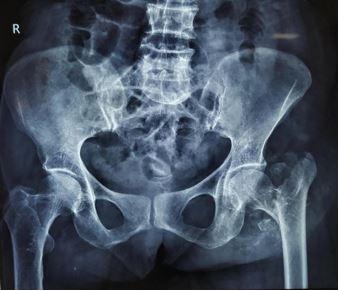
Background: The incidence of inter-trochanteric fractures of the femur is very high in the elderly population. (1) Anatomic restoration of the neck of femur, along with articular congruity is the goal of management.

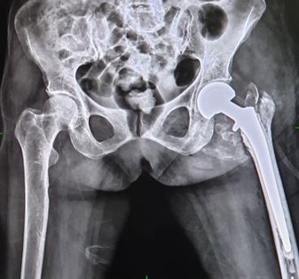
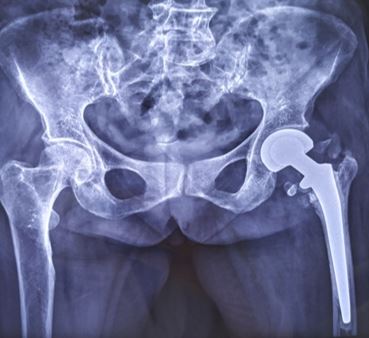
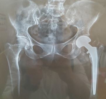
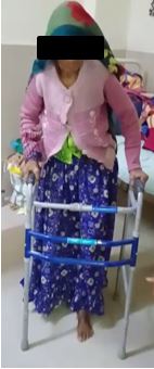
Materials and Methods: Thirty patients with unstable Intertrochanteric fractures were operated by Bipolar Hemiarthroplasty. Follow-ups were taken at 2, 6, 10 and 16 weeks. Harris Hip Score and FIM score were assessed.

Results: Mean Harris Hip score achieved was 87 at 16 weeks and Mean FIM score achieved was 78.9 at 16 weeks indicating good functional outcomes. The outcome was excellent in 23.4%, Good in 63.3%, Fair in 10% and Poor in 3.3% as per HHS.

Conclusion: This procedure offered pain free mobile hip with early mobilization, easy rehabilitation and early return to functional level, when standard techniques were used.

Keywords: Harris Hip Score, FIM Score, Bipolar Hemiarthroplasty, Intertrochanteric Femur Fracture

Corresponding Author How to Cite this Article To Browse
Ankit Thora, , Department of Orthopaedics, Mahatma Gandhi Memorial Medical College, Indore, MP, India.
Email:
Thora A, Maurya A, Primary cemented bipolar hemiarthroplasty in elderly unstable intertrochanteric fractures.. ojmpc. 2022;28(1):7-11.
Available From
https://ojmpc.com/index.php/ojmpc/article/view/143
Manuscript Received Review Round 1 Review Round 2 Review Round 3 Accepted
2022-06-06 2022-06-12 2022-06-18 2022-06-24 2022-06-30
Conflict of Interest Funding Ethical Approval Plagiarism X-checker Note
Authors state no conflict of interest. Non Funded. The conducted research is not related to either human or animals use. 14.34 All authors have accepted responsibility for the entire content of this manuscript and approved its submission.

© 2022by Thora A, Maurya Aand Published by Madhya Pradesh Orthopaedic Association. This is an Open Access article licensed under a Creative Commons Attribution 4.0 International License https://creativecommons.org/licenses/by-nc/4.0/ unported [CC BY NC 4.0].

Introduction

The world-wide incidence of all hip fractures is around 402 per 100000 and the incidence of Intertrochanteric fracture is estimated to be around 171 per 100000 in 2019.(2)The annual incidence of hip fractures across Indian population can be estimated roughly as 61,083 men and 81,724 women above the age of 50 years. (3).

The incidence is roughly the same as intra-capsular femoral neck fractures. The female: male ratio is between 2:1 and 8:1. Stable intertrochanteric fracture can be easily treated by osteosynthesis with predictable good result (4,5), whereas the management of unstable intertrochanteric fracture are challenging because of poor bone quality, loss of reduction and prolonged protected weight bearing(6,7).

In past fixed angle devices were used for fixation of these fractures, but complication like implant cut out and fracture displacement were seen(8,9) Subsequently, sliding hip screw fixation was used with much success and became prominent method of fixation of these fractures (10,11) but complications such as head perforation, excessive sliding with subsequent shortening, plate pull out and plate breakage continued to be the problem particularly with the unstable type fractures (12,13).

Intramedullary interlocking devices have shown reduced tendency for cut out in osteoporotic bone(14) and also better results in case of unstable intertrochanteric fractures(15,16).

The duration of surgery is less minimising blood loss. The procedure may sometimes result in screw cut out or loss of reduction in osteoporotic patients. Management of such cases with primary cemented bipolar hemiarthroplasty permits early mobilization and thus avoiding most complication related to fixation and prolonged immobilization (17).

Materials and Methods

This study was conducted on 30 cases with unstable intertrochanteric femur fracture above 60 years of age, treated at our Centre. All the patients with neglected injury, pathological fracture or other associated injuries, psychiatric disorders, neurological disorders were excluded. All Patients were assessed for associated injuries and primarily stabilized with fluids and analgesics. Standard AP and Lateral hip radiographs were done and ankle traction applied.

Prior to study institutional review board approval was obtained and well-informed consents were taken and bipolar Hemiarthroplasty was planned. Patient taken for OT under all aseptic precautions and suitable anesthesia (Spinal Anesthesia or GA) and operated in lateral position with affected limb above. All the patients were operated using standard posterior approach to the hip.

Femoral head was removed and a neck cut is taken roughly about 1-2 cm above lesser trochanter (LT) depending upon the amount of comminution.

In case of greater trochanter (GT) fractures, the gluteus medius, GT and the vastus lateralis apparatus were maintained in continuity as a stable lateral support which was fixed loosely to shaft fragment with ethibond sutures or Stainless Steel (SS) wires. The Femoral Canal was broached in appropriate ante-version.

A bipolar prosthesis was then inserted and trial reduction was done and with the trial prosthesis in situ traction was applied to the leg to compare it with the opposite leg for any Limb Length Discrepancy (LLD). After confirming the leg length the implant was inserted into the femur and prosthesis was reduced. Traction was then applied with implant in situ to achieve the desired limb length by comparing with the opposite limb on table.

Applied traction caused femur to be pulled distally to note the amount of distraction between the prosthesis and the femoral cut so as to mark the level on the prosthesis. This gave an idea of how much the femur implant should sink into the proximal femur so as to achieve the desired limb length at the time of final cementing of the implant.

During the final fixation of the stem, the cemented stem was allowed to sink in the femoral canal up to the mark made on the prosthesis in above stated manner and for the remaining portion a cement mantle was made so that the final limb length was equalized. Once the prosthesis was fixed, the broken trochanter and the calcar were also fixed using Ethibond sutures or SS wires or tension band wiring (if required). The sleeve of Gluteus medius, GT and vastus lateralis if reconstructed was now reattached to the shaft by additional wires.

The short external rotators were then re-sutured using bone tunnels in GT with the closure of the superficial layers, as routine over a suction drain after achieving hemostasis. Standard precautions of hip hemiarthroplasty were followed. Post-operative intravenous antibiotic was given for 3 days. Oral analgesics and antibiotics were given for 5 days.

All patients underwent a routine postoperative physiotherapy protocol that included early gait training in form of walking with the help of a walker which was started second day post-surgery. Patients were followed up at 2week, 6 weeks, 10 weeks and 16 weeks for functional outcome assessment using HHS and FIM score. Suture Removal was done at 2weeks. Case 1- 65 years old female with Left side fracture intertrochanteric femur (Evans Type V)

Results

Results-In our study of 30 patients (15 Males & 15Females) with unstable Intertrochanteric femur fracture, the mean age found was 76 years (range - 61 years to 90 years) with right side preponderance. The mean operative time of 90 minutes with average duration of hospital stay being 7.8 days and average blood loss being 247 ml. The most common comorbidities the patient had were Hypertension and Diabetes Mellitus which led to delay in their surgery. Most common type of Intertrochanteric Fracture was Evans Grade IV.


ojmpc_143_01.jpg
Figure 1: Preop x-ray

ojmpc_143_02.jpg
Figure 2: Post op xray

ojmpc_143_03.jpg
Figure 3: 2.5 months post op xray

The mean Harris Hip Score at 2 weeks postoperative was 62 ± 2.09, and at 4 months it was 87± 4.19. The mean FIM score at 2 weeks postoperatively was 44 and at 4months it was 78.9.

Mean HHS was excellent for 7 patients (23.4%), good for 19 patients (63.3%), fair for 3 patients (10%) and poor for 1 patient (3.3%).

ojmpc_143_04.jpg
Figure 4: 4 moths post op xray

ojmpc_143_05.jpg
Figure 5: walking with walker


ojmpc_143_06.jpg
Figure 6: walking independently

Major complications associated with the procedure were superficial infection in 1 patient (3.3%) and Joint Dislocation in 1 patient (3.3%).

Discussion

In our study around 86% patients had good to excellent results of hemiarthroplasty in management of unstable intertrochanteric fractures. Sanchetti et al reported 71% of good to excellent results according to HHS in their series of 35 patients treated with hemiarthroplasty(18). Rodop et al, in a study of 37 intertrochanteric fractures treated with bipolar hemiarthroplasty achieved 82% of good to excellent results as assessed by HHS (19).

In our study, at 4 months of follow up, we encountered 1 case of superficial infection (which responded to analgesics and antibiotics) and one case of dislocation which was managed with girdlestone arthroplasty subsequently. Grimsurd et al, in a study of 39 patients of unstable intertrochanteric fractures treated with cemented bipolar hemiarthroplasty, reported a relatively low rate of complication (20).

Stern et al, used Leinbach prosthesis for treatment of 22 intertrochanteric factures and found early ambulation and early return to preinjury status as a definitive advantage (21). In our study average blood loss was 334 ml and average operative time was 90 minutes. Sanchetti et al reported average blood loss of 350 ml and operative time of 71 minutes (19).

Conclusion

In this study we come to conclusion that, primary cemented hemiarthroplasty in intertrochanteric femur fractures offered pain free mobile hip with early mobilization, easy rehabilitation and early return to functional level, when standard techniques were used. Bipolar hemiarthroplasty reduced the complications related to prolonged immobilization, need for prolong rehabilitation, residual deformities and need for revision surgeries. The procedure offered faster mobilization, rapid return to preinjury level, improve the quality of life and gave long term solution in elderly patients with unstable intertrochanteric fracture of femur. Functional outcomes in our study were at par with other implants like DHS or PFN with much less complications and early immobilization in geriatric age group patients. From above findings

we conclude that, Primary Cemented Bipolar Hemiarthroplasty can be used as a preferred method of treatment of unstable intertrochanteric femur fractures in elderly age group patients.

References

1. Kannus P, Parkkari J, Sievδnen H, Heinonen A, Vuori I, Jδrvinen M. Epidemiology of hip fractures. Bone. 1996;18:57S–63. [Crossref][PubMed][Google Scholar]

2. Adeyemi, Ayoade PhD, Delhougne, Gary MS, Incidence and Economic Burden of intertrochanteric Fracture. JBJS (OA) March28,2019 volume4. Issue 1 p e0045. . [Crossref][PubMed][Google Scholar]

3. D. K. Dhanwal,1 R. Siwach,3 V. Dixit,1 A. Mithal,2 K. Jameson,4 and C. Cooper4, Arch Osteoporos. Incidence of hip fracture in Rohtak district, North India, Author manuscript PMC 2013 Dec; 8(0): 135. doi:10.1007/s11657-013-0135-2 [Crossref][PubMed][Google Scholar]

4. Marsh JL, Slongo TF, Agel J, Broderick JS, Creevey W, et al. , Fracture and dislocation classification compendium: TraumaAssociation classification, database and outcomes committee. JOrthop Trauma. 2007;21:S1-133. [Crossref][PubMed][Google Scholar]

5. Larsson S. Treatment of osteoporotic fractures. Scand J Surg. 2002:91:140-146. [Crossref][PubMed][Google Scholar]

6. Bannister GC, Gibson AG, Ackroyd CE, Newman JH. The fixation and prognosis of trochanteric fractures: A randomized prospectivecontrolled trial. Clinical Orthop Relat Res. 1990;254:242-6. [Crossref][PubMed][Google Scholar]


7. Chinoy MA, Parker M). Fixed nail plates, versus sliding hipsystems for the treatment of trochanteric femoral fractures: A metaanalysis of 14 studies. Injury 1999;30:157-63. . [Crossref][PubMed][Google Scholar]

8. Flores LA, Harrington II. Martin H. The stability ofintertrochanteric fractures treated with a sliding screw plate. J BoneJoint Surgery Br. 1990:72:37-40 [Crossref][PubMed][Google Scholar]

9. Sernbo 1, Fredin H. Changing methods of hip fractureosteosynthesis in Sweden: An epidemiological enquiry covering46,900cases. Acta Orthop Scand. 1993;64:173-4. [Crossref][PubMed][Google Scholar]

10. Larsson S, Friberg S, Hansson LI. Trochanteric fractures: Mobility,complications, and mortality in 607 cases treated with the sliding-screw plate. Clin Orthop Relat Res. 1990;260:232–41. [Crossref][PubMed][Google Scholar]

11. Bess RJ, Jolly SA. Comparison of compression hip screw and gamma nail for treatment of peritrochanteric fractures. J SouthOrthop Assoc. 1997;6:173-9. [Crossref][PubMed][Google Scholar]

12. Kim WY, Han CH, Park J, Kim JY. Failure of intertrochantericfracture fixation with a dynamic hip screw in relation topreoperative fractures ability and osteoporosis. Intra op. 2001;25:360-2. [Crossref][PubMed][Google Scholar]

13. Jensen JS, Tondevold E, Mossing N. Unstable trochantericfractures treatedthe sliding screw-plate system:biomechanical study of unstable trochanteric fractures. III, ActaOrtho Scand. 1978;49:392-7. [Crossref][PubMed][Google Scholar]

14. Halder SC. The Gamma nail for peritrochanteric fractures. J BoneJoint Surg: A Br. 1992;74:340–4. [Crossref][PubMed][Google Scholar]

15. Davis TR, Sher JL, Horsman A, Simpson M, Porter BB, ChekettsRG. Intertrochanteric femoral fractures. Mechanical failure afterinternal fixation. J Bone Joint Surg Br 1990;72:26-31. [Crossref][PubMed][Google Scholar]

16. Thomas AP. Dynamic hip screws that fail, Injury. 1991;22:45-46. . [Crossref][PubMed][Google Scholar]

17. Silverton CD, Jacobs JJ, Rosenberg AG, Kull L, Conley A, Galante JO. Complications of a cable grip system. J Arthroplasty. 1996;11:400–404. [Crossref][PubMed][Google Scholar]

18. KH Sancheti, PK Sancheti, AK Shyam, S Patil, Q Dhariwal, and R Joshi, Indian J Orthop. 2010 Oct-Dec; 44(4). 428–434. PMCID: PMC2947731,PMID: 20924485. Primary hemiarthroplasty for unstable osteoporotic intertrochanteric fractures in the elderly. A retrospective case series [Crossref][PubMed][Google Scholar]

19. Rodop O, Kiral A, Kaplan H, Akmaz I. Primary bipolar hemiprosthesis for unstable intertrochanteric fractures. Int Orthop. 2002;26:233–7. [Crossref][PubMed][Google Scholar]

20. Grimsrud C, Monzon RJ, Richman J, Ries MD. Cemented hip arthroplasty with a novel cierclage cable technique for unstable intertrochanteric hip fractures. J Arthroplast. 2005;20:337–43. [Crossref][PubMed][Google Scholar]

21. Stern MB, Goldstein TB. The use of the Leinbach prosthesis in intertrochanteric fractures of the hip. Clin Orthop Relat Res 1977. 128:325-331. [Crossref][PubMed][Google Scholar]

Disclaimer / Publisher's Note

The statements, opinions and data contained in all publications are solely those of the individual author(s) and contributor(s) and not of Journals and/or the editor(s). Journals and/or the editor(s) disclaim responsibility for any injury to people or property resulting from any ideas, methods, instructions or products referred to in the content.