Orthopaedic Journal of MP Chapter

Publisher: Madhya Pradesh Orthopaedic Association www.mpioa.com
E-ISSN:2582-7243, P-ISSN:2320-6993
2022 Volume 28 Number 1 Jan-Jun

Primary Osteoarthritis Knee: establishing its cause, pathogenesis and treatment -A Prospective Case-Control Study

Agrawal R1*

1* RC Agrawal, Purva Fountain Square Marathahalli, Bangalore, Karnataka, India.

Background: The objectives of this analytical study were to compare two scores, Western Ontario McMaster Universities Osteoarthritis Index (WOMAC), Euroqol Group Health Status Score through Visual Analog Scale (EQVAS); Deficient Full Flexion (DFF) and Deficient Full Extension of knee at the beginning and end-point in two, Trial (G1) and Control (G2) groups of Primary Osteoarthritis Knee patients.

Material and Methods: In this study total patients were 125, in G1 - 100 and in G2 – 25. G1 group received hypothesized treatment, contracture correction therapy (CCT) while G2 did no therapy. WOMAC determination done by the questionnaire; EQVAS by vertical-scale and deficiencies by goniometer at 0, 6, 12 and 24 weeks. The CCT consisted of eight body postures, aimed to provide passive flexion or passive extension.

Results: The CCT receiving was associated with recovery (P 0.00) while non-receiving with deterioration (P 0.00). In G1, WOMAC improved: 71.70 to 3.68 and EQVAS 22.25 to 91.55 (P 0.00). In G2, WOMAC deteriorated score worsened from 53.00 to 71.88 and EQVAS 58.60 to 11.96 (P 0.00). DFF and DFE showed coinciding changes.

Conclusion: The cause, pathogenesis and treatment are deficient full flexion/deficient full extension; capsular contracture formation and passive flexion or passive extension respectively.

Keywords: Knee Osteoarthritis, Arthroplasty, Deficient Full Flexion, Deficient Full Extension, Contracture Correction Therapy

Corresponding Author How to Cite this Article To Browse
RC Agrawal, , , Purva Fountain Square Marathahalli, Bangalore, Karnataka, India.
Email:
Agrawal R, Primary Osteoarthritis Knee: establishing its cause, pathogenesis and treatment -A Prospective Case-Control Study. ojmpc. 2022;28(1):36-43.
Available From
https://ojmpc.com/index.php/ojmpc/article/view/142
Manuscript Received Review Round 1 Review Round 2 Review Round 3 Accepted
2022-06-06 2022-06-12 2022-06-18 2022-06-24 2022-06-30
Conflict of Interest Funding Ethical Approval Plagiarism X-checker Note
Authors state no conflict of interest. Non Funded. The conducted research is not related to either human or animals use. 15.76 All authors have accepted responsibility for the entire content of this manuscript and approved its submission.

© 2022by Agrawal Rand Published by Madhya Pradesh Orthopaedic Association. This is an Open Access article licensed under a Creative Commons Attribution 4.0 International License https://creativecommons.org/licenses/by-nc/4.0/ unported [CC BY NC 4.0].

Introduction

Primary osteoarthritis knee (OA Knee) is also known as degenerative disease or age-related arthritis. It is found in both the sexes, all over the world and existing since ages. It starts as pain in the knee at about 30 yrs without any evident cause. Pain occurs on joint movement and subsides on rest. Disease progresses over a period of 10-15 yrs when its signs develop at >50 yrs (clinical plus radiographic criteria developed by American College of Rheumatology)1. Despite the age in diagnostic criteria >50, it occurs in adults also- “OA Knee (pain with x-ray evidence) occurs in 12% of persons age ≥60 in United States and 6% of all adults age ≥302. As per the literature, its etiology and pathogenesis are not known- “The exact mechanism for the development of primary OA remains unknown and it is therefore termed idiopathic3. The basic lesion described is degeneration (weakening) of the articular cartilages which progresses with age and aggravates by obesity, diabetes and heredity. OA knee occurs more in those who work in standing or squatting position 4. There are many treatments being used for this disease which are as follows-

A. Non-surgical

1. Physiotherapy, Assistive devices Heat, Electricity5, USG, Laser, Cane, Knee- sleeve
2. Medication NSAIDS, Paracetamol
3. Intra-articular injections1 Hyaluronic Acid, Corticosteroids, Stem cells etc.
B. Surgical

Total knee Arthroplasty (TKA)6,7, Knee Joint Distraction8

There is no cure to this disease9. The treatment is based on pain management and mobility restrictions. All the patients are initially treated with one or more non-surgical options which work for some time. Surgery is advised on failure of this regimen. The most relied operation at present is TKA6,7. This has its own disadvantage as it works for only 15-20 yrs6. So, the question arises what is the specific treatment?

The available literature lacks precise knowledge of its causes, pathogenesis & treatment. This article aims to establish the same and assess the results by a Randomized Controlled Trial (RCT). The hypothesized causes are DFF/DFE/both; pathogenesis is contracture formation in front/back of the capsule and treatment, passive flexion/passive extension knee (CCT). The treatment differs as per the cause & site of contracture.

Material and Methods

Subjects

The total participants included 125 patients (250 joints) of OA Knee. The G1 included 100 and G2, 25. This number depended upon their scanty availability in my setup. Their inclusion criteria consisted of

1. Age 30 to 85 years
2. Knee pain which appeared without any apparent cause, exacerbated by exertion and subsided on rest

3. Limited morning stiffness
4. No past H/O infection, trauma or inflammation (to rule out secondary OA) and
5. Disability in sitting, climbing stairs or walking. The exclusion criteria consisted of
6. Backache
7. Leg pain (e.g. sciatica)
8. Inability to lie supine (e.g. kyphosis)
9. Inability to lie prone (e.g. central obesity)

Exclusion was based on patients’ inability to lie supine and prone, required for the intervention. This trial was based on “Pragmatic Cluster Randomized Controlled Trial”, also known as Cluster Randomized Trial (CRT)13 or Group Randomized Trial. In this variety pre-existing groups, called clusters, of individuals are randomly allocated to treatment arms. CRTs can be used when individual randomization to treatment arm is not possible or the intervention is naturally applied to whole cluster. My patients, who consulted me, were of two types. The type I wanted to avoid surgery, had tried other non-surgical options (e.g. drugs, physiotherapy, and intra-articular injections) and did not want those anymore. In such a situation it was not possible to give them any other intervention except the CCT. So those were included in the trial group. The type II were already using some options and living with disabilities but not convinced to receive CCT. So those were included in control group with “no intervention”.

The proof of control group as valid pre-existing cluster was obtained by following formula:

All primary OA Knee patients = Pre-existing subjects valid for trial

OR

One patient X n (imaginary number) = Pre-existing subjects valid for trial

OR

1 patient X 28 cluster* = Pre-existing subjects valid for trial

OR

All 28 patients cluster = Pre-existing subjects valid for trial hence

My control group (G₂) = Pre-existing subjects valid for trial

The randomization was non-blinding as CCT was unmaskable procedure

The study setting consisted of my clinic, one charitable hospital, free weekly health camps and clinics of two colleagues. The study period was March 2017 - December 2017. This ten month period gave sufficient time of six months for follow up. Informed consent was sought from all patients. The patients with bilateral affection were investigated and treated simultaneously. The procedure consisted of

1. Baseline Data Recording
2. Intervention
3. Data Collection and Monitoring.


Baseline Data Recording

It consisted of recording name, age, sex, complaints, side of affected knee, difficulty in sitting, climbing, walking, history of swelling, crackling sound, lurching, deformity, past history of injury/infection, pain in small joints and local examination of affected knee for flexion deformity or bow-knee (genu varum). Skiagrams taken in standing position and radiological grades (I, II, III, and IV) were decided.

The physical examination within the baseline data recording consisted of physical signs elicitation and calculations with measurements.

Only 28 patients fulfilled inclusion criteria with no consent for CCT, the value of n was 28.

Physical signs (author designed):

ojmpc_142_01.jpg
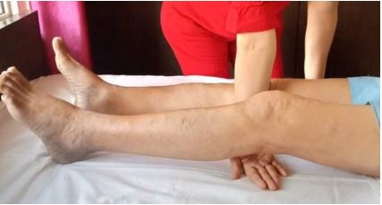
Figure: 1 Hand insinuation sign

1. Inability to sit on soles (indicative of DFF) – Yes/No
2. Palpable crepitus (first and diagnostic sign of OA knee) – Yes/No
3. Hand insinuation (indicates DFE), Figure 1 – Yes/No

Calculations and Measurements:

1. WOMAC osteoarthritis score14 (0-96, 96 meant worst) on 5-point Likert-type scale
2. EQ VAS health status score15 (0-100, 100 meant best) by vertical 20 cm scale
3. DFF/DFE/both – these were measured by goniometry. Normal Range of Motion (ROM) of knee was taken to be 0º to 145º. Their details are as below:
4. DFF = ROM 0º to < 145º
5. DFE = ROM > 0º to 145º
6. Both DFF and DFE = ROM > 0º to < 145º

All the Baseline data recording, measurements and CCT treatments were done by corresponding investigators only.

Intervention

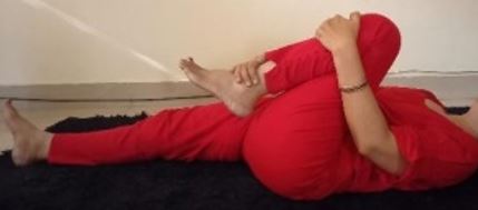
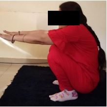
This is the designed specific treatment of OA knee which works by providing passive flexion or passive extension. It consisted of following eight body postures-

ojmpc_142_02a.jpg
Posture 1: Full knee flexion in supine

ojmpc_142_02b.jpg
Posture 2: Knee flexion in prone

ojmpc_142_03.jpg
Posture 3: Full knee flexion by sitting on buttocks

ojmpc_142_04.jpg
Posture 4: Full knee flexion by sitting on legs


ojmpc_142_05.jpg
Posture 5: Full knee flexion by sitting on soles

ojmpc_142_06.jpg
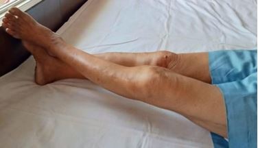
Posture 6: Extension knee in Supine

ojmpc_142_07.jpg
Posture 7 and 8: Extension knee by sitting on buttocks Posture Extension knee by standing on toes

The first five postures (Fig.2-6) were used in DFF and last three (Fig.6-8) in DFE. Every patient was taught to adopt each posture for 20 slow counts (20 secs/20") twice daily. The sequence of postures was adhered to without applying any force to achieve a high degree of flexion or extension.

After each posture, relaxation was permitted by lying flat on back or abdomen for 10 sec. At the outset the therapy was contraindicated in presence of severe pain when oral NSAIDS with rest were given for 5-7days prior to CCT. The duration for posture 2(Fig.3) was 10 sec twice daily. In the initial stage for DFF postures 1&2 (Fig.2&3) and for DFE posture 6(Fig.7) sufficed. Later, if required for DFF postures 3, 4 or 5 (Fig.4, 5, 6) and for DFE postures 7, 8 (Fig.8) were considered. The therapy was taken by the patient as advised and supervised by investigator in weekly visits. Along with this therapy, light physical exercise e.g. walking and taking deep breath (minimum 5 times) were advised for general health.

2.2.3. Data Collection and Monitoring

The monitoring was done during weekly visits and phone conversations. The data were collected at 0,6,12 and 24 weeks by the corresponding investigator.

2.3. Outcome Measures

To assess the results, the primary outcome measure consisted of WOMAC14 and secondary outcome measures consisted of EQ VAS15 score, DFF and DFE at the beginning and end of the follow up. These measures were also used to compare the results between G1 and G2.

2.4. Statistical Analysis

The statistical analyses were performed using IBM SPSS Statistics Version-26. Paired t-test was used to compare the clinical scores of WOMAC, EQ VAS, DFF and DFE at baseline and endpoints while Independent t-test for comparing final outcome data between G1 and G2. The value of P<0.05 was considered statistically significant. For the quantitative data mean tested for two groups using Independent t test while for the categorical and nominal data we applied Chi-square test of association.

Results  

A CONSORT (CON=consolidated, S=Standards, O=Of, R=Reporting, T=Trials) chart has been presented for the trial - Fig.9

The incidence of various ways of spending time (occupation) and professions including- 1. Walking and standing (e. g. doctors, nurses and teachers) 2. Squatting and sitting with folded legs (e.g. some shop-keepers and home confined elderly) and 3. Sitting on chair (e.g. table workers) in G1 was 64 (64%), 28 (28%) and 8 (8%) respectively while the same in G2 was 19 (76%), 5 (20%) and 1 (4%).

The incidence of some important features (in G1) consisted of obesity 22 (22%), Diabetes 7 (7%), Grades of OA I-55 (55%), II-4 (4%), III-4 (4%) and IV-7 (7%).

The incidence of various physical signs of knee including 1. Inability to sit on soles, 2. Presence of crepitus and 3. Hand insinuation in G1 was 76 (76%), 75 (75%), and 31 (31%) respectively, while the same in G2 was 16 (64%), 5 (20%), and 6 (24%).


The incidence of various measurable Knee Movement Deficiencies including -1. DFF, 2. DFE, 3. Both DFF and DFE and 4. None in G1 (n=200 knees) was 120 (60%), 50 (25%), 6 (3%) and 24 (12%) respectively while the same in G2 (n=50) was 33 (66%), 11 (22%), 0 (0%) and 6 (12%).

ojmpc_142_09.jpg
Figure 9 CONSORT Chart

The patients did not differ much in basic demographic and clinical data in two groups (Table1).

Table 1: Some baseline characteristics of OA knee in both groups

S. NoName of CharacteristicsTrial (n=100)Control (n=25)P value
1.Age in years57
Range 30-85
68
Range 50-85
<0.005
2.GenderMale42 (42%)16 (64%)0.048
Female58 (58%)9 (36%)
3.Disease ExtentBilateral66 (66%)18 (72%)0.56
Right34 (34%)7 (28%)
4.Duration (in months)29.8
Range 1-120
66
Range 6-180
0.000

In the statistical analysis all four outcome measures at the baseline were compared with those at the end point (Paired t- test). There was significant improvement in G1 (P-.00) while deterioration in G2 (P-.00). See Table 2.

Table 2: Details of clinical score results in both groups at initial and endpoints

S. No.Outcome MeasuresTrial GroupControl Group
0 weeks24 weeksP value0 weeks24 weeksP value
1WOMAC71.703.68.0053.0071.88.00
2EQ VAS22.2591.55.0058.6011.96.00
3DFF10.71°.00°.009.74°14.47°.00
4DFE10.97°.00°.009.29°10.71°.00

The improvement was indicated by decrease in WOMAC, DFF and DFE; by increasing EQ VAS while opposite changes in case of worsening. The comparison of final outcome scores between two groups (Independent t-test) also showed significant difference (Table 3)

Table 3: Comparison of Final Outcome Scores between Trial and Control Groups

S. No.Outcome MeasuresTrial GroupControl Group
0 weeks 24 weeks P value 0 weeks 24 weeks P value
1WOMAC71.703.68.0053.0071.88.00
2 EQ VAS22.2591.55.0058.6011.96.00
3 DFF10.71°.00°.009.74°14.47°.00
4 DFE10.97°.00°.009.29°10.71°.00

The data recorded at six and twelve weeks in G1, WOMAC 16.90 and 7.90; EQVAS 65.32 and 88.89; DFF 1.7° and 0.1°; DFE 2.5° and 0.00°. The similar data in G2 consisted of 64.52 and 89; 33.80 and 21.40; 9.6° and 12.8°; 9.2° and 10.9° respectively. The clinical improvement in G1 patients was far superior to that in G2 (Table 4)

Table 4: Treatment Results Analysis

S. No.ResultTrial Group(n=100)Control Group(n=25)
No. of Patients Remarks No. of Patients Remarks
1Full Relief98 (98%)-2 (8%)By exercises and drugs
2Partial Relief2 (2%)Pain subsided; deformity persisted--
3No Relief--15 (60%)-
4ComplicationsNIL-8 (32%)Deformity and pain worsened

Discussion

The hypothesis was regarding three parts of OA, viz cause, pathogenesis and treatment. Out of these, treatment was proved through experiment while other two by deducing from its result. When the passive flexion/passive extension as treatment was found to be effective, the related cause and pathogenesis were presumed to be proved.

Synovium is non innervated thin lining of capsule & secretes synovial fluid which traverses from periphery to all around cartilages. Its speed is increased by mutual movements between femur and tibia16. Cartilages are devoid of blood vessels and sensory nerves17, unable to regenerate & conduct pain sensation from knee to brain. They receive their supplies through synovial fluid only. These anatomical facts show that the articular cartilage cannot be the seat of this disease, as it is insensitive to pain. The pain in the condition develops only on joint movement which indicate that responsible structure must be capable of undergoing momentary structural change. This criterion is fulfilled only by the joint capsule.

A body structure, due to any cause, when does not perform its function, loses its functional capacity. For example, the uterus after menopause cannot conceive, knee kept in a plaster cast for some time cannot flex soon exhibiting DFF and shortening anterior part of capsule (Table 5) and an immobilized elbow in semi flexed position neither can fully flex nor extend soon after plaster removal.

Inferences:

  • Denied 140° flexion caused 140° of DFF.
  • Denied degree of flexion = degree of DFF
  • Denied flexion is a functional anomaly represented by DFF

  • 140° of DFF caused 7.5 cms shortening in anterior capsule.
  • It indicates that pathogenesis is contracture formation in capsule due to DFF. As per literature any knee anomaly produces OA Knee often after many years18.

After plaster removal patient regains full flexion by folding knee itself repeatedly, which meant that passive flexion removed the DFF. Similar event was related to the DFE which was removed by passive extension. Thus passive flexion & passive extension are the methods of treatment of DFF and DFE respectively. For hypothesis summary see Fig.10

Improved outcome measures indicate that the unknown factors (causes, pathogenesis and treatment) are discovered & the hypothesis proven (Table 6).

Limitation in this study: The G2 was smaller than G1 due to unavailability of patients fulfilling the inclusion criteria. To the best of my knowledge, this is second study of its kind, first one11 was also conducted by me. The present study is much improved in form of an RCT. As regards the comparison of results in literature Heidari4 has described the incidence of OA Knee to be more in those who work in standing or those who work in squatting. In the present study first group coincided with patients having DFF & second those with DFE. Incidence in first was 60 (60%), in second 28 (28%) but only 12 (12%) in rest. The later observation is coinciding with the former.

Table 5: Showing relation among Denied Knee Flexion, DFF & Length of Anterior Capsule

KneeDenied FlexionROMDFFLength of Femur shaft & Anterior Knee Capsule* (in cms)
Full Flexion Full Extension
NormalNil0° – 145°Nil66†58.5
Plastered (with
50flexion)
140°0° – 05°140°58.558.5

*Presumed to be the distance between anterior superior iliac spine and tibial tuberosity

†Measurement of anterior capsule, in full flexion, was taken in unaffected contralateral leg

The hypothesized treatment (CCT) looks superior to those described in literature.

There is a drawback in this therapy that the elongation in the contracture is short lived which necessitates its frequent sessions. The steps of this therapy look imitation of physiotherapy and exercises which undermine their impression. The future research should be directed to obtain permanent/long-lasting corrections of the contracture so the CCT may not be a lifelong necessity. Other field of research will be to design a lifestyle which will prevent the disease automatically. At present, according to literature the cause and specific treatment of this disease are not known. The results of this study will remove this lacuna.

The addition of these facts will enhance the knowledge of disease and improve its treatments. By the results of this study, there can be vast change in the clinical practice.

Table 6: Showing important aspects of Results

S.No.Result SummaryInterpretationSignificanceWhether proves hypothesis
1.↓*WOMAC
score(71.70 to3.68)
Decreased score signifies measurable improvementQuantitative proof of cure14Yes
2.↑*EQ VAS
score(22.25 to 91.55)
Increased score signifies measurable improvementQuantitative proof of cure15Yes
3.Abolished- DFF
Initial-10.71°
Final-.00°
Average flexion increased from 134.29° to145° by passive knee flexion through anterior contracture correctionProof of cause removalYes
4.Abolished-DFE
Initial-10.97°
Final-.0°
Average extension increased from 10.97° to 0° by passive knee extension through posterior contracture correctionProof of cause removalYes
5.Relief in symptoms
(Table 4)
A.  G1-
full relief-98 (98%)
Partial relief-2 (2%)
Complication- nil
B.  G2-
Full relief-2 (8%)
Partial relief-nil
No relief-15 (60%)
Complications-8
(32%)
CCT cured a large percentage of patients.
Non CCT cured in much less percentage of patients
Proof that CCT is specific treatment
Proof that non CCT was almost ineffective
Yes
Yes

So far, the disease being treated by orthopaedic & general surgeons, physiotherapist and quacks by various surgical operations, medicines, and devices without unfounded basis. Now the treatment shall be easy and would be carried out by orthopaedic & general surgeons and even by general medical practitioners.

Conclusions

  • Cause of Primary Osteoarthritis Knee in a patient is prolonged Deficient Full Flexion /Deficient Full Extension or both.
  • Pathogenesis of Osteoarthritis Knee is formation of contracture in joint capsule. The contracture forms in front when cause is DFF and in back when cause is DFE.
  • The treatment of Primary Osteoarthritis Knee is passive flexion when cause is DFF, passive extension when cause is DFE and both when causes are combined.

Sources of Support - This research did not receive any specific grant from funding agencies in the public, commercial, or not-for-profit sectors Competing Interests statement - No conflict of interests

Submission declaration – This work11 has been published previously which is now a ‘redundant publication’.

Informed consent was obtained from each patient.

Obtained permissions for use of following copyrighted material

1. WOMAC Osteoarthritis Index14

2. EQVAS scale15

3. Indian Journal of Physiotherapy and Occupational Therapy11


References

1. Michelle J Lespasio, Nicolas S Piuzzi, M Elain Husni, George F Muschler, AJ Guarino, Michael A Mont. Knee Osteoarthritis: A Primer. Perm J. 2017;21:16-183. [Crossref][PubMed][Google Scholar]

2. Felson David T, Neogi T. Osteoarthritis. In: Jamson, Fauci, Kasper, Hauser, Longo, Loscalzo. editors. Harrison’s Principles of Internal Medicine. Vol 2. 20th ed. New York: Mc Graw Hill Education; 2018.p.2624-30 [Crossref][PubMed][Google Scholar]

3. Khanduja V, Khan S W. Hip and knee. In: Williams Norman S, O’connell Ronan P, Mc Caskie Andrew W, editors. Baily & Love’s Short Practice of Surgery, Vol 1, 27th ed. Great Britain: CRC Press; 2018.p.513 [Crossref][PubMed][Google Scholar]

4. Heidari Behzad, Knee Osteoarthritis prevalence, risk factors, pathogenesis & clinical features: Part I. Caspian J Intern Med. 2011 Spring; 2(2): 205-212. . [Crossref][PubMed][Google Scholar]

5. Cherian JJ, Jauregui JJ, Leichliter AK, Elmallah RK, Bhave A, Mont MA. The effects of various physical non-operative modalities on the pain in osteoarthritis of knee. Bone Joint J. 2016 Jan; 98B (1 Suppl A): 89-94. [Crossref][PubMed][Google Scholar]

6. Mihalko William M. In: Azar Frederick M, Beaty James H, Canale Terry S. editors. Campbell’s Operative Orthopedics, Vol 1, 13th ed. Philadelphia: Elsevier; 2017.p. 41320 [Crossref][PubMed][Google Scholar]

7. Vaishya R, Vijay V, Agarwal AK. Total Knee Arthroplasty using patient specific blocks after prior femoral fracture without hardware removal. Indian J Orthop 2018;52:154-60. . [Crossref][PubMed][Google Scholar]

8. Van der Woude J. A. D. Wiegant K. Heerwaarden van R.J. Struijt S Emans P.J. Mastheegen S.C. et al. Knee joint distraction compared with total knee arthroplasty. A Randomised Controlled Trial: Bone Joint J 2017;99-B:5-8 [Crossref][PubMed][Google Scholar]

9. Heidari Behzad, Knee Osteoarthritis diagnosis. treatment and associated factors of progression. part II Caspian J Intern Med. 2011 Summer. 2(3) 249–55 [Crossref][PubMed][Google Scholar]

10. Dhaliwal U, Writing Aim and Objectives: Getting Clarity. In: Gupta P, Singh N. How to Write the Thesis and Thesis Protocol: A primer for Medical, Dental and Nursing Courses. First ed. Delhi: Jaypee Brothers Medical Publishers (P) Ltd. 2014. p.64 [Crossref][PubMed][Google Scholar]

11. Agrawal RC, Trial of a treatment on the patients of Primary Osteoarthritis Knee. Indian Journal of Physiotherapy and Occupational Therapy 2016. 10:185-90. . [Crossref][PubMed][Google Scholar]

12. Joshi M. Selecting the Research Design. In: Gupta P, Singh N editors. How to Write the Thesis and Thesis Protocol: A primer for Medical, Dental and Nursing Courses. First ed. Delhi: Jaypee Brothers Medical Publishers (P) Ltd.2014. p.33 [Crossref][PubMed][Google Scholar]

13. Clustered Randomized Trials. https://www. qmul. ac. uk> research (date last accessed 30 August 2020) [Crossref][PubMed][Google Scholar]

14. WOMAC Osteoarthritis Index (Lk3. 1). https://www. xediton. com>GO-ON-Knee-orHip-WOMAC.pdf (date last accessed 30 August 2020) [Crossref][PubMed][Google Scholar]

15. Mandy Van Reenen / Bas Janseen. preparators. EQ-5D-5L User Guide: Basic information on how to use the EQ-5D-5L instrument; Version 2. 1 April 2015. [Crossref][PubMed][Google Scholar]

16. Bulstrode CJK. Diseases of bone and joints: generalised diseases and chronic joint disorders. In: Russell RCG, Williams NS, Bulstrode CJK. editors. Baily & Love’s Short Practice of Surgery, London: Arnold; 2000. p.415 [Crossref][PubMed][Google Scholar]

17. Caroline W, Adams Michael A. Functional Anatomy of the Musculoskeletal system. In: Standring S, Borley Neil R, Collins P, Crossman Alan R, Gatzoulis Micheal A, Healy Jeramiah C et al. editors. Gray’s Anatomy, The Anatomical Basis of Clinical Practice. 40th ed. Elsevier: Churchill Livingstone; 2008. p.99 [Crossref][PubMed][Google Scholar]

18. Hamblen David L, Simpson RW Hamish A. editors. Osteoarthritis (Degenerative Arthritis; arthrosis; osteoarthrosis; hypertrophic arthritis; post-traumatic arthritis). In:. [Crossref][PubMed][Google Scholar]

19. Adams’s Outline of Orthopaedics. 14thed. Edinburgh: Churchill Livingstone;2010. p. 140 [Crossref][PubMed][Google Scholar]


20. Ralstone S. H, McInnes I. B. Rheumatology and bone diseases. In: Walker Brian R, Colledge Nicki R, Ralston Stuart H, Penman Ian D. editors. Davidson’s Principles and Practice of Medicine, 22nd ed. Edinburgh: Churchill Livingstone; 2014. p.1082 [Crossref][PubMed][Google Scholar]

Disclaimer / Publisher's Note

The statements, opinions and data contained in all publications are solely those of the individual author(s) and contributor(s) and not of Journals and/or the editor(s). Journals and/or the editor(s) disclaim responsibility for any injury to people or property resulting from any ideas, methods, instructions or products referred to in the content.