Orthopaedic Journal of MP Chapter

Publisher: Madhya Pradesh Orthopaedic Association www.mpioa.com
E-ISSN:2582-7243, P-ISSN:2320-6993
2021 Volume 27 Number 1 Jan-Jun

Neglected Cauda Equina Syndrome Due To Prolapsed Lumbar Intervertebral Disc In An Adolescent Patient: A Case Report And Review Of Literature

Gawande J1, Verma P2, Mishra S3, Lakhtakia P4

1 Jeetesh Gawande, Assistant Professor, Department of Orthopaedics, Shyam Shah Medical College, Rewa, MP, India.

2 PK Verma, Department of Orthopaedics, Shyam Shah Medical College, Rewa, MP, India.

3 S Mishra, Department of Orthopaedics, Shyam Shah Medical College, Rewa, MP, India.

4 PK Lakhtakia, Department of Orthopaedics, Shyam Shah Medical College, Rewa, MP, India.

Case report: Lumbar disc herniation is very rare in children and adolescent age group. We report a rare case of two months old neglected case of post traumatic L4-L5 disc herniation causing cauda equina syndrome and bilateral foot drop in a 13-year-old patient. He was treated successfully with emergency L4 laminectomy and L4-L5 discectomy, and he recovered fully without any restricted activity. Cauda equina is a surgical emergency, which should be diagnosed and operated as early as possible for good results, even if the patient presents late.

Keywords: Cauda equine syndrome, Lumbar disc herniation, Laminectomy

Corresponding Author How to Cite this Article To Browse
Gawande J, Verma P, Mishra S, Lakhtakia P, Neglected Cauda Equina Syndrome Due To Prolapsed Lumbar Intervertebral Disc In An Adolescent Patient: A Case Report And Review Of Literature. ojmpc. 2021;27(1):46-49.
Available From
https://ojmpc.com/index.php/ojmpc/article/view/113
Manuscript Received Review Round 1 Review Round 2 Review Round 3 Accepted
2021-06-06 2021-06-12 2021-06-18 2021-06-24 2021-06-30
Conflict of Interest Funding Ethical Approval Plagiarism X-checker Note
Authors state no conflict of interest. Non Funded. The conducted research is not related to either human or animals use. 11.11 All authors have accepted responsibility for the entire content of this manuscript and approved its submission.

© 2021by Gawande J, Verma P, Mishra S, Lakhtakia Pand Published by Madhya Pradesh Orthopaedic Association. This is an Open Access article licensed under a Creative Commons Attribution 4.0 International License https://creativecommons.org/licenses/by-nc/4.0/ unported [CC BY NC 4.0].

Introduction

Back problems, as such are rare problems in pediatric and adolescent age group except for congenital deformities and infections [1-2]. These problems can have variable clinical presentation from local or radicular pain, different motor or sensory deficit, with or without involvement of bladder and bowel, depending on the pathology, size, location and relationship of the lesion with the nerve root. Radicular symptoms from caudal nerve root compression may vary from sciatica, sacral or buttock pain, vaginal or penile paraesthesia or sensory changes over the buttocks, perineal area and lower extremity or cauda equina syndrome. When these clinical features are present or are symptomatic, they need detailed evaluation and investigations, including thorough history, proper clinical and neurological examination and radiographic and MRI scans of the affected area. They may require surgical treatment, if they are symptomatic or leading to neurological deficit [3-5].

Delayed presentation of such debilitating neurological deficit problems is further rare, as these hamper the daily activities of the person grossly. We report such a rare case of neglected cauda equina syndrome in an adolescent which was successfully treated by laminectomy. We reported this rare case to create awareness among the surgeons regarding occurrence of cauda equina syndrome even in adolescents and to have high clinical suspicion for such cases and include it in differential diagnosis of back pain even in adolescents and considering them for early MRI scans.

Case Report

A 13 years old male, presented to our OPD with complains of back pain since 2 months. He had a history of lifting a heavy object about 2 months ago after which the pain started, for which he was only prescribed some analgesics by the local practitioner, after investigated to normal, on X-rays and blood investigations.

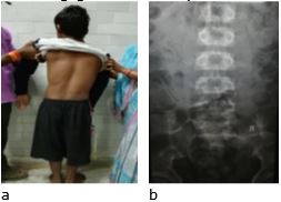
No further treatment or any consultation was taken by the patient for next 2 months and even the complaints did not improve either. Hence patient reported to our tertiary center OPD. He had severe back pain with severe difficulty to pass urine and stools since last 15 days and was unable to pass urine since last two days. He was having severe back ache, radiating down to both legs, inability to stand without support, peri-anal numbness and bilateral foot drop. On examination he was not able to stand erect and was taking support to stand. He preferred to lie down with his knees and hips flexed. Straight leg raise test was painful at 300 on both side, power was 3/5 in ankle dorsi-flexors, 1/5 in extensor hallucis longus and 2/5 in planter flexors on both side. Ankle reflex was absent, planter reflex was mute, peri-anal sensations were absent and anal reflex was absent.

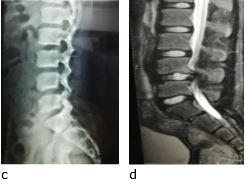
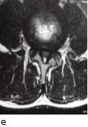
An immediate MRI of lumbar spine with whole spine screening was ordered. MR showed us a big central disc prolapse of L4-L5 disc compressing the dural sac and the transversing nerve roots causing both central canal and lateral recess stenosis with complete cut off sign of CSF.

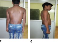
Pre-operative investigations were done and after fitness for surgery patient was planned for surgery on emergency list. Under general anesthesia in prone position, open L4 laminectomy with L4-L5 discectomy was done through posterior approach. The surgery was completed uneventfully. He was given a lumbar corset for 2 months and physiotherapy and mobilization was slowly started as he regained power. Postoperatively, patient reported improvement in radicular pain immediately and was able to walk without support on 5th post-operative day. He was able to pass urine and stools on his own on 7th postoperative day. After a period 2 months of restricted activity, he started going to school. At 6 month follow up his only complaint was mild numbness on dorsal aspect of both feet with full recovery of power in both limbs and bladder and bowel control.

Figure 1:Preoperative clinical photograph (a), anteroposterior (b) and lateral (c) x rays of lumbar spine and sagittal (d) and transverse section of MRI at L4-5 level of the patient showing L4-5 disc herniation with severe nerve compression. Postoperative clinical photograph (f & g) after laminectomy and discectomy at L4-5 showing good recovery.
ojmpc_113_01a.jpg
ojmpc_113_01c.jpgojmpc_113_01e.jpg
ojmpc_113_01f.jpg


Discussion

Disc degeneration and it’s prolapse generally starts around 30 years of age and lumbar disc prolapse causing neurological deficit is very rare in children. Wahren first reported a lumbar disc herniation in a 12-year-old child in 1945 [1]. Report published by Mayo clinics in 1982, on 9991 discectomies showed only 0.5 % were children of the age 16 years and younger [2].

Trauma in the form of sports or lifting any heavy object is the most common cause being reported to precede symptoms of disc prolapsed in children, whereas in adult population disc degeneration leads to disc prolapse [3-5]. Few studies have demonstrated epiphyseal ring separation in lumbar disc prolapse in children [6-8]. High body mass index and genetic predilection have also been associated with increase incidences of lumbar disc herniation in adolescent patients [9].

Most of the patients with disc herniation present with acute onset low back pain radiating down to one or both legs. On examination, there is loss of lumbar lordosis and may be sciatic listing on either side. Child may prefer to lie down on the sides with hip and knee flexed. There can be gross restriction of movement in spine and as such the whole body. Straight leg raise is painful in these patients. It is important to rule out other more common diagnosis like infection, fracture, muscle strain, ligament strain, osteoid osteoma and lytic spondylolysis. Plain radiographs and MRI are the most important investigations to be done. Apophyseal separation should be looked for and reported carefully [9,10].

After confirming the diagnosis, treatment is aimed to relieve the symptoms, neurological improvement and early return to routine life. Conservative treatment is recommended and found to be effective by Zamani et al [9]. While most of other studies suggest excellent results after surgical decompression [2,3,6,11-15]. Kurihara and Kataoka reported only 40% patients responding to conservative treatment and recurrence was common after starting routine activities [3]. The main cause of failure of conservative treatment in adolescent is the well hydrated disc. It does not resorb like the degenerated dehydrated adult disc. Other cause can be the less compliance of children to activity restriction as compared to adults. Symptoms due to separated epiphyseal cartilage seen in few patients are also very difficult to manage conservatively [16].

Cauda equina syndrome, progressive neurological deficit and disabling pain not responding to conservative treatment need surgical treatment [16]. Cauda equina syndrome is a very rare presentation in adolescents but should be diagnosed promptly and should be decompressed on emergency basis. Early surgical intervention promises the best chance for neurological recovery. Percutaneous endoscopic discectomy, micro-discectomy and open discectomy are the techniques which are generally used. Open discectomy with partial or complete laminectomy is the procedure most often performed. Posterolateral disc can be excised with laminotomy and flavotomy. With the central disc bulge and stenotic canal we need to remove the complete lamina to adequately decompress the cauda equine nerve roots.

The protruded annulus and disc should be removed and nucleus should be adequately decompressed. All loose disc fragments should be removed. Complete or overzealous discectomy should be avoided as it does not serve any good in neurological improvement and it may speed up the degeneration of the disc and facet joints. This leads to stenotic changes at that level [17]. For children and adolescents, it is very important to maintain the integrity of the inner part of the annulus where the proteoglycan synthesis is the most active [18]. Ishihara’s et al showed that leaving the inner annulus intact could lead to partial regeneration of the intervertebral disc [14]. Though our case presented very late with typical features of cauda equina syndrome but responded well to the decompression, as prompt surgery was done. This better outcome might be due to his young age. Early post-operative complications found in adolescent patient could be wound hematoma (1-4%) and delayed wound healing (3%), none of which were seen in our case. Post-operative surgical site infection or discitis is rare in children and adolescent patients [16]. Late complications reported are the disc space narrowing, foraminal stenosis and adjacent level disc degeneration [17]. There are chances of recurrent disc herniation (5-10%) which may need revision standard discectomy [10].

Conclusion

Prolapsed intervertebral disc causing symptoms though very rare but should be considered in adolescent patients with significant history of trauma. Cauda equina is a surgical emergency and should be diagnosed and operated as early as possible for good results. Even if the patient presents late, like ours decompression should be offered to the neural tissue whenever possible, especially in young age patients.

References

1. Wahren H. Herniated nucleus pulposus in a child of twelve years. Act Orthop Scand 1945;16:40-2. . [Crossref][PubMed][Google Scholar]

2. DeOrio JK, Bianco AJ Jr. Lumbar disc excision in children and adolescence. J Bone Joint Surg Am 1982;64;991-6. . [Crossref][PubMed][Google Scholar]

3. Kurihara A, Kataoka O. Lumbar disc herniation in children & adolescents – A review of 70 operated cases and their minimum 5 year follow-up studies. Spine 1980;5:443-51. . [Crossref][PubMed][Google Scholar]

4. Durham SR, Sun PP, Sutton LH. Surgically treated lumbar disc disease in the pediatric population: an outcome study. J Neurosurg 2000;92:1-6. . [Crossref][PubMed][Google Scholar]

5. Shillito J Jr. Pediatric lumbar disc surgery: 20 patients under 15 years of age. Surg Neurol 1996;46:14-8. . [Crossref][PubMed][Google Scholar]

6. Papagelopoulus PJ, Shaughnessy WE, Ebersold MJ et al. Long term outcome of lumbar discectomy in children and adolescents sixteen years of age or younger. J Bone Joint Surg Am 1998;80:689-98. . [Crossref][PubMed][Google Scholar]


7. Yang IK, Bahk YW, Choi KH et al. Posterior lumbar apophyseal ring fractures: a report of 20 cases. Neuroradiology 1994;36:453-5. . [Crossref][PubMed][Google Scholar]

8. Parisini P, Di Silvestre M, Greggi T et al. Lumbar disc excision in children and adolescents. Spine 2001;26:1997-2000. . [Crossref][PubMed][Google Scholar]

9. Zamani MH, MacEwen GD. Herniation of lumbar disc in children and adolescents. J Pediatr Orthop 1982;2:528-33. . [Crossref][PubMed][Google Scholar]

10. Lukhele M, Mayet Z, Dube B. Lumbar disc herniation in a 9 year old child: case report and review of literature. SA Orthop J 2011;10:3:93-6. . [Crossref][PubMed][Google Scholar]

11. Borgesen SE, Vang PS. Herniation of the lumbar intervertebral disc in children and adolescents. Acta Orthop Scand 1974;45:540–9. . [Crossref][PubMed][Google Scholar]

12. DeLuca PF, Mason DE, Weiand R, Howard R, Bassett GS. Excision of herniated nucleus pulposus in children and adolescents. J Pediatr Orthop 1994;14:318–22. . [Crossref][PubMed][Google Scholar]

13. Garrido E, Humphreys RP, Hendrick EB, Hoffman HJ. Lumbar disc disease in children. Neurosurgery 1978;2:22–6. . [Crossref][PubMed][Google Scholar]

14. Ishihara H, Matsui H, Hirano N, Tsuji H. Lumbar intervertebral disc herniation in children less than 16 years of age. Long-term follow-up study of surgically managed cases. Spine 1997;22:2044–9. [Crossref][PubMed][Google Scholar]

15. Russwurm H, Bjerkreim I, Ronglan E. Lumbar intervertebral disc herniation in the young. Acta Orthop Scand 1978;49:158–63. . [Crossref][PubMed][Google Scholar]

16. Dang L. Liu Z A review of current treatment for lumbar disc herniation in children and adolescents. Eur Spine J 2010. 19:205–14. [Crossref][PubMed][Google Scholar]

17. Luukkonen M, Partanen K, Vapalahti M. Lumbar disc herniation’s in children: a long-term clinical and magnetic resonance imaging follow-up study. Br J Neurosurg 1997;11:280–5. . [Crossref][PubMed][Google Scholar]

18. Bayliss MT, Johnstone B, O’Brien JP. Proteoglycan synthesis in the human intervertebral disc. Variation with age, region and pathology. Spine 1988;13:972–81. [Crossref][PubMed][Google Scholar]

Disclaimer / Publisher's Note

The statements, opinions and data contained in all publications are solely those of the individual author(s) and contributor(s) and not of Journals and/or the editor(s). Journals and/or the editor(s) disclaim responsibility for any injury to people or property resulting from any ideas, methods, instructions or products referred to in the content.