Orthopaedic Journal of MP Chapter

Publisher: Madhya Pradesh Orthopaedic Association www.mpioa.com
E-ISSN:2582-7243, P-ISSN:2320-6993
2020 Volume 26 Number 1 Jan-Jun

Tarlov Cyst: A Case Report

Verma A1, Jain S2*, Jain M3, Mundra A4

1 A Verma, Department of Orthopaedics, Mahatma Gandhi Memorial Medical College, Indore, Madhya Pradesh, India.

2* Saurabh Jain, Assistant Professor, Department of Orthopaedics, Mahatma Gandhi Memorial Medical College, Indore, Madhya Pradesh, India.

3 M Jain, Department of Orthopaedics, Mahatma Gandhi Memorial Medical College, Indore, Madhya Pradesh, India.

4 A Mundra, Department of Orthopaedics, Mahatma Gandhi Memorial Medical College, Indore, Madhya Pradesh, India.

Case Report: Tarlov cyst is rare perineural cyst, which may be symptomatic and present with with low back pain, sciatica, coccydynia or cauda equina syndrome. Symptomatic tarlov cyst needs to be removed surgically. We present such a rare case of symptomatic sacral cystic mass (tarlov cyst) presenting with severe lower back pain for months which was successfully treated by sacral laminotomy and cyst excision. The aim of reporting this case was to create awareness among the surgeons regarding this rare entity and to include it in differential diagnosis of chronic low back pain.

Keywords: Tarlov cyst, sacral cyst, perineural cyst

Corresponding Author How to Cite this Article To Browse
Saurabh Jain, Assistant Professor, Department of Orthopaedics, Mahatma Gandhi Memorial Medical College, Indore, Madhya Pradesh, India.
Email:
Verma A, Jain S, Jain M, Mundra A, Tarlov Cyst: A Case Report. ojmpc. 2020;26(1):45-48.
Available From
https://ojmpc.com/index.php/ojmpc/article/view/104
Manuscript Received Review Round 1 Review Round 2 Review Round 3 Accepted
2020-06-06 2020-06-12 2020-06-18 2020-06-24 2020-06-30
Conflict of Interest Funding Ethical Approval Plagiarism X-checker Note
Authors state no conflict of interest. Non Funded. The conducted research is not related to either human or animals use. 11.11 All authors have accepted responsibility for the entire content of this manuscript and approved its submission.

© 2020by Verma A, Jain S, Jain M, Mundra Aand Published by Madhya Pradesh Orthopaedic Association. This is an Open Access article licensed under a Creative Commons Attribution 4.0 International License https://creativecommons.org/licenses/by-nc/4.0/ unported [CC BY NC 4.0].

Introduction

Tarlov cyst is a perineural cyst, which was first described by Tarlov, after whom they are named. They are usually asymptomatic and are seen as an incidental finding at autopsy [1].Tarlov described first case of symptomatic perineural cyst and recommended its removal. Very few cases of such Tarlov cyst are reported in the literature [2-4].

Most of the cysts are asymptomatic, but some of them can be symptomatic. The cyst can present as low back pain, sciatica, coccydynia or cauda equina syndrome, which when symptomatic, may require surgical treatment [4]. Diagnosis of cyst can be confirmed on MRI, which shows the fluid filled cystic lesion arising from the sacral nerve root near the dorsal root ganglion [5].

We here report such a rare case of symptomatic sacral Tarlov cyst presenting as back pain and radiculopathy which was successfully treated by surgical excision.

Case Report

A 45 year old male, labourer by occupation presented to outdoor patient department with history of insidious onset pain in lower back since 8 months.

There was no history of trauma, jerk or history of lifting any heavy object. The pain was localized to lower back, dull in nature with no diurnal or postural variations. Pain was initially relieved with rest and NSAIDS but had aggravated since last 2 months affecting the daily activities of the patient.

There was history of claudication and aggravation of pain on long standing. The pain had started radiating to left lower limb upto the ankle and toes. There was no history of bladder bowel dysfunction. The constitutional symptoms like loss of appetite, fever, weight loss or malaise were absent. He had no other complaint related to other body parts with bilateral hip and knees being normal.

On examination, there was mild local tenderness in the sacral area and lower back. Straight leg rising was 40º on left side and 80º on right side. There was weakness of ankle dorsiflexors, toe flexors and extensors (MRC grade 3) on left side with no sensory deficit. Right side was normal. All the reflexes were normal bilaterally.

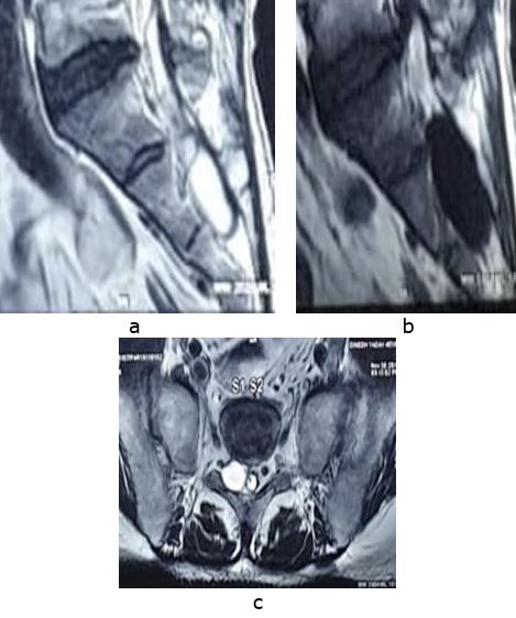
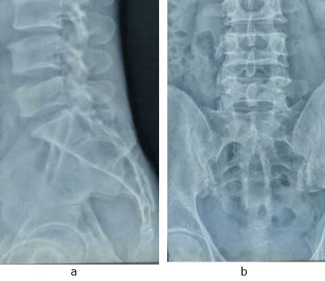
All haematological investigations and radiographs of lumbosacral spine were normal. MRI of the lumbosacral spine revealed a right sided extra dural, 3.8×1.8cm fluid filled cyst at level of S2 vertebra causing medial compression of dural sac.

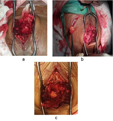
After routine pre-operative fitness patient was planned for surgical excision of cyst. Patient was operated under general anaesthesia in prone position via midline dorsal approach. Sacral laminotomy was done and a window was made in the sacrum. Flaps of ligamentum flavum elevated to expose the cyst (Figure 3a).

The cyst was excised and the dural sac was decompressed and a free fat graft taken from right gluteal region (Figure 3b) was placed over the after the plication of cyst wall.

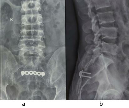
Finally osteosacral flap was reinforced using a Recon plate. The excised cyst was sent for histopathological examination which conformed presence of nerve cells in the cyst confirming the diagnosis of Tarlov cyst.

Figure 1:Lumbosacral X rays spine AP (a) and lateral (b) view with no obvious lesion
ojmpc_104_01.jpg

Post-operatively, there was no deterioration of neurology, and patient had immediate relief from radicular pain after the surgery. His motor power increased by grade one at 2 weeks of the surgery with no complains of the pain at back or radiculopathy. At 2 weeks post-surgery, he was able to walk without any claudication.

Discussion

Tarlov cysts are most commonly found in sacral region between perineurium and endoneurium of nerve root, although they can be present at any spinal segment. Small, asymptomatic Tarlov cysts are not uncommon and may be seen in 5 to 9% of the general population but large cysts that cause symptoms are relatively rare [2]. Cyst is common in females (86%) and in age group of 31 to 60 years (80%) [3]. Our case was a 45 years male who was labourer by profession.

Clinical presentation of the cyst can be variable. It can present with local or radicular pain, or different motor or sensory deficit, with or without involvement of bladder and bowel, depending on the size, location and relationship with the nerve root. Radicular symptoms from caudal nerve root may vary from sciatica, sacral or buttock pain, vaginal or penile paraesthesia or sensory changes over the buttocks, perineal area and lower extremity. Rare, but a larger cyst can even result in cauda equina syndrome [3,4].

Radiographs and CT scans are usually normal except for bone erosion of the spinal canal or neural foramina.


Figure 2:MRI of sacral region saggital T2WI (a), saggital T1WI (b) and axial (c) views showing fluid filled cyst overlying second sacral vertebra
ojmpc_104_02.jpg

Figure 3:Intra operative photos (a to c) showing laminotomy done at S2, showing cyst and free fat graft placed in the spinal canal
ojmpc_104_03.jpg

With the advent of MRI, myelography is usually not needed now, but if done shows delayed filling of the meningocele sac.

Figure 4:Post-operative lumbosacral X rays spine AP (a) and lateral (b)view showing plate over the sacrum
ojmpc_104_04.jpg

MRI is the investigation of choice which shows cyst lesion along the nerve root, which is low signal on T-1 weighted images and high signal on T-2 weighted images, similar to CSF [5,6].

Tarlov described the formation of cyst in space between the arachnoid which covers the root i.e. the perineurium (covers the root) and the endoneurium (outer layer of the pial cover of the root). It begins in one portion of the circumferential perineural space and if large enough can cause compression of the nerve root to one side, like in our case the cyst was compressing the nerve root causing radicular symptoms typically on left side. In later stages, cyst occupies the posterior root and abuts the proximal part of dorsal ganglion, which is bordered by reticulum or by nerve fibers [1].

The cause for genesis of these perineural cysts is not clear, but sacro-coccygeal trauma has been suspected, which causes hemorrhage into subarachnoid space and accumulation of red cells. This causes impediment of drainage of veins in the perineurium and epineurium, leading to rupture and subsequent cyst formation. Tarlov, Schreiber and Haddad all supported trauma as the cause of cyst formation but Fortuna et al believed that the perineural cysts were congenital, caused by arachnoidal proliferations within the root sleeve [6,7,9].

Treatment of cyst ranges from wait and watch, excision, percutaneous aspiration to excision and duraplasty. Asymptomatic cysts are left as such. Tarlov advised sacral laminectomy and complete excision of single perineural cysts together with the posterior root and ganglion whereas Paulsen advised CT-guided percutaneous aspiration of cyst [3,7]. Recently Caspar advocated microsurgical cyst excision combined with duraplasty or plication of the cyst wall, leaving the parent nerve root intact [10]. We in our case, completely excised the cyst via sacral laminectomy, followed by free fat graft over cyst wall and the repositioning of osteosacral flap using a Recon plate, so that we can mobilize patient early.


Conclusion

Symptomatic Tarlov cyst is a rare finding, which can cause radicular or other pain syndromes. We reported this rare case to create awareness among the surgeons regarding this rare entity and to have clinical suspicion and include it in differential diagnosis of chronic low back pain, considering early MRI scans.

References

1. Tarlov IM. Perineural cysts of the spinal nerve roots. Arch Neural Psychiatry. 1938;40:1067–74. [Crossref][PubMed][Google Scholar]

2. Prashad B, Jain AK, Dhammi IK. Tarlov cyst: Case report and review of literature. Indian J Orthop. 2007;41(4):401–403. [Crossref][PubMed][Google Scholar]

3. Paulsen RD, Call GA, Murtagh FR. Prevalence and percutaneous drainage of cysts of the sacral nerve root sheath (Tarlov cysts) AJNR Am J Neuroradiol. 1994;15:293–9. . [Crossref][PubMed][Google Scholar]

4. Acosta FL, Quinones-Hinojosa A, Schmidt MH, Weinstein PR. Diagnosis and management of sacral Tarlov cysts: Case report and review of the literature. Neurosurg Focus. 2003;15:E15. [Crossref][PubMed][Google Scholar]

5. Rodziewicz GS, Kaufman B, Spetzler RF. Diagnosis of sacral perineural cysts by nuclear magnetic resonance. Surg Neurol. 1984;22:50–2. [Crossref][PubMed][Google Scholar]

6. Tarlov IM. Cysts (perineurial) of the sacral roots. J Am Med Assoc. 1948;138:740–4. [Crossref][PubMed][Google Scholar]

7. Tarlov IM. Spinal perineurial and meningeal cysts. J Neural Neurosurg Psychiatry. 1970;33:833–43. [Crossref][PubMed][Google Scholar]

8. Schreiber F, Haddad B. Lumbar and sacral cysts causing pain. J Neurosurg. 1951;8:504–9. [Crossref][PubMed][Google Scholar]

9. Fortuna A, La Torre E, Ciappetta P. Arachnoid diverticula: A unitary approach to spinal cysts communicating with the subarachnoid space. Acta Neurochir (Wien) 1977;39:259–68. . [Crossref][PubMed][Google Scholar]

10. Caspar W, Papavero L, Nabhan A, Loew C, Ahlhelm F. Microsurgical excision of symptomatic sacral perineurial cysts: A study of 15 cases. Surg Neurol. 2003;59:101–6. [Crossref][PubMed][Google Scholar]

Disclaimer / Publisher's Note

The statements, opinions and data contained in all publications are solely those of the individual author(s) and contributor(s) and not of Journals and/or the editor(s). Journals and/or the editor(s) disclaim responsibility for any injury to people or property resulting from any ideas, methods, instructions or products referred to in the content.