Orthopaedic Journal of MP Chapter

Publisher: Madhya Pradesh Orthopaedic Association www.mpioa.com
E-ISSN:2582-7243, P-ISSN:2320-6993
2020 Volume 26 Number 1 Jan-Jun

Giant cell tumor of proximal radius: A rare case report and review of literature

Jain T1, Bharadwaj L2, Tiwari DA3*, Verma R4

1 T Jain, Department of Orthopaedics, Gandhi Medical College, Bhopal, MP, India.

2 L Bharadwaj, Department of Orthopaedics, Gandhi Medical College, Bhopal, MP, India.

3* Dr Anurag Tiwari, Assistant Professor, Department of Orthopaedics, Gandhi Medical College, Bhopal, MP, India.

4 R Verma, Department of Orthopaedics, Gandhi Medical College, Bhopal, MP, India.

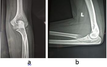
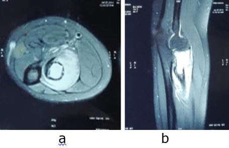
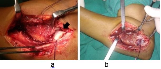
Case report: Giant-cell tumor (GCT) of the bone is benign bone tumor which usually arises from the epiphysis of long bones. Distal femur and proximal tibia are the most common sites of this tumor. The proximal radius is extremely rare site of this particular tumor. We present a rare case of Giant cell tumor of proximal radius in a twenty-one year old girl, for which wide margin resection was performed successfully with no recurrence, complications or disability seen at one year follow up.

Keywords: Giant cell tumor, proximal radius, resection

Corresponding Author How to Cite this Article To Browse
Dr Anurag Tiwari, Assistant Professor, Department of Orthopaedics, Gandhi Medical College, Bhopal, MP, India.
Email:
Jain T, Bharadwaj L, Tiwari DA, Verma R, Giant cell tumor of proximal radius: A rare case report and review of literature. ojmpc. 2020;26(1):40-44.
Available From
https://ojmpc.com/index.php/ojmpc/article/view/103
Manuscript Received Review Round 1 Review Round 2 Review Round 3 Accepted
2020-06-06 2020-06-12 2020-06-18 2020-06-24 2020-06-30
Conflict of Interest Funding Ethical Approval Plagiarism X-checker Note
Authors state no conflict of interest. Non Funded. The conducted research is not related to either human or animals use. 14.44 All authors have accepted responsibility for the entire content of this manuscript and approved its submission.

© 2020by Jain T, Bharadwaj L, Tiwari DA, Verma Rand Published by Madhya Pradesh Orthopaedic Association. This is an Open Access article licensed under a Creative Commons Attribution 4.0 International License https://creativecommons.org/licenses/by-nc/4.0/ unported [CC BY NC 4.0].

Introduction

Giant cell tumour (GCT), also known as Osteoclastoma, is a locally aggressive benign bone tumour, arising from epiphysis of long bones [1]. It commonly occurs in 20 to 40 year of age group and is more common in females [2]. Most common locations are distal femur, proximal tibia, distal radius and spine [3]. The proximal radius is an extremely rare site for giant cell tumours, in contrast to the distal radius. The prevalence of GCT proximal radius is reported to be 0.16%- 0.5% of all GCT cases [4].

We here thus present a rare such case of this tumour i.e. GCT in proximal radius in a twenty one year old female who was successfully managed by wide marginal resection of proximal radius.

Case report

A 21-year old female with right hand dominance presented to orthopaedic department with complain of pain and swelling over non-dominant left elbow and upper forearm since 1 month. Constitutional symptoms were absent. She had taken no treatment, except some analgesics and had never consulted a doctor.

Pain was insidious in onset, gradually progressive, localized to left lateral elbow and forearm, dull aching in character which was aggravated with supination-pronation and relieved temporarily by taking rest and oral analgesics. The pain was associated with mild to moderate swelling over the forearm and elbow which was more on the lateral aspect. Family history and past history were unremarkable. There was no other swelling in any other body parts.

On examination, there was localised moderate tenderness at radial head. On palpation, swelling was mild localized and was hard in consistency and immobile, whereas due to the swelling the deeper radial head could not be palpated. Bleeding or ulceration was absent. Range of motion (ROM) in the left elbow joint was 0° -140° of flexion, 40° of pronation, and 45° of supination (Figure 1). Distal neurovascular status was normal. All laboratory investigations were within normal limit.

Plain radiograph of affected elbow and forearm showed a relatively well-defined expansile lytic lesion in epiphysio-metaphyseal region of proximal radius with geographical bone destruction and loss of contours of radial head and neck (Figure 2).

Magnetic resonance imaging (MRI) T1 axial and sagittal image showed relatively well defined hyper-intense osteolytic destructive lesion involving proximal radius with cortical breach at anterolateral and postero-medial aspect (figure 3).

The radiological and MRI features showed the typical features of giant cell tumor, but to confirm the diagnosis needle biopsy was taken which showed stromal cells, numerous osteoclastic giant cells and scattered lymphocytes, which confirmed the diagnosis of Giant cell tumor of proximal radius.

Because of the marked destruction and expansion of the radius, the decision was made to perform wide margin resection of proximal radius.

Figure 1: Clinical photograph of the patient showing restricted supination (a) and pronation (b) on the left side.
ojmpc_103_01.jpg

Figure 2: Pre-operative radiograph AP (a) and lateral (b) views of left elbow showing expansile lesion in proximal radius with no periosteal reaction.
ojmpc_103_02.jpg

Figure 3: MRI axial (a) and sagittal (b) image of the patient showing hyperintense lesion in proximal radius, intraosseous as well as extra osseous component.
ojmpc_103_03.jpg

Under brachial block in supine position, Kocher approach was used to approach the proximal radius (figure 4). The annular ligament was incised along with the joint capsule. The posterior interosseous nerve was identified and was protected. An osteotomy of the radius was performed 2 cm from the distal margin of the lesion and closure was done in layers after achieving haemostasis. The specimen was sent for histo-pathological examination, which confirmed the presence of giant cell tumor, by presence of multinucleate giant cells.


Figure 4: Intra-operative photograph (a & b) showing the lesion involving the neck and proximal shaft of radius. Note the cartilage of radial head visible (black arrow).
ojmpc_103_04.jpg

Post-operatively, patient’s limb was kept in above elbow slab, which was removed after suture removal. She did not have any wound problems post-operatively. At final follow of 12 months, she had nearly full range of flexion/extension at elbow and supination/pronation with no clinical or radiological symptoms and signs of recurrence of tumour (figure 5).

Figure 5: Post-operative radiograph AP (a) and lateral (b) view and clinical photo (c & d) at 1 year post surgery. There were no signs of recurrence and complete range of pronation and supination.
ojmpc_103_05.jpg

Discussion

Giant cell tumour is a bone tumor that is most common in third to fourth decade of life, though rarely seen in younger age group also. It is benign but locally aggressive tumour involving epiphyseal region of skeletally mature long bones [1,2].

Patients may present with pain, swelling or pathological fracture. Radiologically, it presents as eccentric, expansile, lytic lesion in the epiphyses of long bones [1,2]. Distal femur, proximal tibia and distal radius are the usual sites involved [3,4].

The common differential diagnosis includes aneurysmal bone cyst, chondroblastoma, chondromyxoid fibroma, brown tumor, non-ossifying fibroma, and osteosarcoma [5,6].

The proximal radius is extremely rare site for giant cell tumours. To best of our knowledge, only, ten cases of GCT involving proximal radius have been reported so far in the literature [7-17] (table 1). Methods of treatment ranged from curettage with or without bone grafting, resection with or without reconstruction to even above elbow amputation [17,18].

Mir NA et al and Khan KM et al performed marginal resection [8,14] and Lewis MM et al, Akmaz I et al and Novais EN et al treated the tumor with curettage and bone grafting [7,9,12]. Singh AP et al in 2009 performed above elbow amputation for GCT proximal radius [11].

Table 1:Comparison of our case with reported literature

SrYearStudyAge(yr)GenderClinical presentationSideDuration(mths)TreatmentFollow up(mths)Outcome
11985Lewis MM735FemaleCurettage + Bone grafting
22003Mir NA835MalePain elbowR18Excision36No recurrence
32004Akmaz I921MaleDiscomfort proximal forearmLCurettage and autogenous iliac crest bone graft36No recurrence
No infection
No fracture
42006Sakayama K1073FemaleElbow painLEn bloc resection and reconstruction with radial head prosthesis72No recurrence
No loosening
No dislocation
52009Singh AP 1152FemalePain and progressively increasing swelling around elbowR8Above elbow amputation60No recurrence
62011Novais EN1213MaleMulticentric GCT
Pain and swelling elbow
RCurettage + Burr + Phenol + Bone graftLack terminal 150 of supination and pronation
72011Song WS 1333MaleElbow and forearm painL8En bloc resection and reconstruction with polyethylene insert and bone cement36No recurrence
82014Khan KM1448FemaleMass elbowR2Wide margin excision with sacrifice of PIN7No recurrence
92017Dahuja A 1550FemaleSwelling around elbow with wrist dropRWide margin excision with non-vascularised fibula with TENS fixation24No recurrence
Nerve recovered at 2 months post-surgery
102018Nayar SK1623MalePain in elbowL6Resection and reconstruction with osteoarticular allograft54No recurrence
Union at 17 months
112019Present study21FemalePain elbowL1Wide margin excision12No recurrence

Dahuja A et al performed resection and reconstruction with fibula autograft [15], Nayar SK et al performed resection and reconstruction with osteoarticular allograft [16]. Song WS et al performed reconstruction with polyethylene insert; pins, screws and bone cement [13], whereas Sakayama K et al used floating radial head prosthesis [10].

Resection and reconstruction with radial head prosthesis is a good option, when the tumor is small. Further loosening of the prosthesis is one of the most serious complications, but the major limitation with the use of radial head prosthesis (RHP) is that only short to midterm results are known and long term results of RHP are not known and the cost of RHP is a big issue especially in developing countries [19,20].

Osteoarticular allografts have been used for reconstruction after resection of giant cell tumor, which is cost effective, restores the bone stock and promotes biological bone union. But the drawbacks are that it is technically demanding surgery, long healing time, risk of fracture, risk of disease transmission, and surgical site infection [21-24]. Considering the benign nature of tumor, amputation per se has little place in the treatment of giant cell tumours.

In our case, the patient presented to us late, by that time, the disease had advanced to the extent where, curettage and bone grafting was not an advisable option, because of fear of recurrence [25,26]. Hence we planned the patient for extensive marginal resection. Resection alone, in our case appeared to be completely effective in eradicating the disease and providing excellent functional outcome. In our patient on last follow-up, she had nearly full range of flexion/extension at elbow, and supination/pronation after extensive supervised physiotherapy.

Conclusion

GCT of proximal radius is very rare tumor, for which, wide margin resection of tumour is a good, cost effective treatment with no recurrence, minimal complications, and disability.

References

1. Campanacci M, Baldini N, Boriani S, Sudanese A. Giant-cell tumour of bone. J Bone Joint Surg Am 1987;69(1):106-14. . [Crossref][PubMed][Google Scholar]

2. Saglik Y, Yildiz Y, Karakas A, Ogut H, Erekul S. Giant cell tumor of bone. Bull Hosp Jt Dis. 1999;58(2):98–104. [Crossref][PubMed][Google Scholar]

3. Mc Gough RL, Rutledge J, Lewis VO, Lin PP, Yasko AW. Impact severity of local recurrence in giant cell tumour of bone. Clin Orthop Relat Res. 2005;438:116-22. [Crossref][PubMed][Google Scholar]

4. Turcotte RE. Giant cell tumor of bone. Orthop Clin North Am. 2006;37(1):35-51. [Crossref][PubMed][Google Scholar]

5. Dahlin DC. Caldwell Lecture. Giant cell tumor of bone: highlights of 407 cases. Am J Roentgenol. 1985;144(5):955–960 [Crossref][PubMed][Google Scholar]

6. Murphey MD, Nomikos GC, Flemming DJ, Gannon FH, Temple HT, Kransdorf MJ. From the archives of AFIP. Imaging of giant cell tumor and giant cell reparative granuloma of bone: radiologic-pathologic correlation. Radiographics 2001;21(5):1283-309. [Crossref][PubMed][Google Scholar]

7. Lewis MM, Kaplan H, Klein MJ, Ferriter PJ, Legouri RA. Giant cell tumour of the proximal radius. Clin Orthop Relat Res 1985;201:186-9. . [Crossref][PubMed][Google Scholar]

8. Mir NA, Bhat JA, Halwai MA. Giant cell tumours of proximal radius and patella—an unusual site of presentation: case report. JK Sci. 2003;5:35–37. [Crossref][PubMed][Google Scholar]

9. Akmaz I, Arpacioglu MO, Pehlivan O, Solakoglu C, Mahirogullari M, Kiral A, et al. An infrequent localization of giant cell tumour: proximal radius: case report. J Arthroplast Arthroscop Surg. 2004;15:174–177. [Crossref][PubMed][Google Scholar]

10. Sakayama K, Sugawara Y, Kidani T, Miyawaki J, Yamamoto H. Bipolar-type floating radial head prosthesis for the treatment of giant cell tumor of the proximal radius: a case report. J Shoulder Elbow Surg. 2006;15(5):9-12. [Crossref][PubMed][Google Scholar]

11. Singh AP, Mahajan S, Singh AP. Giant cell tumour of the proximal radius. Singap Med J. 2009;50:388–390. [Crossref][PubMed][Google Scholar]

12. Novais EN, Shin AY, Bishop AT, Shives TC. Multicentric giant cell tumor of the upper extremities: 16 years of ongoing disease. J Hand Surg Am. 2011;36(10):1610-3. [Crossref][PubMed][Google Scholar]

13. Song WS, Cho WH, Kong CB, Jeon DG. Composite reconstruction after proximal radial giant cell tumour resection. Arch Orthop Trauma Surg. 2011;131:627–630. [Crossref][PubMed][Google Scholar]

14. Khan KM, Minhas MS, Khan MA, Bhatti A. Giant cell tumour of proximal radius in a 48 years old lady. J Coll Physicians Surg Pak. 2014;24(1):S48-9. [Crossref][PubMed][Google Scholar]

15. Dahuja A, Kaur R, Bhatty S, Garg S, Bansal K, Singh M. Giant-cell tumour of proximal radius in a 50-year-old female with wrist drop: a rare case report. Strategies Trauma Limb Reconstr. 2017;12(3):193–196. [Crossref][PubMed][Google Scholar]

16. Nayar SK, Dein EJ, Spiker AM, Bernard JA, Zikria BA, Weber KL. Resection of a Giant-Cell Tumor of the Proximal Aspect of the Radius with Osteoarticular Allograft Reconstruction: A Case Report. JBJS Case Connect. 2018;8(1):e7. [Crossref][PubMed][Google Scholar]

17. Lackman RD, Hosalkar HS, Ogilvie CM, Torbert JT, Fox EJ. Intralesional curettage for grades II and III giant cell tumours of bone. Clin Orthop Relat Res 2005;438:123-7. . [Crossref][PubMed][Google Scholar]


18. Bi Z, Pan Q, Fu C, Han X. Wrist joint reconstruction with vascularized fibular head graft after resection of distal radius giant cell tumour. Zhongguo Xiu Fu Chong Jian Wai Ke Za Zhi 2010;24:1416-8. . [Crossref][PubMed][Google Scholar]

19. Pribyl CR, Kester MA, Cook SD, et al. The effect of the radial head and prosthesis radial head replacement on resisting valgus stress at the elbow. Orthop 1986;9(5):723-6. . [Crossref][PubMed][Google Scholar]

20. Kodde IF, Kaas L, Flipsen M, van den Bekerom MPJ, Eygendaal D. The current concepts in management of radial head fractures. World J Orthop. 2015;6(11):954–960. [Crossref][PubMed][Google Scholar]

21. Enneking WF, Campanacci DA. Retrieved human allografts: a clinico-pathological study. J Bone Joint Surg Am. 2001;83(7):971-86. [Crossref][PubMed][Google Scholar]

22. Muscolo DL, Ayerza MA, Aponte-Tinao LA. Survivorship and radiographic analysis of knee osteoarticular allografts. Clin Orthop Relat Res. 2000;373:73-9. [Crossref][PubMed][Google Scholar]

23. Thompson RC Jr, Pickvance EA, Garry D. Fractures in large-segment allografts. J Bone Joint Surg Am. 1993;75(11):1663-73. [Crossref][PubMed][Google Scholar]

24. Muscolo DL, Ayerza MA, Aponte-Tinao LA, Ranalletta M. Use of distal femoral osteoarticular allografts in limb salvage surgery. J Bone Joint Surg Am. 2005;87(11): 2449-55. [Crossref][PubMed][Google Scholar]

25. O'Donnell RJ, Springfield DS, Motwani HK, Ready JE, Gebhardt MC, Mankin HJ. Recurrence of giant-cell tumors of the long bones after curettage and packing with cement. J Bone Joint Surg Am. 1994;76(12):1827–1833. [Crossref][PubMed][Google Scholar]

26. Yip KMH, Leung PC, Kumta SM. Giant cell tumor of bone. Clin Orthop 1996;323:60-4. . [Crossref][PubMed][Google Scholar]

Disclaimer / Publisher's Note

The statements, opinions and data contained in all publications are solely those of the individual author(s) and contributor(s) and not of Journals and/or the editor(s). Journals and/or the editor(s) disclaim responsibility for any injury to people or property resulting from any ideas, methods, instructions or products referred to in the content.