Introduction
Paediatric forearm fractures are amongst the most common injuries encountered in childhood which makeup 40% of all pediatric fractures [1]. Amongst fractures of forearm i.e. radius-ulna fractures, fractures occur in distal third in 60%, 20% occurs in middle third, 14% in distal physis and approximately 4% in proximal third of radius ulna [2].
Closed reduction followed by application of well molded plaster cast is the standard treatment for pediatric forearm fractures, whereas operative treatment is reserved for unstable fractures, failure to achieve closed acceptable reduction and open fractures or those associated with compartment syndrome [3-5].
Loss of fracture reduction, re-displacement or late displacement is the most common complication of manipulated forearm fractures & cast application, which also may need surgery [6-9].
The cast index (CI) is a simple and quick method of predicting the re-displacement after cast application in radius ulna fractures in paediatric patients, particularly distal radius fractures [10].
Ideal cast index is 0.7 or less for distal radius ulna fractures for reduced risk of re-displacement, whereas a cast index of 0.8-0.84 is associated with significant risk of subsequent re-displacement [2,3,9]. Hence we conducted this study to predict the re-displacement in forearm fractures at all levels by calculating of cast index.
Material and Methods
The study was a prospective series conducted at our center in 30 cases of paediatric fracture forearm by calculating the cast index and correlating it with re-displacement rate. The study was approved by the institutional ethical committee and written informed consent from the guardian was taken before including them in the study.
All children between 2 – 12 years of age with close fracture of both radius ulna were included in study. Patients less than 2 years or more than 12 years, single bone forearm, open, pathological, segmental or intra-articular fractures were excluded from study.
All case included in the study, were given initial symptomatic treatment with slab, Ice fomentation and limb elevation for 3 to 5 days for decreasing the swelling. When swelling subsided, under supine position under short general anaesthesia, the fracture was manipulated and closed reduced to anatomical position under image intensifier guidance.
Once proper acceptable reduction was achieved, an above elbow plaster of paris cast was applied after sufficient uniform padding with elbow flexed to 900 with forearm in supination for proximal third fracture and mid prone position for all other fractures (table 1) [1].
Table 1: Acceptable criteria for forearm fractures
| Age(years) | Saggital plane(in degree) | Frontal plane(in degree) |
|---|
| 4-9 | 20 | 15 |
| 9-11 | 15 | 5 |
| 11-13 | 10 | 0 |
| >13 | 5 | 0 |
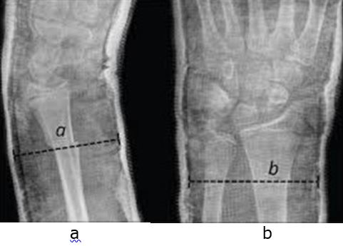
After the plaster had dried up, true antero-posterior and true lateral radiograph were taken and cast index was calculated using software ‘IMAGEJ’. Cast index is calculated by measuring the internal anterio-posterior (AP) diameter of the cast (excluding padding) at the level of the fracture taken on lateral view and dividing it by the internal medio-lateral diameter of the cast (excluding padding) taken on AP view (fig 1). Both measurements are made on the first proper radiograph taken after closed reduction [10].
Figure 1: Calculation of cast index by measuring internal anterio-posterior (AP) diameter on true lateral view (a) and internal medio-lateral diameter on true AP view (b) of the cast (excluding padding) at the level of the fracture.

Post reduction all patients were advised light finger grip and finger extension exercises along with analgesics. Patients were followed up regularly at 2, 4 and 6 weeks after cast application and radiographs were obtained in true antero posterior and true lateral views at each visit and were assessed for re-displacement as well as cast index was calculated.
Patients showing re-displacement were re-manipulated under image intensifier guidance and still not having satisfactory reduction were considered for intramedullary nailing. Cast removal was done at 6 weeks when radiological callus bridging three cortexes was seen. Functional outcomes such as final range of motion were not studied.
Results
A total of 30 cases of both bone forearm fracture with mean age 7.06 year (range 2 to 12 year) were included. 17 were male and 13 were female. Mean follow period in study was 7.2 weeks (6 to 12 week). The fracture was seen at proximal, middle and distal third forearm in seen in 6, 15 and 9 cases respectively.


