Orthopaedic Journal of MP Chapter

Publisher: Madhya Pradesh Orthopaedic Association www.mpioa.com
E-ISSN:2582-7243, P-ISSN:2320-6993
2024 Volume 24 Number 2 Jul-Dec

A rare case of Posterior Subtalar Dislocation

Jamoria R1, Pathak A2*

1 R Jamoria, Department of Orthopaedics, Gandhi Medical College, Bhopal, Madhya Pradesh, India.

2* Abhishek Pathak, Associate Professor, Department of Orthopaedics, Gandhi Medical College, Bhopal, Madhya Pradesh, India.

Case report: Posterior subtalar dislocations are extremely rare injuries which are caused by high energy trauma, which is generally managed by closed reduction, if present early. We present such a rare case of neglected posterior subtalar dislocation, which was successfully treated with open reduction. For satisfactory outcome, early diagnosis, anatomical reduction, stable fixation of peritalar joint, and the resection of small, free osteochondral fragments for the prevention of early posttraumatic arthrosis, is necessary.

Keywords: Posterior subtalar dislocation, Talocalcanealnavicular dislocation, peritalar dislocation

Corresponding Author How to Cite this Article To Browse
Abhishek Pathak, Associate Professor, Department of Orthopaedics, Gandhi Medical College, Bhopal, Madhya Pradesh, India.
Email:
Jamoria R, Pathak A, A rare case of Posterior Subtalar Dislocation. ojmpc. 2024;24(2):87-90.
Available From
https://ojmpc.com/index.php/ojmpc/article/view/95
Manuscript Received Review Round 1 Review Round 2 Review Round 3 Accepted
2018-12-07 2018-12-13 2018-12-19 2018-12-25 2018-12-31
Conflict of Interest Funding Ethical Approval Plagiarism X-checker Note
Authors state no conflict of interest. Non Funded. The conducted research is not related to either human or animals use. 13.32 All authors have accepted responsibility for the entire content of this manuscript and approved its submission.

© 2024by Jamoria R, Pathak Aand Published by Madhya Pradesh Orthopaedic Association. This is an Open Access article licensed under a Creative Commons Attribution 4.0 International License https://creativecommons.org/licenses/by-nc/4.0/ unported [CC BY NC 4.0].

Introduction

Subtalar joint dislocations are rare injuries, which were first described by Judey in 1811 [1]. In 1853, Broca classified these subtalar joint dislocations into three types according to the direction of displacement of the foot in relation to the talus: medial, lateral, and posterior [2]. Later, in 1855, Malgaine and Burger added subgroup of anterior subtalar dislocation to it [3]. Among all these type of dislocations, posterior subtalar dislocation is a very rare kind of injury which is also known as talocalcaneal navicular (TCN) dislocation or peritalar dislocation and is characterized by simultaneous dislocation of talocalcaneal and talonavicular joints while tibiotalar and calcaneocuboid articulations remain intact [4,5]. These types of dislocations are caused by high energy trauma such as a fall from height or road traffic accident. Prompt diagnosis followed by closed reduction and immobilization in plaster cast is the recommended treatment but when closed reduction is failed or in cases of neglected dislocations, open reduction and internal fixation may be required to minimize further soft tissue and neurovascular compromise [4,5]. We report such a rare case of neglected posterior subtalar dislocation with associated fracture of posterior process of talus which was successfully managed by open reduction with k- wire fixation followed by immobization in plaster slab for 6 weeks.

Case report

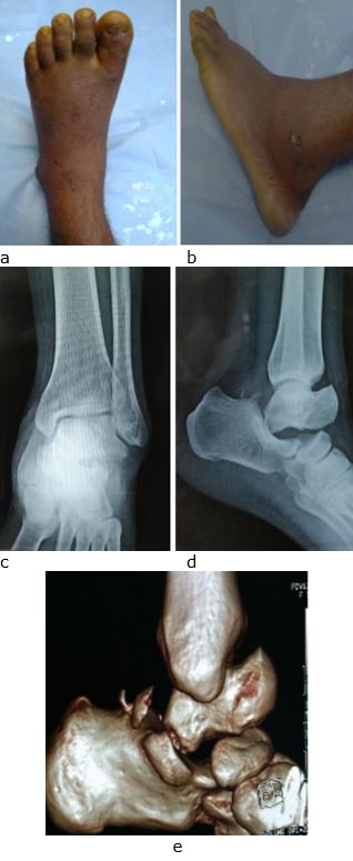
A 40-year male laborer injured his left ankle due to fall while painting over roof. The mode of injury of injury was fall from height of around 10 feet, with patient landing on the left foot, with contact of the dorsum of the foot to the ground, with the position of the foot to be inversion and plantar-flexion during strike on floor. Except of foot injury, he had no other injury. Immediately patient went to local quack for treatment where suspected ankle dislocation without being investigation was tried to reduce by manipulation and massage. Following this massage by bonesetter, patient went back to home without splinting. Since pain, swelling and deformity persisted, along with the patient’s inability to bear weight on affected limb, patient presented to our center for consultation and management, 3 weeks after the injury. On physical examination, the foot was in plantar flexion with deformity and swelling over anterior aspect of ankle and foot. Sensation and vascularity was intact with saturation of toes to 98%. The ankle range of dorsiflexion and plantarflexion were grossly restricted and severely painful with associated stiffness. But all the toes movements were normal (fig 1). Radiograph of the ankle demonstrated talocalcaneal and navicular joint dislocation with posterior displacement, which was further confirmed by doing 3D CT reconstruction. The CT scan showed talocalcaneal-navicular dislocation in which the calcaneus was displaced posteriorly, perching of the head of the talus on the dorsal margin of the navicular, and the impingement of the posterior process of talus on the posterior subtalar facet of calcaneus and multiple fragments of posterior process of talus on the top of calcaneus (Fig. 1).

Figure 1:Pre-operative clinical photo (a & b), AP (c) and lateral (d) x rays and CT scan sagittal view (e) showing posterior subtalar dislocation.
ojmpc_95_01.jpg

MRI of left ankle additionally demonstrated joint effusion, partial tear in talo-fibular and deep fibers of deltoid ligament and marrow edema in subtalar articular surfaces. As the dislocation was 3 weeks old, any attempt of closed reduction was supposed to be unsuccessful and the patient was planned for open reduction after pre-anesthetic fitness.


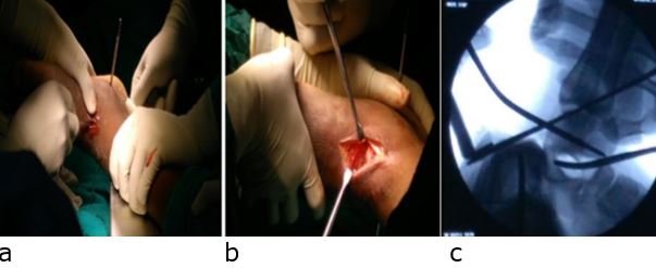
Under anesthesia, initial attempt of closed reduction was made with help of calcaneal skeletal traction and another counter traction pin inserted from posterior facet into talus to lift the body of talus. This maneuver of closed reduction with application of longitudinal manual traction and counter traction along with application of digital pressure over head of talus anteriorly failed, following with open reduction with dorsal approach was done. Talus was explored through incision of approx. 1.5 cm made over anterior aspect of left ankle and a blunt long bone spike passed under talus to lift the impacted body of talus from posterior facet of calcaneum (fig 2).

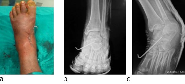
The reduction was successful by lifting the body of talus, with help of traction and counter-traction of previously passed skeletal pins which was confirmed under C-arm. One the reduction was achieved; it was stabilized in position by inserting a 2.5mm Kirschner wire from the navicular bone into the talus to hold the reduction (fig 3). After primary closure and sterile dressing, below knee slab was applied. After ankle immobilization of about 6 weeks, slab and k-wire were removed and gradual range of motion exercises started. Some ankle stiffness and minimal limitation of dorsiflexion and plantar flexion were found at final follow up but patient was able to weight bearing and was satisfied with the result.

Figure 2:Intraoperative photograph (a & b) and fluoroscopic lateral view (c) showing open reduction of posterior subtalar dislocation with exposure and reduction.
ojmpc_95_02.jpg

Figure 3:Immediate post-operative clinical photo (a) and AP (b) and oblique (c) x rays of patient showing reduced posterior subtalar dislocation and fixed with k wire.
ojmpc_95_03.jpg

Discussion

Subtalar dislocations are a rare form of dislocation and accounts for less than 1% of all traumatic dislocations [1-3]. According to available literature, medial dislocation is the most common type accounting for 72%-80% followed by lateral dislocation (17%-22%) and anterior dislocation (1%).

Posterior dislocations, in particular, are extremely rare, and amount to a mere 0.8 % of all subtalar dislocations [2,3]. Owing to gross deformity, pain and severe limitation of motion and functional impairment, along with awareness for treatment these case present early for treatment. But in developed country like our, it is common that these case present late because of either primary treatment by bone setters/quacks, not seeking treatment at all or inability to identify injury.


Our case was also a case of 3 week old neglected case, which was due to the primary treatment done by bonesetter.Subtalar dislocations mostly occur in young adults after a high energy trauma such as a fall from height or road traffic accidents [4,5].

Various reports hypothesize mechanism of injury of posterior subtalar joint dislocation as forced hyper-plantar flexion of foot which leads to a progressive subtalar ligament weakening resulting in a complete tear of ligament if the plantar flexion force is prolonged [4-8]. Our case was also an active middle aged male with no comorbid condition sustaining injury due to fall from height with landing on dorsum of inverted and plantarflexed foot.

Diagnosis of posterior subtalar joint dislocation is easy with anterior-posterior and lateral radiographs. Inokuchi et al, defined the posterior subtalar dislocation on a lateral radiograph, when the head of the talus is seen perched on the posterior margin of the navicular and the posterior portion of the talus resting in the posterior subtalar facet of the calcaneum in the absence of any significant displacement or rotation of the foot in frontal view radiograph [5,9].

Recommended treatment to avoid further damage to skin, soft tissue, neurovascular structures and to reduce the chances of avascular necrosis of the talus, is prompt closed reduction as soon as possible under sedation or general anesthesia with constant counter-traction and flexion at knee so the gastrocnemius muscle is relaxed [6-9].

For reduction initially, the force is applied in the same direction as the existing deformity, then traction is applied, and at the same time a force in opposite direction of the dislocation is applied by a firm digital pressure over the head of the talus from anterior to posterior, passing through plantar flexion to dorsiflexion. The reduction is usually associated with an audible clunk [4,5,6-9]. Post reduction immobilization is done in non-weight bearing cast but the period of immobilization is controversial [10,11].

A delayed presentation, soft tissue interposition, interposed bony fragments, severe swelling or capsulo-ligamentous retraction renders the closed reduction difficult and which requires open reduction, which is required in 10 to 20% cases [12].

Since our case was also a 3 weeks neglected case with history of maltreatment and massage present, the closed reduction attempt failed and we could reduce it only after open reduction.

Conclusion

Posterior subtalar dislocations are extremely rare injuries which require early diagnosis, anatomical reduction, stable fixation of peritalar joint fractures, and the resection of small, free osteochondral fragments for the prevention of early posttraumatic arthrosis which, in turn, may cause pain, joint stiffness, and an unsatisfactory final result.

References

1. Judey P. Observation dune luxation metatarsienne. Bull Fac Med Paris. 1811;11:81–6. [Crossref][PubMed][Google Scholar]

2. Broca P. Memories sur les luxations sous-astragaliennes. Mem Soc Chir 1853;3:566–656. . [Crossref][PubMed][Google Scholar]

3. Malgaigne JF, Buerger CG. Die Knochenbrüche und Verrekungen. Rieger, Stuttgart, 1853. pp 820. [Crossref][PubMed][Google Scholar]

4. Prada-Cañizares A, Auñón-Martín I, Vilá Y Rico J, Pretell-Mazzini J. Subtalar dislocation: management and prognosis for an uncommon orthopaedic condition. Int Orthop. 2016;40(5):999-1007. [Crossref][PubMed][Google Scholar]

5. Inokuchi S, Hashimoto T, Usami N. Posterior subtalar dislocation: case report. J Trauma 1997;42(2):310–13. . [Crossref][PubMed][Google Scholar]

6. Bibbo C, Anderson RB, Davis WH. Injury characteristics and the clinical outcome of subtalar dislocations: a clinical and radiographic analysis of 25 cases. Foot Ankle Int. 2003;24(2):158–63. [Crossref][PubMed][Google Scholar]

7. Fortin PT, Kou JX. Commonly missed peritalar injuries. J Am Acad Orthop Surg. 2009;17(12):775–86. [Crossref][PubMed][Google Scholar]

8. Garofalo R, Moretti B, Ortolano V, Cariola P, Solarino G, Wettstein M, Mouhsine E. Peritalar dislocations: a retrospective study of 18 cases. J Foot Ankle Surg 2004;43(3):166–72. . [Crossref][PubMed][Google Scholar]

9. Siddiqui YS, Zahid M, Bin Sabir A et al. Neglected peritalar dislocation: a case report with review of literature. J Clin Diagn Res. 2011;5(4):849–52. [Crossref][PubMed][Google Scholar]

10. Bali K, Kumar V, Bhagwat K et al. Closed posterior subtalar dislocation without any associated fracture: a case report and review of the literature. Foot Ankle Surg. 2011;17(3):40–2. [Crossref][PubMed][Google Scholar]

11. Camarda L, Martorana U, D’Arienzo M. Posterior subtalar dislocation. Orthop. 2009;32(7):530. [Crossref][PubMed][Google Scholar]

12. Budd H, Wimhurst J, Davis B et al. Irreducible posterior subtalar dislocation with incarceration of a fracture of the anterior process of the calcaneum. J Bone Joint Surg Br. 2010;92(7):1025–7. [Crossref][PubMed][Google Scholar]

Disclaimer / Publisher's Note

The statements, opinions and data contained in all publications are solely those of the individual author(s) and contributor(s) and not of Journals and/or the editor(s). Journals and/or the editor(s) disclaim responsibility for any injury to people or property resulting from any ideas, methods, instructions or products referred to in the content.