Orthopaedic Journal of MP Chapter

Publisher: Madhya Pradesh Orthopaedic Association www.mpioa.com
E-ISSN:2582-7243, P-ISSN:2320-6993
2019 Volume 25 Number 2 Jul-Dec

Sprengel Shoulder treated with modified Green’s procedure

Jain S1*, Jain M2, Ajmera A3

1* Saurabh Jain, Assistant Professor, Department of Orthopaedics, Mahatma Gandhi Memorial Medical College, Indore, Madhya Pradesh, India.

2 M Jain, Department of Orthopaedics, Mahatma Gandhi Memorial Medical College, Indore, Madhya Pradesh, India.

3 A Ajmera, Department of Orthopaedics, Mahatma Gandhi Memorial Medical College, Indore, Madhya Pradesh, India.

Case report: Sprengel shoulder or congenital elevation of the scapula is a rare condition of unknown aetiology that results from the abnormal termination of the caudal migration of the scapula. The main clinical changes are the hypoplasia and abnormal positioning of the scapula, causing cosmetic problems and limit the movements of the shoulder girdle. We report such a similar case of 6 years old child presenting with high ridging scapula, who had pain, difficulty in caring school bag and cosmetic deformity. He was successfully treated by modified Green’s procedure.

Keywords: Sprengel shoulder, Scapula, congenital elevation of scapula

Corresponding Author How to Cite this Article To Browse
Saurabh Jain, Assistant Professor, Department of Orthopaedics, Mahatma Gandhi Memorial Medical College, Indore, Madhya Pradesh, India.
Email:
Jain S, Jain M, Ajmera A, Sprengel Shoulder treated with modified Green’s procedure. ojmpc. 2019;25(2):85-88.
Available From
https://ojmpc.com/index.php/ojmpc/article/view/90
Manuscript Received Review Round 1 Review Round 2 Review Round 3 Accepted
2019-12-07 2019-12-13 2019-12-19 2019-12-25 2019-12-31
Conflict of Interest Funding Ethical Approval Plagiarism X-checker Note
Authors state no conflict of interest. Non Funded. The conducted research is not related to either human or animals use. 13.32 All authors have accepted responsibility for the entire content of this manuscript and approved its submission.

© 2019by Jain S, Jain M, Ajmera Aand Published by Madhya Pradesh Orthopaedic Association. This is an Open Access article licensed under a Creative Commons Attribution 4.0 International License https://creativecommons.org/licenses/by-nc/4.0/ unported [CC BY NC 4.0].

Introduction

Sprengel shoulder, which is characterized by congenital high riding scapula, is a rare complex deformity of shoulder griddle [1]. It is diagnosed as upward elevation of scapula in relation to thoracic cage or abnormally high placed scapula. Eulenberg, first described this condition of undescended scapula in 1863, and claimed its cause as traumatic dislocation of the scapula [2]. Later, it was Otto Sprengel who described the pathology and proposed a theory of its existence in 1891. The scapular embryonic primordium appears in 5th intrauterine week at the level of 4th to 5th cervical vertebrae & acquires its final morphology by 8th week of gestation & descends till 12th week over the upper 5 ribs to reach the correct anatomical position. This failure to descend leads to sprengel deformity [3].

The exact cause of the interruption of the normal caudal migration of the scapula during fetal development is unknown, but different theories have been postulated, like cerebrospinal fluid leak through the membranes of the roof of the fourth ventricle into the adjacent tissues of the neck, increased intrauterine pressure, and abnormal articulation of the scapula to the cervical vertebrae with abnormal muscle formation (fibrous, cartilaginous, or osseous omovertebral) [4].

Females are more affected and it is bilateral in about 10-30% with a predilection to left side. It can be part of other congenital anomaly. It is associated with severe cosmetic deformity and functional leads to limitation of shoulder movements [5]. We report such a rare case of isolated congenital sprengel shoulder with was treated by Green’s procedure successfully.

Case Report

A 6 year old male child visited to us in outdoor patient department with complains of visible deformity of left shoulder. The child was delivered by full term normal vaginal delivery. He had full immunization completed. Parent and child denied any other complain present, other than the deformity.

Familial history was unremarkable for any congenital disorder. On detailed history, the deformity and asymmetry of shoulder folds was observed by the parents at the age of one year of the child but they did not take any treatment. Gradually, the deformity is increasing and has grown to its current size which is not cosmetically accepted to the parents as well as the child.

There is also difficulty in dressing up himself as well as difficulty in carrying his school bag. Child would complain of pain in left shoulder after returning from school. On clinical examination, there was asymmetrical elevation of the left shoulder fold compared to the right side, which was visible both from front and back.

Since the deformity was severe, initial inspection gave impression of congenital short neck with torticollis as well, but careful examination after proper exposure showed no torticollis although slight deviation of head towards left side was noted. His neck and its movements were normal.

Inspection from back showed that inferior angle of both the scapula was at different level with left side higher up than right. Also the inferior angle of left scapula was medially displaced. Deformity was firm hard in consistency and mobile. Range of motion on left side, particularly, overhead abduction, external rotation and overhead elevation of shoulder was reduced grossly, compared to contralateral side and rest of the shoulder was normal. He had weakness pectoralis major muscle and serratus anterior muscle as seen as winging of the right scapula. Neurological examination as well as all other systemic examination was found to be absolutely normal.

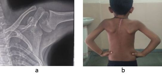
Routine radiographs were done including a chest radiograph PA view, axillary view and a scapular view. Radiographs showed high riding of the left scapula with superior angle of left scapula reaching almost level of C4 cervical spinal level (fig 1). Also the scapula was medially rotated and adducted. No abnormality was noted in left clavicle or head of humerus. Cervical rib or fusion of cervical vertebrae was absent.

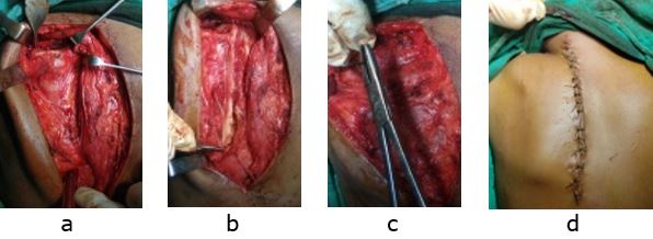
After routine haematological investigations and pre-anaesthetic work up patient was planned for scapulopexy by modified Green’s procedure. In general anaesthesia, in prone position the scapula was opened by dorsal midline approach extending from base of neck to inferior angle of scapula. Soft tissue dissected to undermine laterally toward the medial border of scapula. Lateral border of

trapezius was identified and was separated from underlying latissimus dorsi by blunt dissection. Using an osteotome trapezius is released from spine of scapula and retracted. Rhomboideus major and minor are released from their insertion at lateral border of scapula. Omovertebral bar was identified and resected, and contracted levator scapulae muscle was released from supraspinous part of the scapula.

Figure 1: Pre operative radiographs AP view (a) of chest and both shoulder showing high riding scapula on left side and clinical photographs (b to d) showing deformity & restricted movements.
ojmpc_90_01.jpg


Figure 2: Intraoperative photographs (a to d) of modified Green’s procedure
ojmpc_90_02.jpg

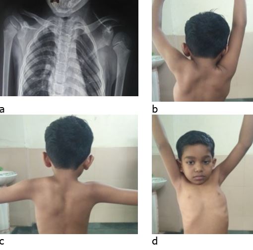
Figure 3: Post-operative AP (a) X ray of shoulder gridle & clinical photograph (b) showing good cosmetic correction & range of motion
ojmpc_90_03.jpg

Scapula was displaced distally into the pocket in latissimus dorsi along the attached muscles and holding scapula in this position trapezius, rhomboideus major and minor are reattached to spine of scapula and medial border of scapula respectively using non absorbable sutures (fig 2). Immediate post-operative radiograph of patient showed descended scapula with its superior angle at C7 level. Post-operatively, patient was given a shoulder immobiliser for 2 weeks, following which suture removal was done and shoulder range of motion was started. Patient showed good recovery with a 2 month follow up showing both the inferior angle of scapula at same level as well as significantly increased shoulder range of motion (fig 3).

Discussion

Sprengel shoulder occurs due to inadequate caudal movement of scapula during development resulting in an abnormally high placed scapula, which can be unilateral or bilateral [1-4]. This high position of the scapula in the process of skeletal development leads to changes in axial rotation, shape, and size and other musculoskeletal defects including hypoplasia, medialization, and adduction of the scapula, prominence of its upper angle, distal rotation and lateral angulation of the glenoid cavity, changes in the position of the clavicle, omovertral bar (ossified fibrous band between cervical spine with the undescended scapula), anomalies of the cervicothoracic vertebrae and ribs, and muscular hypoplasia or atrophy of the shoulder musculature [6]. Thus although, the deformity commonly occurs sporadically, but it can occur in combination with other congenital anomalies, such as congenital scoliosis, spina bifida, fusion of cervical vertebrae, like Klippel-Feil syndrome, kidney abnormalities and cleft palate [7,8]. But we did not find any associated anomaly with our patient.

This dysplasia of the pectoral girdle results in cosmetic and functional disability and each case needs to be individualized depending on the degree of anomaly as well as associated defects. The cosmetic classification of Sprengel deformity, based on deformity grade was given by Cavendish in 1972, whereas the radiographic classification based on superio-medial angle was given by Rigault [9,10]. Cavendish grade 1 is a very mild deformity that is not noticeable when the patient is dressed. Grade 2 is a mild deformity that is visible as a lump in the web of the neck when the patient is dressed. Grade 3 is a moderate deformity described as an easily visible deformity with the shoulder joint elevated 2–5 cm. Grade 4 is a severe deformity with shoulder joint elevation greater than 5 cm or evidence of the superior angle of the scapula near the occiput with or without webbing. Radiographic Rigault grade 1, is superio-medial angle lower than T2 but above T4 transverse process, in grade 2 the superio-medial angle is located between C5 and T2 transverse process and in grade 3, the superio-medial angle is above C5 transverse process. Our case was Cavendish type 4 and Rigault type 3, which was severe deformity. The CT scan, particularly with 3D reconstruction, is a more useful diagnostic tool for thorough assessment and preoperative planning of the surgical strategy, but in our case we could not do a CT scan [11].

The treatment for Sprengel deformity is done for improvement in appearance and function and depends on the severity of the abnormality. For mild deformities as Cavendish Grades 1 and 2, nonsurgical options including physical therapy, stretching, and continued observation are done to prevent torticollis and increase range of motion. Moderate and severe deformities i.e. higher Cavendish classification grades are candidates for surgical intervention. The optimal age for surgical correction is recommended to be usually between 3 and 8 years [9,12]. Many procedures are described for treatment of Sprengel shoulder like osteotomies, bone resections, muscle releases with repositioning of the scapula, or a combination of these etc, but the hallmark techniques involve caudal relocation of the scapula and resection of the omovertebral bone, if present [12].

Two of the most popular procedures are the Green’s and Woodward procedures. Both procedures involve detaching muscles of scapular origin, resection of the elevated portion of the scapula, removal of the omovertebral bone, and mobilization of the scapula to a more caudal position and reattachment of the muscles distally to help secure the lowered scapula. The Woodward procedure adds an osteotomy of the clavicle, whereas the Mears procedure also releases the long head of the triceps and part of the origin of the teres minor muscle, and progressive resection of the inferio-medial portion of the scapula to achieve abduction [13-15].

We contemplated the modified Green’s procedure by Leibovic et al in which the scapula is kept lowered by sutures in pocket made in the latissimus dorsi muscle [16]. This method is usually the treatment of choice which provides excellent repositioning of the scapula to a more normal position and achieves good postoperative results, even with limited supervised physical therapy.


Surgical complications described include scar-related complications, brachial plexus palsy, brachial neuritis, winging of the scapula, re-growth of the resected omovertebral bar, recurrence of the deformity and prominence of the sternoclavicular joint [12-16]. To prevent the complications of brachial plexus palsy, either osteotomy of clavicle or avoiding excessive correction is advised. Our patients at the last follow-up had got excellent cosmetic correction and increased range of motion without any complications.

Conclusion

Sprengel shoulder is a rare congenital deformity, which occurs due to failure of the scapula to descend during intrauterine development. It can lead to cosmetic problems and limitation of movements, but is accurately diagnose by plain x rays. Milder forms can be treated conservatively, whereas surgical correction is needed for severe variety.

References

1. Harvey EJ, Bernstein M, Desy NM, Saran N, Ouellet JA. Sprengel deformity: pathogenesis and management. J Am Acad Orthop Surg 2012;20(3):177–86. . [Crossref][PubMed][Google Scholar]

2. Eulenberg M: Beitrag zur Dislocation der Scapula. Amtliche Berichte u ¨ber die Versammlungen deutscher Naturforscher und A ¨rzte fu ¨r die Jahre 1863 (On the dislocation of the scapula. Official report of the meetings of German bioresearchers and physicians for the year 1863;37:291–4. . [Crossref][PubMed][Google Scholar]

3. Sprengel O. Die angeborene vershiebung der schulterblattes nach oben. Archiv fur Klinische Chirurgie. 1891;42:545-49. [Crossref][PubMed][Google Scholar]

4. O’Brien SJ, Voos JE, Nevasier AS, Drakos MC. Developmental anatomy of the shoulder and anatomy of the glenohumeral joint. In: Rockwood CA, Matsen FA, editors. Th e Shoulder. 4th ed. Philadelphia, PA: Saunders/Elsevier; 2009. p. 1-12 [Crossref][PubMed][Google Scholar]

5. Carson WG, Lovell WW, Whitesides TE. Congenital elevation of the scapula. Bone Joint Surg Am 1981;2013:1199–207. . [Crossref][PubMed][Google Scholar]

6. Samilson RL. Congenital and developmental anomalies of the shoulder girdle. Orthop Clin N Am. 1980;11:219-31. [Crossref][PubMed][Google Scholar]

7. Dhir R, Chin K, Lambert S. The congenital undescended scapula syndrome: Sprengel and the cleithrum: a case series and hypothesis. J Shoulder Elbow Surg. 2018;27:252-9. [Crossref][PubMed][Google Scholar]

8. Solanki C, Bhat DI, Devi BI. “Os” omovertebrale variants prove it to be a misnomer. Neurol India. 2016;64:984-7. [Crossref][PubMed][Google Scholar]

9. Cavendish ME. Congential elevation of the scapula. J Bone Joint Surg Br 1972;54:395–408. . [Crossref][PubMed][Google Scholar]

10. Rigault P, Pouliquen JC, Guyonvarch G, et al. Congenital elevation of the scapula in childhood. Rev Chir Orthop. 1976;62:5-26. [Crossref][PubMed][Google Scholar]

11. Cho TJ, Choi IH, Chung CY, Hwang JK: The Sprengel deformity. Morphometric analysis using 3D-CT and its clinical relevance. J Bone Joint Surg Br 2000; 82(5): 711–8. . [Crossref][PubMed][Google Scholar]

12. Reginaldo SS, Macedo RR, Andrade AR, et al. Sprengel's deformity: Surgical correction by a modified green procedure. Rev Bras Ortop. 2015;44:208-13. [Crossref][PubMed][Google Scholar]

13. Woodward JW. Congenital elevation of the scapula: correction by release and transplantation of the muscle origin: a preliminary report. J Bone Joint Surg. 1961;43:219-28. [Crossref][PubMed][Google Scholar]

14. Green WT. The surgical correction of congenital elevation of scapula (Sprengel deformity). Proceedings of the American Orthopedic Association. J Bone Joint Surg Am. 1957;(39):1439 [Crossref][PubMed][Google Scholar]

15. Mears DC. Partial resection of the scapula and a release of the long head of triceps for the management of Sprengel’s Deformity. J Pediatric Orthop. 2001; 21:242-5. [Crossref][PubMed][Google Scholar]

16. Leibovic SJ, Ehrlich MG, Zaleske DJ. Sprengel deformity. J Bone Joint Surg Am. 1990;72:192-7. [Crossref][PubMed][Google Scholar]

Disclaimer / Publisher's Note

The statements, opinions and data contained in all publications are solely those of the individual author(s) and contributor(s) and not of Journals and/or the editor(s). Journals and/or the editor(s) disclaim responsibility for any injury to people or property resulting from any ideas, methods, instructions or products referred to in the content.