Orthopaedic Journal of MP Chapter

Publisher: Madhya Pradesh Orthopaedic Association www.mpioa.com
E-ISSN:2582-7243, P-ISSN:2320-6993
2019 Volume 25 Number 1 Jan-Jun

Scurvy: An Important Differential Diagnosis Of Sepsis In Young Children

Ajmera A1*, Solanki M2

1* Anand Ajmera, Professor, Department of Orthopaedics, Mahatma Gandhi Memorial Medical College and Maharaja Yashwantrao Hospital, Indore, MP, India.

2 M Solanki, Department of Orthopaedics, Mahatma Gandhi Memorial Medical College and Maharaja Yashwantrao Hospital, Indore, MP, India.

Background: Scurvy a disease, particularly in people with peculiar dietary habits, is cause by Vitamin C deficiency, which is required for collagen synthesis. Impaired collagen synthesis affects skin, cartilage, dentine, osteoid and capillary blood vessels. We present a case of a three year old child having scurvy who presented like sepsis with aim to create awareness among the clinicians about the disease.

Case report: 3 year old female child presented to us with pseudoparalysis, which was misdiagnosed and mistreated as bilateral thigh sepsis for which surgery was also attempted. Radiographs and MRI of the child showed widening of epiphysis, sclerosis of epiphyseal rim, osteopenia and subperiosteal hemorrhages respectively, confirming diagnosis of Scurvy. She was healed fully on Vitamin C supplementation in therapeutic doses.

Conclusion: Malnutrition is rampant in young children in our country and hence nutritional scurvy should be kept in mind as a differential diagnosis of sepsis. This case report also tries to create awareness among the clinicians about the disease and ways to prevent Vitamin C deficiency.

Keywords: Scurvy, Vitamin C Deficiency, Children

Corresponding Author How to Cite this Article To Browse
Anand Ajmera, Professor, Department of Orthopaedics, Mahatma Gandhi Memorial Medical College and Maharaja Yashwantrao Hospital, Indore, MP, India.
Email:
Ajmera A, Solanki M, Scurvy: An Important Differential Diagnosis Of Sepsis In Young Children. ojmpc. 2019;25(1):42-46.
Available From
https://ojmpc.com/index.php/ojmpc/article/view/77
Manuscript Received Review Round 1 Review Round 2 Review Round 3 Accepted
2019-06-06 2019-06-12 2019-06-18 2019-06-24 2019-06-30
Conflict of Interest Funding Ethical Approval Plagiarism X-checker Note
Authors state no conflict of interest. Non Funded. The conducted research is not related to either human or animals use. 14.64 All authors have accepted responsibility for the entire content of this manuscript and approved its submission.

© 2019by Ajmera A, Solanki Mand Published by Madhya Pradesh Orthopaedic Association. This is an Open Access article licensed under a Creative Commons Attribution 4.0 International License https://creativecommons.org/licenses/by-nc/4.0/ unported [CC BY NC 4.0].

Introduction

Scurvy (Barlow’s disease, Moeller’s disease, Cheadle’s disease) is a disease resulting from deficiency of Vitamin C (Ascorbic acid) which is required for collagen synthesis in humans [1].

This disease, described as early as in 480 – 360 B.C. by Hippocrates and an important cause of deaths on long sea voyages, is common in sailors and people who remain on ships for long time. It may also be seen in people with prolonged peculiar dietary habits, who don’t have access to perishable fruits [2]. Treatment is by consuming fresh citrus fruits [3,4].

Most animals are able to synthesis their own vitamin C and hence Scurvy does not occur in animals, but humans and a few other species lack an enzyme necessary for such synthesis, and hence rely on dietary vitamin C [5,6].

In modern scenario, malnutrition is the most common cause of scurvy in children. Vitamin C is essential for collagen synthesis, and hence typical pathologic manifestations of vitamin C deficiency are noted due to impaired collagen synthesis seen in collagen-containing tissues and organs such as skin, cartilage, dentine, osteoid, and capillary blood vessels.

Pathologic changes are a function of the rate of growth of the affected tissues; hence, the bone changes are often observed only in infants during periods of rapid bone growth [6].

We present this case report of a three year old child having scurvy who presented like sepsis with aim to create awareness among the clinicians about the disease and ways to prevent Vitamin C deficiency.

Case Report

A 3 year old female child was referred to our center with parents complaining of swelling thighs and knees, restricted lower limb movements, nonspecific history of fever and excess crying on any attempt to move the child for past 10 days. The child was initially treated elsewhere with a diagnosis of malnutrition and multifocal sepsis and had received multiple antibiotics without relief.

An ultrasonography of thigh showed collection both thighs and knees more on right side. The blood counts, ESR and CRP values were normal. There was also history of an incision and drainage done of right thigh, considering it to be an abscess, but notes mentioned that there was no pus. On our examination, the child was still in acute agony, reluctant to move both the lower limbs.

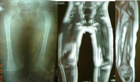
A wound of previous incision and drainage operation was present over right thigh without any pus. Repeat blood counts and ultrasound abdomen were normal. Radiographs of thighs with knee showed some widening of epiphysis with sclerosis of epiphyseal rim and a generalized osteopenia (Figure 1a).

An MRI scan was done which showed subperiosteal hemorrhages in both thighs and legs without any bone erosion (Figure 1b & c).

Figure 1: X ray of pelvis c both thigh AP view (a) and MRI view (b & c) showing subperiosteal haemorrhages
ojmpc_77_01.jpg

Generalized signal alterations were seen in marrow suggestive of edema and increased cellularity with widening and sclerosis of metaphysis. Minor gum bleeding with poor dental health was observed.

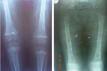
A diagnosis of Scurvy was made and the patient was put on Vitamin C supplementation in therapeutic doses. After 1 week of therapy, there was gross improvement in symptoms. At 3 weeks the child appeared near normal, voluntarily moving all four limbs with significant reduction in pain on movement. The x rays done at this time showed calcification of the subperiosteal hematomas.

At 8 months follow up the child was able to walk without support albeit with some limp. X rays done at this time showed ossified subperiosteal hematomas (Fig 2).

Figure 2: Follow-up X ray at 6week (a) and at 8 months (b) showing healing
ojmpc_77_02.jpg

Discussion

International incidence of scurvy is unknown. Only few series with very little number of cases are published [7-15].


Ratanchu from Thailand reviewed 28 cases of scurvy in children over a 7-year period (1995-2002) and noted prolonged consumption of heated milk and inadequate intake of vegetables and fruits as the risk factors for the development of scurvy [7].

Epidemic scurvy has been reported among refugee populations with incidence of 5% in women and 12% of men, and in those older than 65 years, this proportion increased to 15% of women and 20% of men [8].

Data from the Third National Health and Nutrition Examination Survey (NHANES III) that assessed the prevalence of vitamin C deficiency in the United States among a sample of 15,769 children and adults (12-74 y) found that 14% of males and 10% of females were vitamin C deficient [9].

Scurvy can occur at any age. Most cases of Scurvy occur when the child is aged between 6 to 24 months. Clinical manifestations develop after an infant has lacked Vitamin C for 6 -10 months. Hence, Scurvy is uncommon in neonates as human breast milk contains sufficient Vitamin C, provided the mother has adequate intake. Vitamin C is destroyed by the process of pasteurization, so babies fed with ordinary bottled milk sometimes suffer from Scurvy if they are not provided with adequate Vitamin C supplements. Virtually all commercially available baby formulas contain added Vitamin C for this reason, but heat and storage destroy Vitamin C [10].

Vitamin C deficiency leads to defective collagen synthesis resulting in defective dentine formation, gum bleeding, and loss of teeth. Hemorrhaging is a hallmark feature of Scurvy and can occur in any organ. Hair follicles are one of the common sites of cutaneous bleeding. There are reports of a purpuric rash on trunk and legs which responded to Vitamin C supplementation [11].

Hemorrhages of the gums usually involve the tissue around the upper incisors, which have a bluish-purple hue and spongy feel. Gum hemorrhage occurs only if teeth have erupted. Petechial hemorrhage of the skin and mucous membranes can occur. Rarely, hematuria, hematochezia, and malena are noted.

Proptosis of the eyeball secondary to orbital hemorrhage can occur. Besides being essential for collagen synthesis, ascorbic acid is important for biosynthesis of carnitine and neurotransmitters and in hematopoiesis by promoting iron absorption. Risk factors for development of scurvy include male, having a low dietary intake of vitamin C, not taking vitamin supplements, and smoking [12].

Initial symptoms of a child with scurvyare nonspecific and include loss of appetite, peevishness (ill-tempered), poor weight gain, diarrhea, tachypnoea, fever. Specific symptoms include irritability, pain and tenderness of the legs, pseudoparalysis,

swelling over the long bones and hemorrhage. Costochondral beading or scorbutic rosary is a common finding. The scorbutic rosary is distinguished from rickety rosary (which is knobby and nodular) by being more angular and having a step-off at the costochondral junction. The sternum is typically depressed.

Although, hyperkeratosis, corkscrew hair, and sicca syndrome are typically features of adult scurvy and are rarely seen in infantile scurvy, Mc Kenna reported an infant with these features of adult scurvy showing diffuse, nonscarring scalp alopecia with radiologic features of scurvy was reported in 2008 [13].

Bony involvement is typical for infantile scurvy, which occurs at the junction of the diaphysis end and the growth cartilage. Osteoblasts fail to form osteoid (bone matrix), resulting in cessation of endochondral bone formation, but normal calcification of the growth cartilage continues, leading to thickening of the growth plate. The typical invasion of the growth cartilage by the capillaries does not occur.

Preexisting bone becomes brittle and undergoes resorption at a normal rate, resulting in microscopic fractures of the spicules between the shaft and calcified cartilage. With these fractures, the periosteum becomes loosened, resulting in the classic subperiosteal hemorrhage at the ends of the long bones. Intra-articular hemorrhage is rare because the periosteal attachment to the growth plate is very firm [14].

Subperiosteal hemorrhage is a typical finding of infantile scurvy, which is often palpable and tender in the acute phase causing excruciating pain and is seen at lower ends of the femur and tibia which are the most frequently involved sites.

Plasma ascorbic acid level may help in establishing the diagnosis, but the best confirmation of the diagnosis remains its resolution following vitamin C administration. Plasma ascorbic acid level tends to reflect the recent dietary intake rather than the actual tissue levels of vitamin C. Signs of scurvy can occur with low-normal serum levels of vitamin C.

Fasting serum ascorbic acid level greater than 0.6 mg/dl rules out scurvy. Serum ascorbic acid levels of less than 0.2 mg/dl are deficient and Scurvy occurs at levels less than 0.1 mg/dl. The earliest radiologic manifestations are seen at growth cartilage-shaft junction of bones with rapid growth like distal ends of radius or femur, where fuzziness of the lateral aspects of the cortices is present with slight rarefaction of the neighboring cancellous bone.

As the disease progresses, the cortex becomes thin and the trabecular structure of the medulla atrophies and develops a ground-glass appearance. The zone of provisional calcification becomes dense and widened, and this zone is referred to as the white line of Fränkel. The epiphysis also shows cortical thinning and the ground-glass appearance.


Key imaging features show osteoporosis. Metaphyseal spurs or marginal fractures (Pelkan spur), a transverse band of radiolucency in the metaphysis (scurvy line or Trümmerfeld zone), which is subjacent to the zone of provisional calcification; ring of increased density surrounding the epiphysis (Wimberger ring); and periosteal elevation can also be noted.

As scurvy becomes Infections in childhood can also present in a similar manner, like in our case due to which the child was misdiagnosed and mistreated in lines of sepsis. On physical examination the infant is apprehensive, anxious, and progressively irritable. The child may have low grade fever, anaemia and poor wound healing.

Severe tenderness over the thighs is noted during change of diapers. The excruciating pain results in pseudoparalysis and the child assumes frog leg posture (i.e. keeping hips and knees slightly flexed and externally rotated) for comfort. advanced, a zone of rarefaction occurs at the metaphysis under the white line.

The zone of rarefaction typically involves the lateral aspects of the white line, resulting in triangular defects called the corner sign of Park. This area has multiple microscopic fractures and may collapse with impaction of the calcified cartilage onto the shaft. The lateral aspect of the calcified cartilage can project as a spur.

Subperiosteal hemorrhages are not visualized in the active phase. With healing, they become calcified and are readily observed. Sudden death due to cardiac failure is reported in infants and adults with scurvy. Predominant morbidity is a result of hemorrhage into various tissues. Recent laboratory data suggest that the neonatal brain is particularly susceptible to vitamin C deficiency and that this condition may adversely affect early brain development [15].

Treatment is to prescribe Vitamin C supplementation in therapeutic doses as per age which ranges from 40 mg to 90 mg per day. Prevention is by taking a balance diet with enough Vitamin C.

Conclusion

This case report highlights the fact that in our country where malnutrition is rampant in young children, nutritional scurvy should be kept in mind as a differential diagnosis of sepsis. We also try to create awareness among the clinicians about the disease and ways to prevent Vitamin C deficiency.

References

1. Evans PR. Infantile Scurvy: The centenary of Barlow’s disease. British Med J (Clin Res Ed) 1983;287(6408):1862-3. . [Crossref][PubMed][Google Scholar]

2. Bown SR. Scurvy: How a surgeon, a mariner and a gentleman solved the greatest medical mystery of the age of sail. J Clin Inv. 2004;114(12):1690. [Crossref][PubMed][Google Scholar]

3. Bartholomew M. James Lind’s Treatise of the Scurvy (1753). Postgrad Med J. 2002;78:695-696. [Crossref][PubMed][Google Scholar]

4. Martini E. Jacques Cartier witnesses a treatment for scurvy. Vesalius 2002;8(1):2–6. . [Crossref][PubMed][Google Scholar]

5. Leger D. Scurvy: reemergence of nutritional deficiencies. Can Fam Physician. 2008;54(10):1403-6. [Crossref][PubMed][Google Scholar]

6. Ellis CN, Vanderveen EE, Rasmussen JE. Scurvy. A case caused by peculiar dietary habits. Arch Dermatol. 1984;120(9):1212-4 [Crossref][PubMed][Google Scholar]

7. Ratanchu EK. Scurvy in paediatric patients: a review of 28 cases. J Med Assoc Thailand. 2003;86(3):734-740. [Crossref][PubMed][Google Scholar]

8. Hercberg S, Preziosi P, Galan P. Vitamin status of a healthy French population: dietary intakes and biochemical markers. Int J Vitam Nutr Res. 1994;64(3):220-32. [Crossref][PubMed][Google Scholar]

9. Hampl JS, Taylor CA, Johnston CS. Vitamin C deficiency and depletion in the United States: the third national health and nutrition examination survey, 1988 to 1994. Am J Public Health. 2004;94(5):870-5. [Crossref][PubMed][Google Scholar]

10. Gan R, Eintracht S, Hoffer LJ. Vitamin C deficiency in a university teaching hospital. J Am Coll Nutr. 2008;27(3):428-33. [Crossref][PubMed][Google Scholar]

11. Mc Kenna KE, Dawson JF. Scurvy occurring in a teenager. Clin Exp Dermatol. 1993;18(1):75-7. [Crossref][PubMed][Google Scholar]

12. Ragunatha S, Inamadar AC, Palit A. Diffuse nonscarring alopecia of scalp: an indicator of early infantile scurvy? Pediatr Dermatol. 2008;25(6):644-6. . [Crossref][PubMed][Google Scholar]

13. Jenny C. Evaluating infants and young children with multiple fractures. Pediatrics. 2006;118(3):1299-303. [Crossref][PubMed][Google Scholar]

14. Mosdol A, Erens B, Brunner EJ. Estimated prevalence and predictors of vitamin C deficiency within UK's low-income population. J Public Health. 2008;30(4):456-60. [Crossref][PubMed][Google Scholar]

15. Tveden-Nyborg P, Lykkesfeldt J. Does Vitamin C deficiency result in impaired brain development in infants? Redox Rep. 2009;14(1):2-6. . [Crossref][PubMed][Google Scholar]


Disclaimer / Publisher's Note

The statements, opinions and data contained in all publications are solely those of the individual author(s) and contributor(s) and not of Journals and/or the editor(s). Journals and/or the editor(s) disclaim responsibility for any injury to people or property resulting from any ideas, methods, instructions or products referred to in the content.