Orthopaedic Journal of MP Chapter

Publisher: Madhya Pradesh Orthopaedic Association www.mpioa.com
E-ISSN:2582-7243, P-ISSN:2320-6993
2019 Volume 25 Number 1 Jan-Jun

Eight Plate Hemiepiphysiodesis In Genu Valgum: A Retrospective Study

Choubey R1*, Jain R2, Gupta A3

1* Raghvendra Choubey, Associate Professor Department of Orthopaedics, Bundelkand Medical College, Sagar, MP, India.

2 R Jain, Associate Professor Department of Orthopaedics, Bundelkand Medical College, Sagar, MP, India.

3 A Gupta, Associate Professor Department of Orthopaedics, Bundelkand Medical College, Sagar, MP, India.

Background: Angular deformities of distal femur can be treated with corrective osteotomies and skeletal fixation. In children, this major intervention can be avoided with temporary hemiepiphysiodesis. Recently, a new implant called the eight-Plate, consisting of a two-hole plate and two screws, is popular as an alternative to the Blount staple to perform temporary hemiepiphysiodesis in children.

Materials and Methods: Fifteen patients (16 physes, 16 limbs) were identified retrospectively in between November 2012 to September 2017 who underwent eight plate surgical growth modulation with average follow-up after plate insertion of 2 years 2 months (range, 1 year 6 months to 2 years 6 months).

Results: Average age at eight-Plate implantation was eleven years three months (age range, 9 years 8 months to 13 years 6 months). Eight-plates were inserted for an average 14.0 months (range, 11.0–20.4 months). No growth disturbance was observed. Mechanical lateral distal femoral angle changed an average 10.00 degrees (range, 7–18 degrees) or 0.4 degrees/month.

Conclusions: The eight-Plate effectively treats angular deformities in growing children and is less likely to extrude spontaneously than the Blount staple. We have not observed growth disturbance or other complications related to this device.

Keywords: Eight-Plate, Guided growth, Hemiepiphysiodesis

Corresponding Author How to Cite this Article To Browse
Raghvendra Choubey, , Associate Professor Department of Orthopaedics, Bundelkand Medical College, Sagar, MP, India.
Email:
Choubey R, Jain R, Gupta A, Eight Plate Hemiepiphysiodesis In Genu Valgum: A Retrospective Study. ojmpc. 2019;25(1):30-33.
Available From
https://ojmpc.com/index.php/ojmpc/article/view/74
Manuscript Received Review Round 1 Review Round 2 Review Round 3 Accepted
2019-06-06 2019-06-12 2019-06-18 2019-06-24 2019-06-30
Conflict of Interest Funding Ethical Approval Plagiarism X-checker Note
Authors state no conflict of interest. Non Funded. The conducted research is not related to either human or animals use. 15.32 All authors have accepted responsibility for the entire content of this manuscript and approved its submission.

© 2019by Choubey R, Jain R, Gupta Aand Published by Madhya Pradesh Orthopaedic Association. This is an Open Access article licensed under a Creative Commons Attribution 4.0 International License https://creativecommons.org/licenses/by-nc/4.0/ unported [CC BY NC 4.0].

Introduction

Angular deformities around knee, varus or valgus, is a common growth problem encountered in pediatric population, which may often be managed expectantly with parental reassurance as most of them are physiological, which peaks between 3 and 6 years and resolve spontaneously [1,2].

Pathological angular deformities can be either idiopathic, metabolic or due to congenital syndromes such as skeletal dysplasia and when these pathological deformities of the lower extremities are of more than 100, they can cause activity-related pain, altered gait mechanics, joint instability and early degenerative arthritis [3].

Temporary hemiepiphysiodesis or guided growth by eight-Plate results in equal limb lengths, neutral mechanical axes, and horizontal knees by skeletal maturity without destruction of any part of the physis and allows the entire physis to resume growth after hardware removal. Hence we conducted this retrospective study is to evaluate the effectiveness of hemiepiphysiodesis with the eight-Plate to correct genu valgus deformity in paediatric patients.

Materials and Methods

We retrospectively reviewed cases of genu valgum deformity, which were operated with eight plate hemiepiphysiodesis at our centre during 2012 to 2017. All patients of genu valgus deformity which were operated with eight-plate on medial surface between age 8 to 14 years for which plate removal has been done were retrospectively identified from the records and included in the study.

Patients operated for genu valgus correction for skeletal dysplasia and in which plate removal had not been done were excluded from the study. Patients treated by other methods or whose medical records were incomplete were also excluded from the study.

The medical record was assessed for patient demographics, diagnosis, age, gender, follow up duration, duration of plate, removal time etc. Patients pre-operative, immediate post-operative and final follow-up full length AP x-rays were reviewed for measurement of deformity.

These radiographs were evaluated for magnitude of the deformity by the measuring the lateral distal femoral angle (LDFA), medial proximal tibial angle (MPTA), mechanical axis deviation and the tibio-femoral (TFA) angles both before and after the deformity correction and implant removal.

Results

Sixteen limbs in 15 patients fitted the inclusion criteria whose complete record was available and included in the study. There were 8 male and 7 female. The average age at the time of plate implantation was 11.3 years (range 9 to 13 years). The average time between insertion and removal of the plate was 14 months (range 11 to 23 months).

Average follow-up period after the removal was 26 months (range 12 to 36 months). In all sixteen eight, plates were removed without any complications.

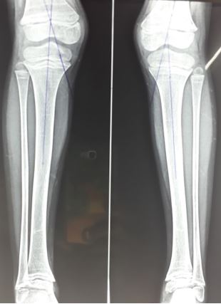
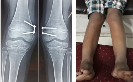
The mean pre-operative TFA was 22.41° (range 12° to 39°) which was corrected to mean angle of 6.7° (range 2° to 11°) at final follow up. The mean pre-operative LDFA was 73.17° (range: 63° to 84°) which was corrected to mean angle of 88.2° (range 83° to 91°) at final follow up. The mean pre-operative MPTA was 96.19° (range 53° to 107°) to mean angle of 90.1° (range 88° to 93°) at final follow up (fig 1 & 2). All cases were successfully treated; reaching full deformity correction with average speed of correction was 0.9°/month. Overcorrection was seen in 1 limb.

Figure 1: Preoperative X rays of both limbs (a & b) showing genu valgus bilaterally
ojmpc_74_01.jpg

Figure 2: Post-operative X rays (a & b) and clinical photos (c) after correction
ojmpc_74_02.jpg


Discussion

Angular deformities of the knee alter the biomechanics of the knee by causing a distorted stress distribution on the weight-bearing surface of the knee joint and various methods have been proposed to address this problem [1-3]. Surgical treatment is recommended only after 8 years of age when it was clear that physiological recovery had failed and when the deformity is more than 10° [4].

Treatment of these angular deformities ranges from osteotomies to hemiepiphysodesis. Osteotomies typical done in adults, involve extensive soft-tissue dissection, complications of wound closure, infection, delayed union, malunion and prolonged immobilization which increase the morbidity of the patients [4-6].

Hemiepiphysiodesis either permanent with physeal ablation or temporary using staple (s) or a tension band plate across one side of the physis are options done in growing child [7,8]. Permanent hemiepiphysodesis or staples which may cause permanent iatrogenic physeal arrest prevent their use in under the age of eight, due to fear of growth disturbances and shortening [9-11].

Less invasive method of hemiepiphysiodesis by application of eight-plate and growth modulation to restore alignment has advantage of lower cost and fewer complications. The only contraindication for this guided growth is physeal closure due to damage or to skeletal maturity [12-18]. We retrospectively reviewed our results of genu valgus correction by eight-plate in 16 limbs in 15 patients and found that we were able to correct the deformity all the patients, whereas overcorrection was seen in one patient.

Peter Stevens, the inventor of the eight-Plate, believes the eight-Plate works faster than the Blount staple because rigid staple produced more of a compressive effect [7,8]. The time for deformity correction is dependent on the width of the growth plate, with narrow growth plates correcting faster than wide ones [12].

The rigid Blount staple causes a technical narrowing of the growth plate, whereas the eight-Plate creates an angulation correction axis outside the growth plate, thereby “widening” the physis [13]. The speed of correction seems to decrease with age, and as a patient approaches skeletal maturity, the physis grows at a slower rate; hence this should be considered and taken into account during the timing of surgery [6].

This explains the mean slow correction rate (0.9°/month) achieved in our study compared to mean correction in others studies (1.53°/month) as the mean age in our study was higher (11.3 year) than mean age in other series (8.2 years) [14-18].

The shortcoming our study being retrospective and less number of cases can be overcome with a large, long-term, prospective study which would give the real incidence of rebound deformity from plate removal.

Conclusion

We consider the temporary hemiepiphysodesis with eight-Plate as very reliable solution for the treatment of pediatric angular deformities when the greater than 10° and there is time left before the physeal arrest.

References

1. Stevens P. Guided growth: 1933 to the present. Strategies Trauma Limb Reconstr. 2006;1(1):29–35. [Crossref][PubMed][Google Scholar]

2. Brooks WC, Gross RH. Genu varum in children: diagnosis and treatment. J Am Acad Orthop Surg. 1995;3(6):326–335. [Crossref][PubMed][Google Scholar]

3. Fabry G, MacEwen GD, Shands AR. Torsion of the femur. A follow-up study in normal and abnormal conditions. J Bone Joint Surg Am. 1973;55(8):1726–1738 [Crossref][PubMed][Google Scholar]

4. Kulkarni GS. Angular deformities in lower limb in children. In: Kulkarni GS, editor. Text Book of Orthopedics and Trauma. 2nd ed. New Delhi: Jaypee Publishers; 2008. p. 3650-7 [Crossref][PubMed][Google Scholar]

5. Mycoskie P. Complication of osteotomies about the knee in children. Orthopedics 1981;4:1005-15. 7. Steel HH, Sandrow RE, Sullivan PD [Crossref][PubMed][Google Scholar]

6. Complications of tibial osteotomy in children for genu varum or valgum. Evidence that neurological changes are due to ischemia. J Bone Joint Surg Am 1971;53:1629-35. . [Crossref][PubMed][Google Scholar]

7. Blount WP, Clarke GR. Control of bone growth by epiphyseal stapling: a preliminary report. J Bone Joint Surg Am 1949;31: 464–78. . [Crossref][PubMed][Google Scholar]

8. Stevens PM. Guided growth for angular correction: a preliminary series using a tension band plate. J Pediatr Orthop 2007;27:253–9. . [Crossref][PubMed][Google Scholar]

9. Zuege RC, Kempken TG, Blount WP. Epiphyseal stapling for angular deformity at the knee. J Bone Joint Surg Am 1979;61:320-9. . [Crossref][PubMed][Google Scholar]

10. Bylski-Austrow DI, Wall EJ, Rupert MP, Roy DR, Crawford AH. Growth plate forces in the adolescent human knee: A radiographic and mechanical study of epiphyseal staples. J Pediatr Orthop 2001;21:817-23. . [Crossref][PubMed][Google Scholar]

11. Frantz CH. Epiphyseal stapling: A comprehensive review. Clin Orthop Relat Res 1971;77:149-57. . [Crossref][PubMed][Google Scholar]

12. Bowen JR, Leahey JL, Zhang ZH, MacEwen GD. Partial epiphysiodesis at the knee to correct angular deformity. Clin Orthop Relat Res. 1985;198:184–90. [Crossref][PubMed][Google Scholar]


13. Paley D. Frontal plane mechanical and anatomic axis planning. In: Paley D. Principles of deformity correction. 1st ed, corrected 3rd printing. Berlin Tokyo Heidelberg New York: Springer-Verlag; 2005:61–97 [Crossref][PubMed][Google Scholar]

14. Kulkarni RM, Ilyas Rushnaiwala FM, Kulkarni GS, Negandhi R, Kulkarni MG, Kulkarni SG. Correction of coronal plane deformities around the knee using a tension band plate in children younger than 10 years. Indian J Orthop 2015;49:208-18. . [Crossref][PubMed][Google Scholar]

15. Wiemann JM 4th, Tryon C, Szalay EA. Physeal stapling versus 8-plate hemiepiphysiodesis for guided correction of angular deformity about the knee. J Pediatr Orthop 2009;29:481-5. . [Crossref][PubMed][Google Scholar]

16. Ballal MS, Bruce CE, Nayagam S. Correcting genu varum and genu valgum in children by guided growth: Temporary hemiepiphysiodesis using tension band plates. J Bone Joint Surg Br 2010;92:273-6. . [Crossref][PubMed][Google Scholar]

17. Schroerlucke S, Bertrand S, Clapp J, Bundy J, Gregg FO. Failure of Orthofix eight-Plate for the treatment of Blount disease. J Pediatr Orthop 2009;29:57-60. . [Crossref][PubMed][Google Scholar]

18. Burghardt RD, Herzenberg JE. Temporary hemiepiphysiodesis with the eight-Plate for angular deformities: Midterm results. J Orthop Sci 2010;15:699-704. . [Crossref][PubMed][Google Scholar]

Disclaimer / Publisher's Note

The statements, opinions and data contained in all publications are solely those of the individual author(s) and contributor(s) and not of Journals and/or the editor(s). Journals and/or the editor(s) disclaim responsibility for any injury to people or property resulting from any ideas, methods, instructions or products referred to in the content.