Orthopaedic Journal of MP Chapter

Publisher: Madhya Pradesh Orthopaedic Association www.mpioa.com
E-ISSN:2582-7243, P-ISSN:2320-6993
2018 Volume 24 Number 1 Jan-Jun

Salter-Harris type II fracture of the femoral bone in an 8-year old boy- A Case Report

Gaur TNS1*, Lashkare D2, Moolchandani D3, Rao H4, Singh S5

1* Tribhuwan Narayan Singh Gaur, Associate Professor, Department of Orthopedics, PCMS, Bhopal, Madhya Pradesh, India.

2 D Lashkare, Department of Orthopedics, PCMS, Bhopal, Madhya Pradesh, India.

3 D Moolchandani, Department of Orthopedics, PCMS, Bhopal, Madhya Pradesh, India.

4 H Rao, Department of Orthopedics, PCMS, Bhopal, Madhya Pradesh, India.

5 S Singh, Department of Orthopedics, PCMS, Bhopal, Madhya Pradesh, India.

Introduction: Distal femur epiphyseolysis, i.e. separation of physis at the distal femur in the immature skeleton of children consists of physis displacement or fracture or both is rarely seen and is often associated with complications. Physeal fractures or dislocation (also known as knee dislocation) should be treated as a medical emergency usually within 6 hours of trauma by adequate reduction, stabilization and vascular injury repair in order to prevent pre-mature physeal closure, deformity and growth arrest thereby leading to limb length discrepancy.

Case Report: Reported here is a rare case of Salter Harris type II fracture dislocation of the distal femur in an 8-year-old boy who presented to our hospital with swelling and flexion deformity of the left knee 15 days after trauma and was previously treated with slab application and immobilization at a rural health centre. Open reduction and internal fixation was performed with Kirschner wires. These were removed after 6 weeks. Post-operative evaluation after 4 months showed no deformity or limb length discrepancy. Radiograph was suggestive of adequate alignment and no bony abnormality.

Conclusion: Although rare, distal femur epiphyseal injuries of the childhood are of a great concern because of the degree of bone deformities, growth disturbances and significant disabilities caused by them. A detailed study of the these type of injuries helps in deciding the treatment methods and procedures to be opted, which eventually affects the prognosis and outcome in such cases.

Keywords: Salter Harris, distal femur fracture, knee dislocation, vascular injury, growth plate

Corresponding Author How to Cite this Article To Browse
Tribhuwan Narayan Singh Gaur, Associate Professor, Department of Orthopedics, PCMS, Bhopal, Madhya Pradesh, India.
Email:
Gaur TNS, Lashkare D, Moolchandani D, Rao H, Singh S, Salter-Harris type II fracture of the femoral bone in an 8-year old boy- A Case Report. ojmpc. 2018;24(1):42-44.
Available From
https://ojmpc.com/index.php/ojmpc/article/view/67
Manuscript Received Review Round 1 Review Round 2 Review Round 3 Accepted
2018-06-06 2018-06-12 2018-06-18 2018-06-24 2018-06-30
Conflict of Interest Funding Ethical Approval Plagiarism X-checker Note
Authors state no conflict of interest. Non Funded. The conducted research is not related to either human or animals use. 14.14 All authors have accepted responsibility for the entire content of this manuscript and approved its submission.

© 2018by Gaur TNS, Lashkare D, Moolchandani D, Rao H, Singh Sand Published by Madhya Pradesh Orthopaedic Association. This is an Open Access article licensed under a Creative Commons Attribution 4.0 International License https://creativecommons.org/licenses/by-nc/4.0/ unported [CC BY NC 4.0].

Introduction

The distal femur epiphysis, being the fastest growing epiphysis grows at an approximate rate of 1cm/year. It contributes in 70% growth of femur and in total accounts for 35% growth of the lower extremity [1-3].

Fractures of the distal end of the femur in childhood are classified by Salter Harris which is helpful in the radiological evaluation and predicting the outcome. As epiphysis is the most fragile part, it is prone to injury [4-5].

Due to higher physiological load, shortenings & deformities are poorly tolerated in the lower limb. As compared to the upper limb; prognosis of the lower limb fractures is poorer [6].

Case Report

An eight-year-old boy visited our institution’s outpatient department with chief complaints of pain, swelling, deformity and inability to bear weight on the left lower limb for 15 days. The patient gave a history of fall from the farming machine.

He had received primary treatment at a district hospital in the form of slab immobilization and analgesics. On examination, fixed flexion deformity of the left knee and limb length discrepancy was noted. There was no neuro vascular deficit.

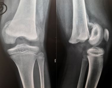
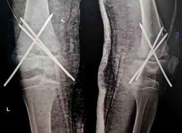
On radiographic examination, displaced Salter Harris type 2 distal femur epiphiseal injury was seen (Figure-1) and operative correction was planned. The patient was operated under spinal anesthesia by open reduction and internal fixation using kirschner wires and limb was immobilized using plaster slab (Figure-2).

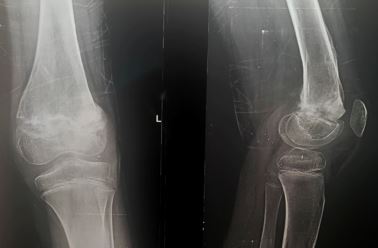
The Kirschner wires were removed after 4 weeks and physiotherapy was started. On a 4 month follow up visit, the patient was walking full weight bearing with no deformity or limb length discrepancy. The 4 month post operative radiograph showed proper bone alignment and absence of any deformity (Figure-3).

Figure: 1 AP and lateral radiographs
ojmpc_67_01.jpg

Figure: 2 Post-Op AP and lateral radiographs
ojmpc_67_02.jpg

Figure: 3 4-month Post-Op radiographs
ojmpc_67_03.jpg

Discussion

The growth plate, contains germinal cells that create longitudinal bone growth, are cartilaginous in nature. Physes, being the weakest point of the immature skeleton, account for only 20% of all fractures in children. Distal femur epiphyseal injuries are rare and contribute about 1% to 6% of all growth plate injuries [7].

Out of all the epiphyseal injuries around the knee, most common are the distal femur injuries, a reason being the stress to the knee ligaments causing traction thereby resulting in physeal-epiphyseal injuries. Sports injuries and road traffic accidents constitute the most common causes of distal epiphyseal injuries. The treatment of these injuries is of utmost importance as this zone of growth contributes to 40% of lower extremity length and 70% of the entire femur. Previously, Neer in his work reported a limb length discrepancy of 42% [8]. Cassebaum and Patterson reported the discrepancy of about 25% [9].

MRI, apart from X-ray, an investigation of choice in such cases for evaluation of post injury disturbances and clinically significant angulation. Many studies and experiments aim at eliminating risk of formation of bone bridge In previous studies, autologous fat was used fill growth plate defect. [10]


A modern experimental approach include excision of the bone bridge and defect compensation with tissue alternatives such as mesenchymal stem cells [10] or autologous chondrocytes. [11]

Conclusion

Although rare, distal femur epiphyseal injuries of the childhood are of a great concern because of the degree of bone deformities, growth disturbances and significant disabilities caused by them. A detailed study of these type of injuries helps in deciding the treatment methods and procedures to be opted, which eventually affects the prognosis and outcome in such cases

References

1. Sponceller PD, Stanitski CL. Distal femoral epiphyseal frac¬tures. In: Beaty JH, Kasser JR, eds. Rockwood and Wilkins’ Fractures in Children. 5th ed. Philadelphia, PA: Lippincott Williams & Wilkins; 2001:982-1010 [Crossref][PubMed][Google Scholar]

2. Pritchett JW. Longitudinal growth and growth-plate activity in the lower extremity. J Pediatr Orthop. 1992;2(4):557. [Crossref][PubMed][Google Scholar]

3. Zionts LE. Fractures around the knee in children. J Am Acad Orthop Surg. 2002;10:345-355. [Crossref][PubMed][Google Scholar]

4. Brown JH, DeLuca SA. Growth plate injuries. Salter-Harris classification. Am Fam Physician. 1992; 46: 1180–84 [Crossref][PubMed][Google Scholar]

5. Ilharreborde B, Raquillet C, Morel E et al. Long-term prognosis of Salter-Harris type 2 injuries of the distal femoral physis. J Pediatr Orthop B. 2006. 15: 433–38 [Crossref][PubMed][Google Scholar]

6. Chadwick CJ, Bentley G. The classification and prognosis of epiphyseal injuries. Injury. 1987; 18: 157–68. [Crossref][PubMed][Google Scholar]

7. Ogden JA. Skeletal injury in the child. 3rd ed. New York: Springer, 2000: 1198 pp. [Crossref][PubMed][Google Scholar]

8. Neer CS. Separation of the lower femoral epiphysis. Am J Surg 1960; 99: 756–761. . [Crossref][PubMed][Google Scholar]

9. Cassebaum WH, Patterson AH. Fractures of distal femoral epiphysis. Clin Orthop 1965; 41: 79–91. . [Crossref][PubMed][Google Scholar]

10. Plánka L, Nečas A, Gál P, et al. Prevention of bone bridge formation using transplantation of the autogenous mesenchymal stem cells to physeal defects: an experimental study in rabbits. Acta Vet Brno 2007; 76: 257–266. . [Crossref][PubMed][Google Scholar]

11. Gál P, Nečas A, Adler J, Teyschl O, Fabián P, Bibrová Š. Transplantation of the autologous chondrocyte graft to physeal defects: an experimental study in pigs. Acta Vet Brno 2002; 71: 327–332. . [Crossref][PubMed][Google Scholar]

Disclaimer / Publisher's Note

The statements, opinions and data contained in all publications are solely those of the individual author(s) and contributor(s) and not of Journals and/or the editor(s). Journals and/or the editor(s) disclaim responsibility for any injury to people or property resulting from any ideas, methods, instructions or products referred to in the content.