Orthopaedic Journal of MP Chapter

Publisher: Madhya Pradesh Orthopaedic Association www.mpioa.com
E-ISSN:2582-7243, P-ISSN:2320-6993
2018 Volume 24 Number 1 Jan-Jun

Comparative Study on Outcome of Early Aggressive and Standard Rehabilitation after Anterior Cruciate Ligament Reconstruction

Tantuway V1, Mustafa Johar SA2, Banerjee T3, Narware S4, Prajapati A5, Murtuza R6*

1 V Tantuway, Department of Orthopedics, Index Medical College Hospital and Research Centre, Indore, Madhya Pradesh, India.

2 S A Mustafa Johar, Department of Orthopedics, Index Medical College Hospital and Research Centre, Indore, Madhya Pradesh, India.

3 T Banerjee, Department of Orthopedics, Index Medical College Hospital and Research Centre, Indore, Madhya Pradesh, India.

4 S Narware, Department of Orthopedics, Index Medical College Hospital and Research Centre, Indore, Madhya Pradesh, India.

5 A Prajapati, Department of Orthopedics, Index Medical College Hospital and Research Centre, Indore, Madhya Pradesh, India.

6* R Murtuza, Department of Orthopaedics and Traumatology, Index Medical College Hospital and Research Centre, Indore, Madhya Pradesh, India.

Background: Anterior cruciate ligament rupture is one of the most common debilitating knee injuries that can result in significant functional impairment. Although ACL reconstruction (ACL-R) is a commonly practiced surgical intervention, controversy still lingers with regard to graft selection and rehabilitation protocol, both of which are largely influenced by surgeon preference. The post-operative restrictions are largely based on the theory of graft and fixation vulnerability, with concerns related to compromising the biological healing process of the reconstructed graft during the first 12 weeks postoperatively. To date, controversy still lingers in evaluating the effects that aggressive rehabilitation has on clinical outcomes with semitendinosus graft. The aim of this study was to investigate whether immediate full weight bearing combined with aggressive rehabilitation in ACL-R significantly altered postoperative outcome over one year, relative to a program that included partial weight bearing and standard rehabilitation protocol in the immediate post-operative period.

Methods: The study was a prospective randomized clinical trial, with all patients being recruited by a single senior orthopedic surgeon at our institute after a confirmed diagnosis of an isolated ACL rupture by clinical examination and magnetic resonance imaging. Seventy patients were enrolled in the study from June 2015 to August 2017. Informed consent was taken. Clearance from ethical committee of the institute was taken. Patients were evaluated pre operatively and post operatively at the end of 1, 3, 6 months and 1 year for outcomes.

Results: In this prospective study conducted with seventy patients, we found better results in group 2 (full weight bearing) as compared to group 2 ( partial weight bearing) in terms of IKDC scoring, range of motion (ROM), ROM difference from opposite knee and fixed flexion deformity. The results were statistically significant.

Conclusion: We conclude that aggressive rehabilitation to be superior to standard rehabilitation after isolated ACL-R using STG .

Keywords: ACL, ACL-R, Rehabilitation

Corresponding Author How to Cite this Article To Browse
R Murtuza, , Department of Orthopaedics and Traumatology, Index Medical College Hospital and Research Centre, Indore, Madhya Pradesh, India.
Email:
Tantuway V, Mustafa Johar SA, Banerjee T, Narware S, Prajapati A, Murtuza R, Comparative Study on Outcome of Early Aggressive and Standard Rehabilitation after Anterior Cruciate Ligament Reconstruction. ojmpc. 2018;24(1):25-35.
Available From
https://ojmpc.com/index.php/ojmpc/article/view/65
Manuscript Received Review Round 1 Review Round 2 Review Round 3 Accepted
2018-06-06 2018-06-12 2018-06-18 2018-06-24 2018-06-30
Conflict of Interest Funding Ethical Approval Plagiarism X-checker Note
Authors state no conflict of interest. Non Funded. The conducted research is not related to either human or animals use. 13.35 All authors have accepted responsibility for the entire content of this manuscript and approved its submission.

© 2018by Tantuway V, Mustafa Johar SA, Banerjee T, Narware S, Prajapati A, Murtuza Rand Published by Madhya Pradesh Orthopaedic Association. This is an Open Access article licensed under a Creative Commons Attribution 4.0 International License https://creativecommons.org/licenses/by-nc/4.0/ unported [CC BY NC 4.0].

Introduction

Anterior cruciate ligament rupture is one of the most common debilitating knee injuries that can result in significant functional impairment.[1-3] Surgical reconstruction of a ruptured ACL is advocated as the treatment of choice, particularly for individuals who intend to resume competitive sporting activities[4-6].

Although ACL reconstruction (ACL-R) is a commonly practiced surgical intervention, controversy still lingers in regard to graft selection and rehabilitation protocol, both of which are largely influenced by surgeon preference [7].

Traditional postoperative restrictions such as bracing for immobilization, delayed weight bearing, and limiting early hyperextension motion (beyond 0° of extension) have all been used throughout rehabilitation in hopes of preventing excessive loads on the healing graft [8].

The implications of these restrictions are largely based on the theory of graft and fixation vulnerability, with concerns related to compromising the biological healing process of the reconstructed graft during the first 12 weeks postoperatively [9-11].

These concerns have most appropriately been justified for Semitendinosus (ST) grafts due to the slower incorporation rate of the soft tissue into the bone tunnel and concerns of excessive graft-tunnel motion leading to increased laxity [12].

Granted that these theories are still valid concerns in postoperative management, advancements in surgical technique and fixation have warranted re-evaluation of the use of restrictions after ACL-R with gathering evidence showing that restrictions may not be necessary.

Early aggressive rehabilitation has shown no adverse effects with respect to future injury rate, Antero-Posterior laxity, Range Of Motion deficits, or ability to return patients back to their previous level of function [12,13].

Although a significant body of literature has shown that aggressive rehabilitation defined as early unrestricted motion, immediate weight bearing, and eliminating the use of immobilizing braces to be appropriate after ACL-R using BPTB grafts conclusions are unclear when evaluating the effects of early aggressive rehabilitation on ST autografts [13-15].

To date, controversy still lingers in evaluating the effects that aggressive rehabilitation has on clinical outcomes with this particular graft. Some studies have shown that aggressive rehabilitation immediately after surgery tends to increase knee laxity [8,10], while others have found no difference in subjective outcomes or functional stability [16,17].

Furthermore, Wright et al conducted a systematic review confirming that the available evidence on postoperative bracing, immediate weight bearing, and unrestricted ROM has largely been performed addressing BPTB grafts, demonstrating the lack of attention in comparison with ST grafts [14].

Therefore, a need exists to evaluate the effects of early aggressive rehabilitation of ST grafts on mobility, strength, and self-reported outcome scores. The aim of this study was to investigate whether immediate full weight bearing combined with aggressive rehabilitation in ACL-R significantly altered postoperative outcome over one year, relative to a program that included partial weight bearing and standard rehabilitation protocol in the immediate post-operative period.

Materials and Methods

Seventy patients were enrolled in the study from June 2015 to August 2017. The study was a prospective randomized clinical trial, with all patients being recruited by a single senior orthopedic surgeon at our institute after a confirmed diagnosis of an isolated ACL rupture by clinical examination and magnetic resonance imaging. Two experienced physical therapists who were not involved in data collection treated all patients in both allocated groups. A single research assistant who was blinded to the treatment allocation measured all outcome variables. Patients were evaluated pre operatively and post operatively at the end of 1, 3, 6 months and 1 year for outcomes.

Inclusion criteria:

1. Age 18 to 55 years
2. Grade II or III isolated ACL tear confirmed by orthopedic surgeon
3. Demonstration of full knee extension and at least 85% knee flexion preoperatively compared with the contralateral knee
4. Ability to comply with a 24-week rehabilitation program.
5. Unilateral pathology
6. No previous knee surgery


Exclusion criteria:

1. Any previous ACL-R to either knee
2. Concurrent injury to the posterior cruciate ligament
3. Grade III tear of either (medial or lateral) collateral ligament
4. Meniscus tears ≥5 mm or meniscus repairs
5. Pregnancy
6. Neurological disorders (multiple sclerosis, cerebral palsy, etc) affecting participation

Patients with age above 50 years were excluded as age related degenerative changes are common after 50 years. After subjects provided consent for participation, the research assistant randomized them into one of two treatment groups by computer software randomization.

A simple randomization technique was performed, and to conceal the treatment allocation, the randomization scheme was computer generated before initiation of the study. Patients were randomized into either the aggressive or the nonaggressive group.

Surgical Procedure

All patients underwent a single-bundle ipsilateral 4-strand ST autograft reconstruction. Confirmation of a complete ACL tear was accomplished arthroscopically, followed by preparation of the femoral notch and tibial footprint. The STGs were harvested in standard fashion through a 3-cm incision over the anteromedial tibia, then the graft was prepared with #5 Ethibond whip stitching the free ends of the graft.

A tibial tunnel was reamed entering the anatomic center of the tibial ACL insertion. An accessory medial portal was created through which the femoral tunnel was placed in the anatomic center of the femoral ACL footprint. The tibial and femoral tunnels were sized to the diameter of the graft. The ACL fixation consisted of femoral button suspension fixation. Tibial fixation was accomplished using an interference screw with the knee positioned in 15° knee flexion with force applied as in doing posterior drawer test.

Rehabilitation

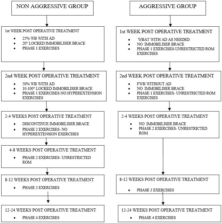
The aggressive group underwent a protocol that was largely derived from previous work described by Biggs et al 18 (Figure 1).

Patients were not required to wear a postoperative knee brace and began exercises to restore full passive motion without restrictions on hyperextension immediately after surgery.

Patients in this group were informed to begin weight bearing as tolerated immediately after surgery and to only use the bilateral axillary crutches for comfort.

During the first postoperative week, patients underwent a rehabilitation regimen consisting of keeping activity to a minimum and remaining in a supine position while the leg was elevated at least 12 inches above the chest for at least 18 hours of the day, and beginning phase I exercises to initiate early motion and muscle activation.

The nonaggressive group were required to wear a ROM brace locked at 20° of flexion for the first week and unlocked 10° to 100° for an additional 3 weeks after surgery.

Patients were instructed to only remove the brace to perform the phase I exercises, and during physical therapy visits. They were required to wear the brace at night to sleep for the first week.

After 4 weeks of postoperative bracing, the brace was discontinued and the treating physical therapists instructed patients to begin full passive knee-flexion motion, but they were restricted to no hyperextension stretching for an additional 2 weeks postoperatively. Hyperextension exercises were defined as any active or passive stretch beyond 0° of knee extension.

Patients in this group also used bilateral axillary crutches for 2 weeks postoperatively at 25% weight bearing for the first week and 50% weight bearing for the second week.

During the first postoperative week, they underwent the same rehabilitation regimen as the aggressive group. All patients performed a standard postoperative physical therapy protocol, and compliance was tracked through weekly logbook entry.

Exceptions to the protocol for the nonaggressive group were as follows: They were confined to a postoperative brace for 4 weeks and not allowed to perform any hyperextension exercises for the first 6 weeks, and they were instructed to ambulate with a modified weight-bearing status for the first 2 weeks.


Figure 1: Treatment protocol for each group. Abbreviations: WB, weight-bearing; AD, assistive device; CPMM, continued passive-motion machine; ROM, range-of-motion; WBAT, weight-bearing as tolerated; FWB, full weight-bearing.
ojmpc_65_01.jpg

Standard post-operative physiotherapy protocol for both groups was as follows:-

1. Phase I (0–4 wk) of the rehabilitation protocol included passive, active-assist, and active ROM exercises; stationary bicycling; muscle-activation exercises; and inflammation reduction.
2. Phase II (4–8 wk) of the protocol emphasized progressive ROM exercises, muscle strengthening, neuromuscular-control training, and functional activities.
3. Phase III (8–12 wk) of the protocol consisted of restoring full symmetrical passive ROM, increased muscle strengthening, higher level neuromuscular-control tasks, and running.
4. Phase IV (12–24 wk) of the protocol involved progressive muscle strengthening, sport-specific neuromuscular-control training, plyometrics, sprinting, and cutting drills.

Patients were scheduled for the same number of physical therapy visits and established time periods for exercise progression.

Outcome Measures

The primary outcomes was subjective IKDC scores at 12 weeks, 24 weeks and 1 year. The secondary outcomes were the difference in ROM at the same duration. The IKDC Subjective Knee Form was used to assess the patient’s opinion about his or her knee function and possible associated problems.

19 The IKDC is based on a 0-to-100 cardinal scale and a knee-specific subjective measure of symptoms, function, and sport activity.19 The IKDC has been shown to be a reliable and valid instrument in measuring patient-oriented clinical outcomes in daily and sport function.


15,19 A dual-arm goniometer was used to measure knee flexion and -extension ROM in both knees, which has shown high reliability.20 The mobility of the knee was measured as described by Shelbourne et al.21 Knee extension was measured with the patient’s heel positioned on a bolster to allow the examiner to measure the amount of extension, or hyperextension if present, with the patient in a seated position.

Knee flexion was measured by instructing the patient to bend the affected knee as far as possible toward the buttocks in a seated position. The outcome score used for analysis was expressed as the difference in ROM between the surgical and nonsurgical knees for both flexion and extension.

Statistical Analysis

Group 1 was defined as patients undergoing the standard rehabilitation protocol or the non-aggressive group, while group 2 was defined as patients undergoing aggressive rehabilitation. The data was initially captured in the customized proforma designed for the study.

Then the data from this customized proforma was entered into the Microsoft Excel for analysis and online software were used for calculating the statistical significant. Unpaired ‘t’ test was used to compare the difference of mean between the two groups, Pearson chi-square of 2x2 was used to compare the two groups.

A p value of < 0.05 was taken as statistically significant. The final data was presented in the form of tables and graphs

Results

Seventy patients were enrolled in the study from June 2015 to August 2017. 56 men and 19 women initially met the inclusion criteria for the study. Four men and one woman were later excluded after operative findings indicated the need for meniscal repair.

Thus, 70 subjects underwent randomization and began the postoperative rehabilitation protocol. 35 patients each were randomized to the aggressive and nonaggressive group respectively.

Baseline characteristics were not different between groups. No subject was lost to follow-up.

The above table shows the distribution of patients according to age in both the partial weight bearing and full weight bearing groups.

Table: 1 Distribution of patients according to age in the partial weight bearing and full weight bearing groups

(N=70)

Age GroupPartial Weight Bearing(n=35)Full Weight Bearing(n=35)
No.%No.%
16-20 years411.4617.1
21-30 years1131.41851.4
31-40 years2057.11131.4
Total35100.035100.0
Mean ± SD (age, years)30.00 ± 6.0127.77 ± 5.98
‘t’ value, df1.555, df=68
P value0.125, NS

Unpaired ‘t’ test applied. P value < 0.05was taken as statistically significant

Table: 2 Distribution of patients according to gender in the partial weight bearing and full weight bearing groups

(N=70)

GenderPartial Weight Bearing(n=35)Full Weight Bearing(n=35)
No.%No.%
Female925.7720.0
Male2674.32880.0
Total35100.035100.0

c2 test applied (2x2). c2 value = 0.324, df=1, P value = 0.569, Not significant

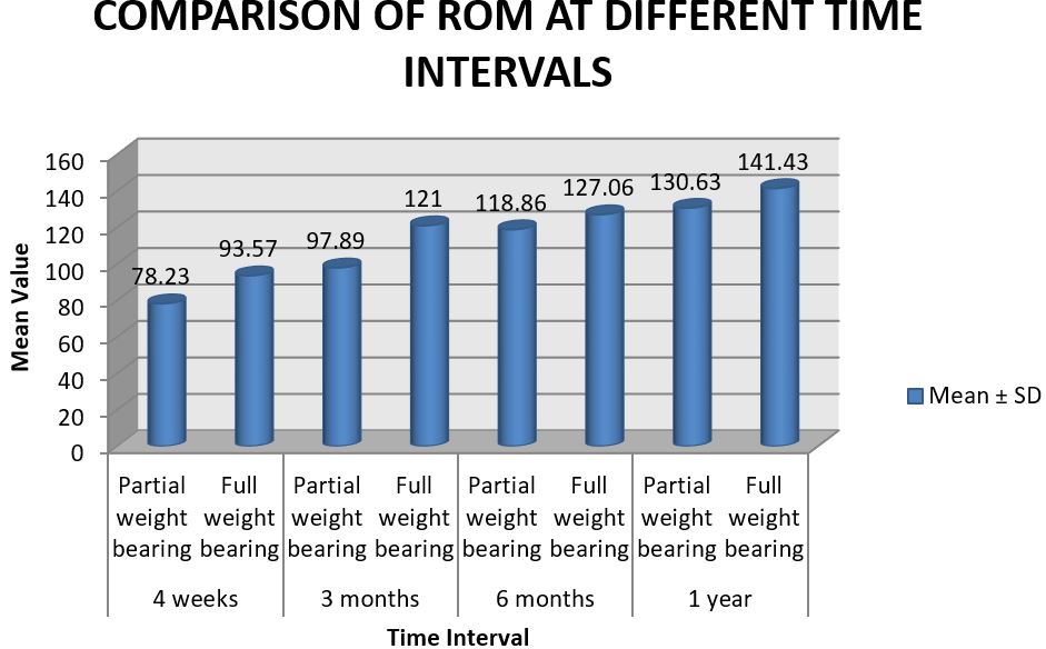
Table: 3 Comparison of mean ROM at different time intervals between the partial weight bearing and full weight bearing groups

ParameterTime IntervalGroupMean ± SD‘t’ valueP value
ROM4 weeksPartial weight bearing78.23 ± 10.42-6.877, df=680.000*
Full weight bearing93.57 ± 8.09
3 monthsPartial weight bearing97.89 ± 11.14-9.629, df=680.000*
Full weight bearing121.00 ± 8.81
6 monthsPartial weight bearing118.86 ± 10.00-4.186, df=680.000*
Full weight bearing127.06 ± 5.85
1 yearPartial weight bearing130.63 ± 6.15-9.573, df=680.000*
Full weight bearing141.43 ± 2.59

Unpaired ‘t’ test applied. P value < 0.05was taken as statistically significant


Figure: 2 Bar diagram showing comparison of ROM at different time intervals between the partial weight bearing and full weight bearing groups
ojmpc_65_02.jpg

Table: 4 Comparison of mean FFD at different time intervals between the partial weight bearing and full weight bearing groups

ParameterTime IntervalGroupMean ± SD‘t’ valueP value
FFD4 weeksPartial weight bearing3.43 ± 3.166.704, df=680.000*
Full weight bearing1.26 ± 2.67
3 monthsPartial weight bearing1.11 ± 1.990.550, df=680.584, NS
Full weight bearing0.86 ± 1.91
6 monthsPartial weight bearing0.94 ± 1.852.112, df=680.038*
Full weight bearing0.23 ± 0.77
1 yearPartial weight bearing0.37 ± 0.942.333, df=680.023*
Full weight bearing0.00 ± 0.00

Unpaired ‘t’ test applied. P value < 0.05was taken as statistically significant

The above table shows the comparison of mean FFD at different time intervals between the two groups.

Table: 5 Comparison of mean ‘Difference from Opposite Knee’ at different time intervals between the partial weight bearing and full weight bearing groups

ParameterTime IntervalGroupMean ± SD‘t’ valueP value
Difference from opposite knee4 weeksPartial weight bearing-58.91 ± 9.66-5.861, df=680.000*
Full weight bearing-46.43 ± 8.09
3 monthsPartial weight bearing-39.26 ± 10.24-8.871, df=680.000*
Full weight bearing-19.00 ± 8.81
6 monthsPartial weight bearing-18.29 ± 9.07-2.929, df=680.005*
Full weight bearing-12.94 ± 5.85
1 yearPartial weight bearing-6.51 ± 6.29-6.908, df=680.000*
Full weight bearing1.43 ± 2.59

Unpaired ‘t’ test applied. P value < 0.05was taken as statistically significant

The above table shows the comparison of mean ‘difference from opposite knee’ at different time intervals between the two groups.

Table: 6 Comparison of mean IKDC Score at different time intervals between the partial weight bearing and full weight bearing groups

ParameterTime IntervalGroupMean ± SD‘t’ valueP value
IKDC Score4 weeksPartial weight bearing30.02 ± 2.80-8.318, df=680.000*
Full weight bearing34.68 ± 1.77
3 monthsPartial weight bearing51.98 ± 1.54-66.018, df=680.000*
Full weight bearing72.27 ± 0.97
6 monthsPartial weight bearing80.50 ± 0.000.960, df=680.340, NS
Full weight bearing79.84 ± 4.07
1 yearPartial weight bearing88.43 ± 4.06-8.917, df=680.000*
Full weight bearing96.11 ± 3.09

Unpaired ‘t’ test applied. P value < 0.05was taken as statistically significant

Overall, we found better results in group 2 (full weight bearing) as compared to group 2 ( partial weight bearing) in terms of IKDC scoring, range of motion (ROM), ROM difference from opposite knee and fixed flexion deformity. The results were statistically significant.

Adverse events relative to motion limitations were present in both groups. Three patients (aggressive group n = 2 and nonaggressive group n = 1) required additional visits due to motion limitations, which were deemed necessary by the treating orthopedic surgeon. No other complication was noted.

Discussion

This randomized clinical trial evaluated the effects of early aggressive rehabilitation on patients recovering from ACL-R using STG autograft, while observing the relationship between clinical measures that are paramount in determining a successful outcome.


Early aggressive rehabilitation compared with nonaggressive rehabilitation was found to be significantly better in this cohort of patients in relation to primary outcomes of subjective IKDC scores. Subjects in the aggressive group did demonstrate a significant difference in ROM compared with the nonaggressive group.

Our findings support the current body of literature as it pertains to BPTB grafts. Previous studies have compared the effects of early aggressive rehabilitation protocols on outcomes after ACL-R using BPTB grafts, indicating it to be appropriate to proceed through postoperative management without immobilizing the knee, restricting early hyperextension motion, or delaying weight bearing [9,14,15,21].

The primary goal after ACL-R is to restore knee stability and function in preparation for patients to return to their previous level of activity. However, even with advancements in surgical techniques and rehabilitation protocols, there is strong evidence that deficits in lower extremity strength, neuromuscular control, and proprioception are continually present as patients are released back to unrestricted sport function[22,23].

Paterno et al reported that female athletes who had been cleared for full unrestricted sport activity still present with significant landing and jumping asymmetry during a vertical drop-jump task that has been used to predict ACL injury risk [24].

This evidence demonstrates that higher-level rehabilitation methods need to be emphasized in later stages of recovery after ACL-R in hopes of reducing the residual limb asymmetries and potentially decreasing the risk of future re injury. Furthermore, future research is needed to establish an objective criterion based on functional testing and outcomes before returning patients back to unrestricted sport after rehabilitation.

Early aggressive rehabilitation has been established for years, but there are discrepancies in the literature relative to overemphasis on BPTB grafts and lack of postoperative management on STG grafts. Our findings are clinically relevant since STG autografts have gained popularity in comparison with other graft choices, and limited research has been conducted evaluating the effects of early aggressive rehabilitation on functional outcomes.

This evidence is important for guiding clinicians in making appropriate decisions on postoperative rehabilitation and restrictions after surgery, because there still appears to be conflicting evidence [14].

The current study appears to indicate that an early aggressive postoperative protocol is superior to a nonaggressive rehabilitation protocol after an isolated ACL-R using STG autograft. The current study had certain limitations. First, outcomes like A-P knee laxity were not gathered. Second, no independent blinded data collector was used in this study, leading to potential performance bias.

Third, our small sample size was small and composed of active subjects, and we were unable to stratify groups by activity level.

Conclusion

We found early aggressive rehabilitation to be superior to nonaggressive rehabilitation after isolated ACL-R using STG autograft for the primary outcomes of knee ROM, FFD and subjective IKDC score. The study emphasizes that early aggressive rehabilitation in such cases is better at restoring the normal function of knee when evaluated objectively as well as subjectively.

References

1. Baquie P and Brukner P (1997). Injuries presenting to an Australian sports medicine centre. A 12–month study. Clinical Journal of Sports Medicine 7: 28–31. [Crossref][PubMed][Google Scholar]

2. Miyasaka KC, Daniel DM, Stone ML and Hirshman P (1991). The incidence of knee ligament injuries in the general population. The American Journal of Knee Surgery 4: 3–8. . [Crossref][PubMed][Google Scholar]

3. Seward H, Orchard J, Hazard H and Collinson D (1993). Football injuries in Australia at the elite level. Medical Journal of Australia 159: 298–301. . [Crossref][PubMed][Google Scholar]

4. Barrack RL, Bruckner JD, Kneisl J, Inman WS and Alexander AH (1990). The outcome of nonoperatively treated complete tears of the anterior cruciate ligament in active young adults. Clinical Orthopaedics and Related Research 259. 192–199. [Crossref][PubMed][Google Scholar]


5. Fetto JF and Marshall JL (1980). The natural history and diagnosis of anterior cruciate ligament insufficiency. Clinical Orthopaedics and Related Research 147: 29–38. . [Crossref][PubMed][Google Scholar]

6. Frank CB and Jackson DW (1997). The science of reconstruction of the anterior cruciate ligament. Journal of Bone and Joint Surgery 79A. 1556–1576. [Crossref][PubMed][Google Scholar]

7. Reinhardt KR, Hetsroni I, Marx RG. Graft selection for anterior cruciate ligament reconstruction: a level I systematic review comparing failure rates and functional outcomes. Orthop Clin North Am. 2010;41(2):249–262. [Crossref][PubMed][Google Scholar]

8. Heijne A, Werner S. A 2-year follow-up of rehabilitation after ACL reconstruction using patellar tendon or hamstring tendon grafts: a prospective randomised outcome study. Knee Surg Sports Traumatol Arthrosc. 2010;18(6):805–813. [Crossref][PubMed][Google Scholar]

9. Isberg J, Faxen E, Brandsson S, Eriksson BI, Karrholm J, Karlsson J. Early active extension after anterior cruciate ligament reconstruction does not result in increased laxity of the knee. Knee Surg Sports Traumatol Arthrosc. 2006;14(11):1108–1115. [Crossref][PubMed][Google Scholar]

10. Muneta T, Sekiya I, Ogiuchi T, Yagishita K, Yamamoto H, Shinomiya K. Effects of aggressive early rehabilitation on the outcome of anterior cruciate ligament reconstruction with multi-strand semitendinosus tendon. Int Orthop. 1998;22(6):352–356. [Crossref][PubMed][Google Scholar]

11. Rodeo SA, Arnoczky SP, Torzilli PA, Hidaka C, Warren RF. Tendon-healing in a bone tunnel: a biomechanical and histological study in the dog. J Bone Joint Surg Am. 1993;75(12):1795–1803. [Crossref][PubMed][Google Scholar]

12. Ekdahl M, Wang JH, Ronga M, Fu FH. Graft healing in anterior cruciate ligament reconstruction. Knee Surg Sports Traumatol Arthrosc. 2008;16(10):935–947. [Crossref][PubMed][Google Scholar]

13. Shelbourne KD, Gray T. Minimum 10-year results after anterior cruciate ligament reconstruction: how the loss of normal knee motion compounds other factors related to the development of osteoarthritis after surgery. Am J Sports Med. 2009;37(3):471–480. [Crossref][PubMed][Google Scholar]

14. Wright RW, Preston E, Fleming BC, et al. A systematic review of anterior cruciate ligament reconstruction rehabilitation: part I: continuous passive motion, early weight bearing, postoperative bracing, and home-based rehabilitation. J Knee Surg. 2008;21(3):217–224. [Crossref][PubMed][Google Scholar]

15. Shelbourne KD, Klootwyk TE, Wilckens JH, De Carlo MS. Ligament stability two to six years after anterior cruciate ligament reconstruction with autogenous patellar tendon graft and participation in accelerated rehabilitation program. Am J Sports Med. 1995;23(5):575–579. [Crossref][PubMed][Google Scholar]

16. Holm I, Oiestad BE, Risberg MA, Aune AK. No difference in knee function or prevalence of osteoarthritis after reconstruction of the anterior cruciate ligament with 4-strand hamstring autograft versus patellar tendon-bone autograft: a randomized study with 10-year followup. Am J Sports Med. 2010;38(3):448–454. [Crossref][PubMed][Google Scholar]

17. Poolman RW, Farrokhyar F, Bhandari M. Hamstring tendon autograft better than bone patellar-tendon bone autograft in ACL reconstruction: a cumulative meta-analysis and clinically relevant sensitivity analysis applied to a previously published analysis. Acta Orthop. 2007;78(3):350–354. [Crossref][PubMed][Google Scholar]

18. Biggs A, Jenkins WL, Urch SE, Shelbourne KD. Rehabilitation for patients following ACL reconstruction: a knee symmetry model. N Am J Sports Phys Ther. 2009;4(1):2–12. [Crossref][PubMed][Google Scholar]

19. Irrgang JJ, Anderson AF, Boland AL, et al. Development and validation of the International Knee Documentation Committee subjective knee form. Am J Sports Med. 2001;29(5):600–613. [Crossref][PubMed][Google Scholar]

20. Brosseau L, Balmer S, Tousignant M, et al. Intra- and intertester reliability and criterion validity of the parallelogram and universal goniometers for measuring maximum active knee flexion and extension of patients with knee restrictions. Arch Phys Med Rehabil. 2001;82(3):396–402. [Crossref][PubMed][Google Scholar]


21. Shelbourne KD, Urch SE, Gray T, Freeman H. Loss of normal knee motion after anterior cruciate ligament reconstruction is associated with radiographic arthritic changes after surgery. Am J Sports Med. 2012;40(1):108–113. [Crossref][PubMed][Google Scholar]

22. Barber-Westin SD, Noyes FR. Factors used to determine return to unrestricted sports activities after anterior cruciate ligament reconstruction. Arthroscopy. 2011;27(12):1697–1705. [Crossref][PubMed][Google Scholar]

23. Myer GD, Paterno MV, Ford KR, Quatman CE, Hewett TE. Rehabilitation after anterior cruciate ligament reconstruction:criteria-based progression through the return-to-sport phase. J Orthop Sports Phys Ther. 2006;36(6):385–402. [Crossref][PubMed][Google Scholar]

24. Paterno MV, Ford KR, Myer GD, Heyl R, Hewett TE. Limb asymmetries in landing and jumping 2 years following anterior cruciate ligament reconstruction. Clin J Sport Med. 2007;17(4):258–262. [Crossref][PubMed][Google Scholar]

Disclaimer / Publisher's Note

The statements, opinions and data contained in all publications are solely those of the individual author(s) and contributor(s) and not of Journals and/or the editor(s). Journals and/or the editor(s) disclaim responsibility for any injury to people or property resulting from any ideas, methods, instructions or products referred to in the content.