Orthopaedic Journal of MP Chapter

Publisher: Madhya Pradesh Orthopaedic Association www.mpioa.com
E-ISSN:2582-7243, P-ISSN:2320-6993
2018 Volume 24 Number 1 Jan-Jun

Comparative study of two techniques of percutaneous pinning of displaced supracondylar humerus fracture

Aher D1, Mishra RK2*, Gohiya A3

1 D Aher, Department of orthopedics, Gandhi Medical College, Bhopal, Madhya Pradesh, India.

2* Rajesh Kumar Mishra, Department of orthopedics, Gandhi Medical College, Bhopal, Madhya Pradesh, India.

3 A Gohiya, Department of orthopedics, Gandhi Medical College, Bhopal, Madhya Pradesh, India.

Background: supracondylar fractures of the humerus are very common fractures in paediatric age groups. If displaced, preferred treatment is close reduction with percutaneous K-wire fixation. This study compares the functional outcome of cross K-wires Vs lateral K-wires in Gartland type III supracondylar humerus fractures in paediatric age groups

Methods: 60 cases of supracondylar fractures of the humerus Gartland type III in children operated with closed reduction and pinning of which 30 were of lateral and 30 were of cross K wire groups from January 2015 to June 2017 with minimum 4 months follow-up period were included.

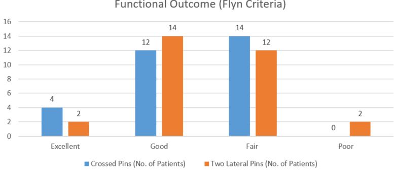
Results: Functional outcome of the patients was assessed by Flynn’s criteria. Results were excellent 13.33 %, good 40% fair 46.6% in cross K wire group and excellent 6.6 %, good 46.6%, fair 40% and poor 6.6 % in lateral K wire group.

Conclusion: Both lateral entry pin fixation and crossed pin fixation are effective in the treatment of Gartland type III extension supracondylar fractures of the humerus in children.

Keywords: Cross k wires, Lateral pinning, Gartland type III Supracondylar fracture humerus

Corresponding Author How to Cite this Article To Browse
Rajesh Kumar Mishra, , Department of orthopedics, Gandhi Medical College, Bhopal, Madhya Pradesh, India.
Email:
Aher D, Mishra RK, Gohiya A, Comparative study of two techniques of percutaneous pinning of displaced supracondylar humerus fracture. ojmpc. 2018;24(1):3-7.
Available From
https://ojmpc.com/index.php/ojmpc/article/view/61
Manuscript Received Review Round 1 Review Round 2 Review Round 3 Accepted
2018-06-01 2018-06-06 2018-06-12 2018-06-24 2018-06-30
Conflict of Interest Funding Ethical Approval Plagiarism X-checker Note
Authors state no conflict of interest. Non Funded. The conducted research is not related to either human or animals use. 13.32 All authors have accepted responsibility for the entire content of this manuscript and approved its submission.

© 2018by Aher D, Mishra RK, Gohiya Aand Published by Madhya Pradesh Orthopaedic Association. This is an Open Access article licensed under a Creative Commons Attribution 4.0 International License https://creativecommons.org/licenses/by-nc/4.0/ unported [CC BY NC 4.0].

Introduction

Supracondylar Humerus Fracture is most common fracture around elbow in pediatric age group. Supracondylar Humerus Fracture has an incidence of 50-70 %, of which extension type is about 98 % and flexion type about 2% [1].

Supracondylar Humerus Fracture can be managed by both non-operative or operative treatment. The decision to operate depends upon the fracture type and stability after reduction. For undisplaced fractures, plaster suffices, but for displaced fractures, fixation with K-wire is preferred [2].

Available treatment options for displaced supracondylar humerus fracture (Gartland Type 3) are Closed reduction and percutaneous pinning or Open reduction and K wire Fixation [3].

Loss of reduction and subsequent malunion is generally seen when type III fractures are treated in plaster, which led to percutaneous pinning as preferred option [4].

There are various techniques of percutaneous pinning with literature supporting one over the other based on biomechanical advantages shown in vitro. This study is aimed to compare functional outcomes and complications between two techniques (crossed pinning Vs lateral pinning) of percutaneous pining of displaced Supracondylar Humerus Fracture in pediatric age group.

Materials and Methods

This was a prospective, randomized study conducted in the Department of Orthopaedics and traumatology at Gandhi Medical College and Hamidia hospital Bhopal from January 2015 to June 2017. Total 60 patients with 30 patients each in cross k wires and lateral pinning group were observed.

Inclusion Criteria

1. Age 2 - 12 years
2. Presenting within 1 week of injury
3. Closed fractures
4. Gartland Type-III supracondylar fracture

Exclusion Criteria

1. Open fractures
2. Floating elbow injuries
3. Previous fracture in the same elbow.

All patients with Supracondylar fracture humerus reporting to emergency department of Hamidia hospital were evaluated by thorough clinico-radiological examination.

Patients who fulfilled the inclusion criteria and whose parents gave the consent to be registered in study, were enrolled in the study as per the detailed proforma. Patients were allocated to one of the groups (Crossed pins Vs Two Lateral Pins) with the help of computer-generated random table. Type lll supracondylar humerus fracture were posted in operation theatre as early as possible after work-up.

Under appropriate anesthesia patient was positioned supine on the operating table, affected limb scrubbed, painted and draped. Limb was placed over the sterile draped C-arm image intensifier. Initially, closed manipulation was performed with linear traction and flexion of elbow, pushing the distal fragment anteriorly.

Assessment of reduction was done under image intensifier in both AP and Lateral views. Following that fixation by one of the two techniques was done depending on the randomization. In crossed pinning technique, lateral pin was inserted first and medial pin was placed with the elbow in less flexion to avoid ulnar nerve injury.

In two lateral pin technique two divergent K-wires were passed from the lateral condyle. After leaving about 2 cm of the pins outside the skin, pins were bent and cut off and well-padded posterior above elbow slab was applied with elbow flexed to 90° or less. The Neurovascular status was again assessed post operatively.

The slab and K wires were removed after 4 weeks, and range of motion exercises and physiotherapy were started. Thus, the patient was followed up at 1-month, 2-month, 4 month, 6 months and final follow-up at 12 months. The functional outcome was assessed by Flynn’s criteria [5]. The results were graded as excellent, good, fair or poor according to the range of motion and loss of carrying angle. Complications if any, were also noted.

Results

The average age was 7 years (range 2-12 years) with a peak incidence in 5-8 years. 40 patients were having Left side and 20 patients were having right side fracture.

Fall on outstretched hand contributed to about 96% cases. In 60 % cases, Postero-medial displacement of distal fragment was seen. The average follow-up duration for patients was of 6 months. Radiological union was seen on an average in 5 weeks. Loss of Baumann’s angle of 5° was seen in 33% lateral pinning cases and in 20% cross k wires cases. Loss of Baumann’s angle of 2° was seen in 33% lateral pinning cases and in 30% cross k wires cases.


Figure 1: Functional Outcome of two groups
ojmpc_61_01

Mean carrying angle loss was 30 in lateral K wire group and 20 in cross K wire group, which was statistically significant(p<0.05).

This loss of carrying angle was more in lateral group probably due to less stable construct. Loss of range of motion was 10° in cross k wires group and 11° in lateral pinning group, which was not statistically significant. In this study, no pin tract infection was noted, no ulnar nerve was injured.

Loss of reduction was seen more in lateral wire group but eventually all achieved radiological union with < 5 angulation. The difference in functional outcome between the two groups was not statistically significant (P=0.69).

Discussion

Fracture that occurs at supracondylar area or metaphysis of distal humerus is labelled as Supracondylar Fracture. For closed reduction and percutaneous pinning, two configuration of K-wires exists either lateral pinning or cross K-wires. In this study, the average age was 7 years (range 2-12 years) with peak incidence in 5-8 years. Other authors reported similar age groups, average age 7 years by Ramsey and Griz [6], 6.4 years by Nacht et al [7].

In this study, there were 90% male, Fowles and Kassab reported 89% [8] and Nacht et al 50% male preponderance [7].

In this study, left sided fractures were more than right sided fractures. Fowles and Kassab showed left (57%) more involved than right [8]. Similar results were seen in study by Nacht et al (55%) [7].

In this study, fall on outstretched hand (96%) was the most common mode of injury, similar findings were shown by Mostafavi and Bhuyan [9,10].

In our study, the average radiological union was seen in 5 weeks (range 3 to 9 weeks). Sudheendra et al reported average radiological union at 7.6 weeks [4] Rijal and Pandey reported radiological union in 6 weeks [11].

In our study, Cross k wires had better stability. Lee SS et al and Zioutset al reported that medial and lateral entry provides greater torsional rigidity than lateral entry pin fixation does [12,13]. Sudheendra et al. in their study noted 82% excellent results and 18% good results in cross k wires case and 71% excellent results and 29% good results in lateral pinning case [4].

Raffi c et al. in their study found 72% excellent results and 28% good results with lateral pinning [14]. Khan obtained 88% excellent, 4% good and 4% poor results in his study [15].

In our series, the functional outcome following cross k wires was excellent in 13.33%, good in 40% of cases, fair 46.6 % and poor in 0 % and lateral pinning showed 6.6 % excellent ,46.6% good results, 40 % fair with 6.6 % poor results.

The difference in functional outcome between the two groups was not statistically significant (P=0.69). No ulnar nerve palsy occurred in our study. Skaggs et al. found no ulnar nerve palsy and no reduction was lost in 124 children managed with only lateral-entry pins [16].

Skaggs et al. noted the incidence of ulnar nerve injury as 4% in patients whom the pins were applied without hyper flexion of the elbow and as 15% in whom the medial pin was applied with the elbow hyper flexed [17]. The rate of ulnar nerve injuries varies in different studies. Lyons et al. have reported this number as 6%, Royce et al. as 3%, Agus et al. as 58% [18,19,20].

No Pin tract infection occurred in our series. In the series by Mostafavi and Sperothe incidence of pin tract infection was 5% [9]. The incidence of infection was 2% in Pirone et al. which was found more compared to our study [21].

No pin migration or significant loss of reduction was seen in our study. Gordon observed pin migration in 6% of cases and Lee noticed the loss of reduction in 7% of cases. [22,12]

Conclusion

Cross K wire or lateral K wire, both are similar in effect for the final functional outcome with no significant difference for treatment of Gartland type III supracondylar humerus fracture in pediatric age group, although taking into consideration the ulnar nerve injury, lateral k wire technique has an upper hand, but at the cost of slight loss of reduction. However, it depends upon the surgeon’s practice and preference which may negate these complications. Hence, in our study, we found lateral k wire and cross k wire equally good in terms of safety and efficacy, but stability is more in cross wires.


References

1. Babal JC, Mehlman CT, Klein G. Nerve injuries associated with pediatric supracondylar humeral fractures: A meta-analysis. J PediatrOrthop 2010;30:253-63. . [Crossref][PubMed][Google Scholar]

2. Yadav UB, Singhal R, Tonk G, Aggarwal T, Verma AN. Crossed pin fixation in displaced supracondylar humerus fractures in children. Indian J Orthop 2004;38:166-9. . [Crossref][PubMed][Google Scholar]

3. Mazda K, Boggione C, Fitoussi F, Penneçot GF. Systematic pinning of displaced extension-type supracondylar fractures of the humerus in children. A prospective study of 116 consecutive patients. J Bone Joint Surg Br 2001;83:888-93. [Crossref][PubMed][Google Scholar]

4. Sudheendra PR, Nazareth EL. A comparative study of outcome of percutaneous lateral and crossed pinning in the treatment of type III supracondylar fractures of humerus in children. Sch J Appl Med Sci 2014;23:959-62. . [Crossref][PubMed][Google Scholar]

5. Attenborough CG. Remodeling of the humerus after supracondylar fractures in childhood. J Bone Joint Surg Br 1953;35:386-95. . [Crossref][PubMed][Google Scholar]

6. Ramsey RH, Griz J. Immediate open reduction and internal fixation of severely displaced supracondylar fractures of the humerus in children. Clin OrthopRelat Res 1973;131-2. . [Crossref][PubMed][Google Scholar]

7. Nacht JL, Ecker ML, Chung SM, Lotke PA, Das M. Supracondylar fractures of the humerus in children treated by closed reduction and percutaneous pinning. ClinOrthopRelat Res 1983;203-9. . [Crossref][PubMed][Google Scholar]

8. Fowles JV, Kassab MT. Displaced supracondylar fractures of the elbow in children. A report on the fixation of extension and flexion fractures by two lateralpercutaneous pins. J Bone Joint Surg Br 1974;56:490-500. [Crossref][PubMed][Google Scholar]

9. Mostafavi HR, Spero C. Crossed pin fixation of displaced supracondylar humerus fractures in children. ClinOrthopRelat Res 2000;56-61. . [Crossref][PubMed][Google Scholar]

10. Bhuyan BK. Close reduction and percutaneous pinning in displaced supracondylar humerus fractures in children. J ClinOrthop Trauma 2012;3:89-93. . [Crossref][PubMed][Google Scholar]

11. Rijal KP, Pandey BK. Supracondylar extension type III fractures of humerus in children: Percutaneous cross-pinning. Kathmandu Univ Med J 2006;4:465-9. . [Crossref][PubMed][Google Scholar]

12. Lee SS, Mahar AT, Miesen D, Newton PO. Displaced paediatric Supracondylar humerus fractures: Biomechanical analysis of percutaneous pinning techniques. J Pediatr Orthop. 2002;22:440–3. [Crossref][PubMed][Google Scholar]

13. Zionts LE, McKellop HA, Hathaway R. Torsional strength of pin configurations used to fix Supracondylar fractures of the humerus in children. J Bone Joint Surg Am. 1994;76:253–6. [Crossref][PubMed][Google Scholar]

14. Raffi c M, MuhammedFazil VV. Percutaneous K-wire fixation of supracondylar fractures in children. J Evid Based Med Healthc 2014;1:2349-562. . [Crossref][PubMed][Google Scholar]

15. Khan AQ, Goel S, Abbas M, Sherwani MK. Percutaneous K wiring for Gartland type III Supracondylar humerus fractures in children. Saudi Med J. 2007;28:603–6. [Crossref][PubMed][Google Scholar]

16. Skaggs DL, Cluck MW, Mostofi A, Flynn JM, Kay RM. Lateral-entry pin fixation in the management of Supracondylar fractures of humerus in children. J Bone Joint Surg Am. 2004;86:702–7. [Crossref][PubMed][Google Scholar]

17. Skaggs DL, Hale JM, Bassett J, Kaminsky C, Kay RM, Tolo VT. Operative treatment of Supracondylar fractures of humerus in children. The consequences of pin placement. J Bone Joint Surg Am. 2001;83:735–40 [Crossref][PubMed][Google Scholar]

18. Lyons JP, Ashley E, Hoffer MM. Ulnar nerve palsies after percutaneous cross-pinning of Supracondylar fractures in children's elbows. J Pediatr Orthop. 1998;18:43–5. [Crossref][PubMed][Google Scholar]

19. Royce RO, Dutkowsky JP, Kasser JR, Rand FR. Neurologic complications after K-wire fixation of Supracondylar humerus fractures in children. J Pediatr Orthop. 1991;11:191–4. [Crossref][PubMed][Google Scholar]

20. Agus H, Kelenderer O, Kayali C. Closed reduction and percutaneous pinning results in children with Supracondylar humerus fractures. Acta Orthop Traumatol Turc. 1999;33:18–22. [Crossref][PubMed][Google Scholar]


21. Pirone AM, Graham HK, Krajbich JI. Management of displaced extension-type supracondylar fractures of the humerus in children. J Bone Joint Surg Am 1988;70:641-50. Of percutaneous pin fixation in displaced supracondylar fractures of the humerus in children. J OrthopSurg (Hong Kong) 2004;12:76-82 [Crossref][PubMed][Google Scholar]

22. Gordon JE, Patton CM, Luhmann SJ, Bassett GS, Schoenecker PL. Fracture stability after pinning of displaced Supracondylar fractures of humerus in children. J Pediatr Orthop. 2001;21:313–8. [Crossref][PubMed][Google Scholar]

Disclaimer / Publisher's Note

The statements, opinions and data contained in all publications are solely those of the individual author(s) and contributor(s) and not of Journals and/or the editor(s). Journals and/or the editor(s) disclaim responsibility for any injury to people or property resulting from any ideas, methods, instructions or products referred to in the content.