Patidar A: Trans-foraminal Lumbar Interbody Fusion (TLIF) in degenerative disc disease


Prospective analysis of clinico-radiological efficacy of Trans-foraminal Lumbar Interbody Fusion (TLIF) in degenerative disc disease - Mid term follow up of 2 years


Patidar A, Mehta R, Sharma S, Singh V, Chauhan A.


Investigation performed at department of orthopedics, R D Gardi Medical College, Ujjain Madhya Pradesh, India


Abstract

Background: Low back pain as a result of degenerative disc disease (DDD) imparts a large socioeconomic impact on the health care system. Correct diagnosis and treatment of DDD is difficult and controversial. Whether inter-body fusion is the treatment of choice in DDD is still a dilemma. The Transforaminal interbody fusion (TLIF) developed by Harms is a modification of posterior lumbar interbody fusion(PLIF). Advantage of the TLIF over PLIF with lesser complications avoidance of epidural scarring, less intra-operative bleeding, and lesser chance of dural injury.

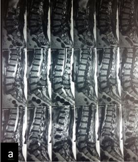
Methods: We evaluated 30 patients operated for DDD with TLIF between 2014 to 2017. Patients > 35 years, both sexes, two level or less involvement, degenerative spondylolisthesis (grade I, II), with predominantly low-back pain, with or without radiculopathy or claudication, disability to perform daily activities and not relived by non-operative treatment for at least 6 months were included. All other cases were excluded. Thorough clinical and radiological examination was done.

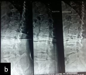
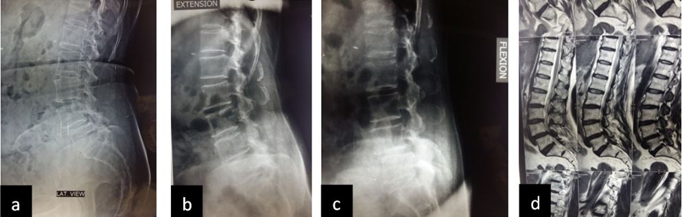
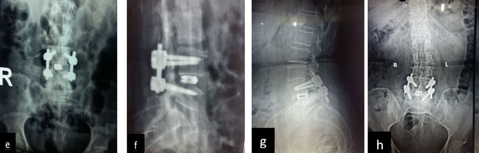
Patient was followed up at 1, 3, 6 and 9 months post-op for X-ray (to see for progress of union), VAS score and ODI index and complications. Bony fusion was assessed by a single radiologist on basis of X-ray only.

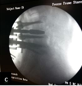
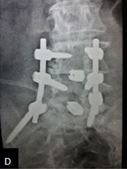
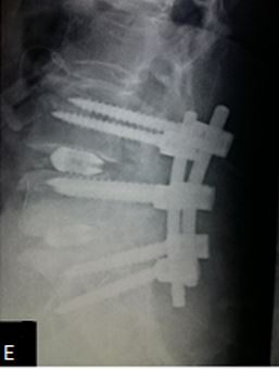
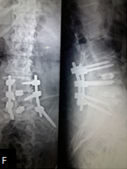
Results: 17 M 13 F patients with 19 patients having instability while 11 not, were evaluated. L4-5 was the most common level. Average pre-operative VAS score was 7.7667 (S.D 1.104) while at last follow-up was 2.133 (S.D 0.434). Average pre-operative ODI was 47.133 (S.D 8.215) while at last follow-up it was 25.533 (S.D 4.191) (Table-2). Mean operative time for one and two level was 97.3 minutes and 143.2 minutes respectively. Average blood loss was 465 ml (390- 580ml). 28 patients had bony fusion at last follow-up (93 %). Two patients who did not show bony fusion were asymptomatic. We encountered intra-operative violation of S1 pedicle in one case, dural puncture in three cases, contra-lateral radiculopathy in one case and asymptomatic adjacent segment disease in 4 cases at final follow-up.

Conclusion: We conclude from our study that TLIF is simpler, easier and safe procedure for Degenerative Disc Disease with good surgical, functional and radiological outcomes.

Keywords: Degenerative disc disease, Interbody Fusion, TLIF

Address for Correspondence: Dr. Ashish Patidar, Assistant Professor, Department of Orthopaedics, R D Gardi Medical College, Ujjain, Madhya Pradesh, India

Email: patidarashish1978@gmail.com


How to site this article: Patidar A, Mehta R, Sharma S, Singh V, Chauhan A. Prospective analysis of clinico-radiological efficacy of Trans-foraminal Lumbar Interbody Fusion (TLIF) in degenerative disc disease - Mid term follow up of 2 years. OrthopJMPC 2017;23(2):3-10.


Introduction

Low back pain as a result of degenerative disc disease (DDD) imparts a large socioeconomic impact on the health care


system. Correct diagnosis and treatment of DDD is difficult and controversial. Patients with DDD and discogenic back pain presents with symptoms that range from mild Low back pain (LBP) to excruciating back pain with lower extremity symptoms. Continued degeneration of the affected disc can lead to secondary problems such as degenerative spondyolisthesis, lumbar canal stenosis and facetal arthropathy. Moreover, diagnostic evaluation of chronic low back pain is complex because of multiple pain generators that are difficult to identify [1]. Various non-surgical and surgical modalities of treatment are available for management of DDD.  Surgical interventions include fusions, nucleoplasty, disc arthroplasty and dynamic stabilization procedures. Whether inter body fusion is the treatment of choice in DDD is still a dilemma. Interbody arthrodesis may improve the clinical results by eliminating the disc as a potential pain generator, improving fusion rate and restoring intervertebral disc height and lumbar lordosis. Reconstruction of anterior column can be performed via anterior approach (trans-peritoneal, retro-peritoneal) posterior fusion and instrumentation to achieve 360-degree fusion. An alternative method to reconstruct anterior column is via posterior lumbar interbody fusion with lesser complications. The Transforaminal interbody fusion (TLIF) developed by Harms is a modification of posterior lumbar interbody fusion (PLIF). Advantage of the TLIF over PLIF with lesser complications avoidance of epidural scarring, less intraoperative bleeding, and lesser chance of dural injury [2]. Over past few years TLIF has gained popularity over ALIF and PLIF. Hence, we studied the clinical, functional and radiological outcomes of TLIF and complications associated with it.

Materials and Methods

After obtaining permission from the Ethical Committee of the Institution, from 2014 to 2107, We studied thirty cases of degenerative disc disease at tertiary care hospital. Aim of the study was to evaluate the role of TLIF in alleviating chronic LBP and leg pain if present in cases of DDD. Patients with chronic LBA (more than 6 months), with failed conservative treatment, wit/without radiculopathy, with reduced inter-vertebral disc space on x-ray and MRI were considered as having DDD. Patient selection was on the basis of following criteria: Age > 35 years, both sexes, less than or equal to two level involvement, degenerative spondylolisthesis (grade I and II) and Patients with predominant symptoms of low-back pain with radicular pain to one or both lower limb or canal stenosis, disability to perform daily activities and not relived by non-operative treatment for at least 6 months. Patients excluded from the study were those with grade III and IV spondylolisthesis, Patients with infection, tumor or revision cases and patients with other co-morbidities making patient unfit for surgery. All patients underwent thorough clinical evaluation. On the basis of displacement in flexion and extension lateral views, grading of listhesis was done. Patient’s pre-operative Visual Analogue Score (VAS) score and Oswestry Disability Index (ODI) were recorded for comparing them with post-operative results. After pre-anesthetic evaluation and obtaining patients consent, patients were posted for TLIF procedure.

Operative technique:

Under general anesthesia, in prone position the level/levels involved were confirmed under image intensifier. A midline incision was made and posterior elements down to the tips of transverse process were exposed sub-periostealy. Pedicle screws were placed bilaterally under fluoroscopic guidance. After interlaminar decompression, unilaterally, on the side of radicular pain, inferior facet of rostral vertebra and the superior facet of the caudal vertebra were resected to expose the disc after fixing a rod to the pedicle screw on the opposite side. Once the working window is created, the exiting and traversing nerve roots were decompressed and protected. Complete disc along with the superior and inferior end plates were removed with straight and angled curettes.  Disc space is increased by distracting the pedicle screws on the opposite side. After confirming the size of disc space with serial dilators and fluoroscopy, appropriate sized banana cage packed with bone graft obtained intra-operatively is inserted and placed antero-centrally.  Area inside the annulus and around the cage is packed with remaining graft. Second connecting rod is connected and the level is compressed. After thorough wash, wound is closed over drain in layers and sterile dressing applied. After 48 hours drain is removed. Patient is mobilized out of bed after day 2 as pain tolerated. I.V antibiotics were given till fifth post-operative day. Post-operative x-rays were done and after suture removal patients were discharged.

Patient was followed up at 1,3, 6 and 9 months post-op. At follow-up, X-ray (to see for progress of union), VAS score and ODI index, complications if any were noted and treated and these variables were used to determine the surgical and functional efficacy of the treatment. Bony fusion was assessed by a single radiologist on basis of X-ray only.

Results

We studied 17male and 13female patients with maximum age 66 years and minimum age 37 years (average: 49 years). Of 30 patients, 19 patients had degenerative disc disease with spondylolisthesis while 11 had only symptoms of degenerative disc disease and no listhesis. L3-L4 was involved in 4 patients, L4-L5 in 11 patients, L5-S1 in 9 patients and L4-L5-S1 in five patients and L3-L4-L5 in one patient (Table-1).


Table-1: vertebral level

Level

Frequency

Percent

Valid

L3-L4

4

13.3

L4-L5

11

36.7

L4-L5-S1

5

16.7

L5-S1

9

30.0

L3-L4-L5

1

3.3

Total

30

100.0


Average pre-operative VAS score was 7.7667 (S.D 1.104), at 3 months post-op was 2.633 (S.D 0.764) while at last follow-up was 2.133 (S.D 0.434). Average pre-operative ODI was 47.133 (S.D 8.215), at 3 months post-op was 26.066 (S.D 5.394) while at last follow-up it was 25.533 (S.D 4.191) (Table-2). Mean operative time for one and two level was 97.3 minutes and 143.2 minutes respectively. Average blood loss was 465 ml (390- 580ml). As assessed by the radiologist, 28 patients had bony fusion at last follow-up (93 %). Two patients who did not show bony fusion were asymptomatic and hence not intervened.  No implant loosening or cage migration was noted in any case. We encountered intraoperatively violation of S1 pedicle in one patient, so for salvage we put iliac screw on that side, Dural puncture was encountered in three cases, which was repaired with 5-0 proline and fat pad was placed over it. Air tight closure was done and post-operatively patient was put on oral Acetazolamide 250 mg BD for 5 days. 4 patients had superficial infection at operative side which was managed with regular dressings and oral antibiotics. One patient had symptoms of contra-lateral radiculopathy post-operatively which was managed conservatively. Adjacent segment DDD was noted in 4 cases radiologically but not clinically. There was no case of cage subsidence till the last follow-up.


Table -2: Paired Samples Statistics


Mean

N

Std. Deviation

t

P


47.1333

30

8.21577

17.665

0.000

21.5333

30

4.19140


7.7667

30

1.10433

25.349

0.000

2.1333

30

0.43417


pic 1.jpg


C:\Users\SPINESURGEON\Desktop\1.png

Figure 1.

(a) MRI image of 60 yr female with degenerated disc L4-L5-S1. (b) X-ray of LS spine flexion extension view showing degenerative listhesis. (c) intraoperative C-arm image showing fixation. (d-e) Post-operative images showing interbody fusion at L4-L5-S1 level. (f)one year follow up X-ray showing consolidation


pic 3.jpg


pic 4.jpg

pic 5.jpg

Figure 2.

(abc) X-ray images of 45/F lateral, flexion and extension views showing instability at L4-L5 level. (d)MRI image. (e-f) Post-operative images showing interbody fusion at L4-L5 Level. (g-h) Two year follow up images showing fusion.

Discussion

The most common symptom with which patients with DDD present is low back pain, although radicular pain and neurogenic symptoms may occur secondary to neural compression. Two major pain generating structures in DDD include Inter Vertebral Disc and facet joint. Back pain arising from spinal IVD is called discogenic pain [3]. Conservative therapy and needle based treatment i.e. injections and rhizotomy, are highly effective in treating back pain from arthritic facets but discogenic pain does not responds well to it [4-7]. Studies in literature conclude that discogenic pain of DDD can be successfully alleviated by surgical intervention in form of fusion, arthroplasty or disc repair [8-10]. In our study of 30 cases of DDD treated with TLIF + Posterior Spinal Fusion we achieved significant improvement in patient’s daily chronic low-back pain. Deng-Lu Yan et al also found that lumbar pain improved in 83.5% of patients in their study [11].

In cases of degenerative listhesis, goal of surgery is stabilization of motion segment by fusion and decompression of neural elements. Interbody fusion by restoring the discs space height indirectly decompresses the neural foramen. As compared to postero-lateral fusion in which the graft is under tensile stress, in interbody fusion the graft is under compression and hence the chances of fusion are increased. Various approaches for lumbar fusion are advocated in literature, TLIF technique has become increasingly popular since introduce by Harms in 1982 and now being in current trend. Humphreys made a comparative study of PLIF with TILF 34 and 40 cases respectively. The authors conclude that the TLIF showed to be good alternative to PLIF with relatively less risk of complications. As compared to PLIF in TILF chances of injury due to retraction of neural elements are minimal, less operating time as well less significant reduction in blood loss. PLIF is limited to levels L3 to S1 since excessive retraction of thecal sac at higher level may cause neurological damage. TLIF can be performed safely from posterior and unilateral approach and hence contralateral side available for revision. Complications like injury to iliac vessels, hypogastric plexus and greater blood loss seen in ALIF are avoided with TLIF [9].

Zhang kai et al (2014) in their study found the mean post-operative VAS score 2.3+0.7 which correlates with our mean post-operative VAS score [10]. In our study we found significant improvement in the VAS and ODI postoperatively which supports the utility of TLIF in DDD as concluded by other studies [12-15].

Lowe and Tahernia in their study on 40 patients underwent TILF surgery reported fusion rate radiologically was 95% of cases [16]. Zhang kai et al (2014) reported solid fusion in 90.29% [10]. Fusion rates in our study 93% are comparable to other studies (89% to 100 %) although criteria of defining fusion may be different [17-19]. France et al looked at instrumented vs un-instrumented fusion in a prospective study and found that instrumentation improved the fusion rate but it did not correlate with clinical outcome [20]. Kanyana et al assessed instrumented and un instrumented lumbar fusion and found that instrumentation resulted in higher fusion rates at 8 weeks compared to the uninstrumented group but at 16 weeks the fusion rates were the same in both groups [21].

We encountered 3 cases of dural puncture intra-operatively which we repaired. Rosenberg and Maummaeni (2001) reported incidental dural tear in one patient that repaired but needed revision surgery for post-operative dural leak [22]. McAfee et al (2005) found 7 cases of dural tear (120 patients) during cage insertion and nerve root decompression [23]. 4 had superficial infection at operative side which was managed with regular dressings and oral antibiotics. One patient had symptoms of contra-lateral radiculopathy post-operatively which was managed conservatively [24]. Adjacent segment DDD was noted in 4 cases radiologically but not clinically. There were no cases of cage subsidence or implant loosening till last follow-up. Adjacent segment DDD was noted in 4 cases radiologically but not clinically.  Adjacent segment disease is a known complication occurring in the level above the fused vertebra due to increased stress and hypermobility. It causes facetal and ligamentum flavum hypertrophy leading to canal and foraminal stenosis at a later stage [25]. The incidence of postoperative Adjacent Segment Disease (up to 30%) is greater following either open or Minimally Invasive Surgery instrumented lumbar fusions (e.g., TLIF/PLIF), while decompressions with non-instrumented fusions led to a much smaller 5.6% risk of ASD [26-27]. In our series, the rate of asymptomatic ASD was 13 %.

Conclusion

We conclude from our study that TLIF is simpler, easier and safe procedure for DDD with good surgical and functional outcomes.


References

1. 

2.                Carragee EJ, Hannibal M (2004) Diagnostic evaluation of back pain. OrthoClin North Am(1):7-16.

3.                Harms J, Jeszenszky D (1998). The unilateral transforaminal approach for posterior lumbar interbody fusion. OrthopTraumatol 6:88-99.

4.                Krock E, Rosenzweig DH, Chabot-Doré AJ, Jarzem P, Weber MH, Ouellet JA, Stone LS, Haglund LJ Cell Mol Med. Painful, degenerating intervertebral discs up-regulate neurite sprouting and CGRP through nociceptive factors.  2014 Jun; 18(6):1213-25.

5.                Hancock MJ, Maher CG, Latimer J, Spindler MF, McAuley JH, Laslett M, Bogduk N. Systematic review of tests to identify the disc, SIJ or facet joint as the source of low back pain. Eur Spine J. 2007 Oct; 16(10):1539-50.)

6.                Shemshaki H, Nourian SM, Fereidan-Esfahani M, Mokhtari M, Etemadifar MR J. What is the source of low back pain?  Craniovertebr Junction Spine. 2013 Jan; 4(1):21-4.)

7.                D'Orazio F, Gregori LM, Gallucci M Eur J Radiol. Spine epidural and sacroiliac joints injections--when and how to perform. 2015 May; 84(5):777-82.)

8.                Tessitore E, Molliqaj G, Schatlo B, Schaller K. Clinical evaluation and surgical decision making for patients with lumbar discogenic pain and facet syndrome. Spine (Phila Pa 1976) 2014;39:1314–24.

9.                Watters WC 3rd, Resnick DK, Eck JC, Ghogawala Z, Mummaneni PV, Dailey AT, Choudhri TF, Sharan A, Groff MW, Wang JC, Dhall SS, Kaiser MG J. Guideline update for the performance of fusion procedures for degenerative disease of the lumbar spine. Part 13: injection therapies, low-back pain, and lumbar fusion. Neurosurg Spine. 2014 Jul; 21(1):79-90.

10.                Humphreys SC, Hodges SD, Patwardhan AG, Eck JC, Murphy RB, Covington LA. Comparison of posterior and transforaminal approaches to lumbar interbody fusion. Spine (Phila Pa 1976) 2001;26:567–71.

11.                Zhang Kai, Sun Wei,Zaho Chang-qing, Li Hua, Ding Wei, Xei You-zhuan, Sun Xiao-jiang & Zaho Jie: International orthopaedics (SICOT)38:111-116.(2014).

12.                Deng-Lu Yan, Fu-Xing Pei, Jian LI, Cheng-long Soo (2008) Comparative study of PILF and TLIF treatment in adult degenerative spondylolisthesis. Eur spine J 17(10):1311-1316.

13.                Sloboda T, Rusnak R: Minimally invasive transforaminal fusion (mitlif) for the treatment of the degenerative lumbar spine (Single centre study). Int Health Journal. 2016 March;1:17-20

14.                MJD Jacobsohn; RN Dunn. Transforaminal lumbar interbody fusion (TLIF): Assessment of clinical and radiological outcome, SA orthop. j. vol.8 n.1 Pretoria Jan. 2009

15.                Seng-Yew Poh, Wai-Mun Yue, Li-Tat John Chen, Chang-Ming Guo, William Yeo, Seang-Beng Tan. Two-year outcomes of transforaminal lumbar interbody fusion, Journal of Orthopaedic Surgery 2011;19(2):135-40)

16.                Pier Paolo Mura, Mauro Costaglioli, Maurizio Piredda, Silvia Caboni , Silvia Casula, TLIF for symptomatic disc degeneration: a retrospective study of 100 patients. Eur Spine J (2011) 20 (Suppl 1):S57–S60)

17.                Lowe T, Tahernia A (2002) Unilateral transforaminal posterior lumbar interbody fusion procedure. Orthopaedics 25: 1179-1183.

18.                Khan NR1, Clark AJ, Lee SL, Venable GT, Rossi NB, Foley KT. Surgical Outcomes for Minimally Invasive vs Open Transforaminal Lumbar Interbody Fusion: An Updated Systematic Review and Meta-analysis.  Neurosurgery. 2015 Dec;77(6):847-74)

19.                Lauber S, SchulteTL, Liljenqvist U, Halm H, Hackenberg L. Clinical and radiologic 2-4-year results of transforaminal lumbar interbody fusion in degenerative and isthmic spondylolisthesis grades 1 and 2. Spine (Phila Pa 1976) 2006;31:1693–8)

20.                Alsebaey AA, Seleem NA (2017) Transforaminal Lumber Interbody Fusion in Management of Degenerative Lumber Disc Disease. Research Article 8(3): 00316. DOI: 10.15406/mojor.2017.08.00316

21.                France JC, Norman TL, Santrock RD, McGrath B, Simon BJ. Randomised prospective study of posterolateral fusion: Outcome with and without pedicle screw instrumentation. Spine 1999 March 15;24(6):553-60.)

22.                Kanyana M, Cunningham BW, Sefter JC, Goldstein JA, Stewart G, Kaneda K. Does spinal instrumentation influence healing process of posterolateral fusion? An in vivo animal model. Spine 1999 June 1;24(11):1058-65.)

23.                Rosenberg WS, MummaneniPV.Transforaminal lumbar interbody fusion: technique, complications, and early results, Neurosurgery.75-48:569;2001

24.                McAfee PC, DeVine JG, Chaput CD, Prybis BG, Fedder IL, Cunningham BW, Farrell DJ, Hess SJ, Vigna FE. The indications for interbody fusion cages in the treatment of spondylolisthesis: analysis of 120 cases. Spine (Phila Pa 1976). 2005 Mar 15;30(6 Suppl): S60-5.

25.                Hunt, T., Shen, F.H., Shaffrey, C.I. et al. Eur Spine J (2007) 16(Suppl 3): 311.

26.                Ghiselli G, Wang JC, Bhatia NN, Hsu WK, Dawson EG. Adjacent segment degeneration in the lumbar spine. J Bone Joint Surg Am 2004; 86:1497–503.

27.                Park P, Garton HJ, GalaVC, Hoff JT, McGillicuddy JE. Adjacent segment disease after lumbar or lumbosacral fusion: review of the literature. Spine (Phila Pa 1976) 2004; 29:1938–44.

28.                Adjacent level disease following lumbar spine surgery: A review. Nancy E. Epstein. SurgNeurol Int. 2015; 6(Suppl 24): S591–S599.