Jati S, Singh P, Jain N: Centralization of Ulna in Treatment of Giant Cell Tumor of Distal Radius - A Case Report


Centralization of Ulna in Treatment of Giant Cell Tumor of Distal Radius - A Case Report


Jati S, Singh P, Jain N


Investigation performed at Department of Orthopaedics, Sri Aurobindo Medical College and Post Graduate Institute, Indore, Madhya Pradesh, India


Abstract

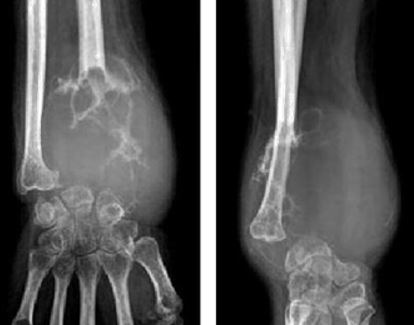
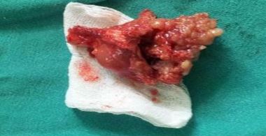
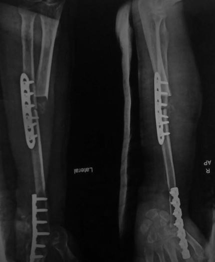
Distal end radius is third most common site for Giant Cell Tumour.  Various Options for reconstructions are available.  In this case report we assessed Functional outcome of centralization of ulna in aggressive giant cell tumour of Distal end radius.

Address for Correspondence:Dr Saket Jati, Dept. of Orthopaedics, SAIMS, Indore-452003, India.

Email:saket555@gmail.com


How to site this article:Jati S, Singh P, Jain N. Centralization of Ulna in Treatment of Giant Cell Tumor of Distal Radius - A Case Report. OrthopJMPC 2016;22(2): 51-53.


Introduction

Giant cell tumour is a solitary benign locally aggressive lesion characterized by osteoclast like multinucleated giant cell mainly involving epiphysis. They are 4-5% of primary bone tumours and 20% of benign bone tumours [1]. Aggressiveness of this lesions is between purely benign and frankly malignant. It occurs in patients 20 to 40 years old. The most common location for this tumour is the distal femur followed closely by the proximal tibia and the distal radius [2]. Patient usually presents late with pain, local swelling and limited range of motion at wrist and associated pathological fracture locally, pulmonary metastases occur in approximately 3% of patients [2]. Treatment of Giant cell tumour in distal radius is difficult, mainly because of their close proximity to multiple tendons, nerve, radial artery and carpal bone. Radiographic findings often are diagnostic. The lesions are eccentrically located in the epiphyses of long bones and usually about the subchondral bone. Management of giant cell tumour of distal end


radius mentioned in literature are, curettage with polymethylmethacrylate cement, en bloc excision and reconstruction with fibular autografts, allografts or ulnar centralization however the aims of treatment are complete removal of the tumour while preserving maximum function of the limb [3,4].

Case Report

35year Normotensive Euglycemic male presented to us with complains of swelling and restricted range of motion at Right wrist since last 6 months for which curettage and cementing was done 4 month back somewhere else. Patient was asymptomatic for next 2 months’ post procedure but gradually swelling reappeared with restricted movement at wrist, since last one month patient complains of pain which increases on performing any day to day activity and relives with rest. Patient had no history of trauma, fever, weight loss or loss of appetite.

On examination old healed scar present on dorso- lateral aspect of right wrist, a gross,

firm and tender swelling of size 8 x 6 cm was seen on dorso- lateral aspect of distal radius with feeling of egg shell cracking, all wrist movement were restricted and painfulwith no Distal neurovascular involvement.

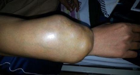
Figure 1: Clinical Picture

Radiograph of wrist revealed an eccentric lytic, multiloculatedlesion in epiphyseo-metaphyseal region of distal radius giving soap bubble appearance without periosteal reaction or soft tissue involvement.

Figure 2: Pre Op Radiograph

Patient was offered wide resection and reconstruction with ulnar centralization. Tumour was exposed by dorsal approach upto 2nd metacarpal a wide resection was the done with margins of normal tissues. Approximately 10 cm of the distal radius along with parts of the radio-scaphoid, radio-lunate capsules and distal radio-ulnar joint capsules was resected, distal ulna was freed of cartilage, the lunate was freshened.Tumour bed was treated with 5% phenol and 3% hydrogen peroxide to take care of the inadvertent spillage. The wrist translocated so as to place the lunate in line with the distal ulna. Stabilization was achieved by means of locking plate at radio ulnar junction and a reconstruction plate applied to ulna across the wrist and over the second metacarpal.

Figure 3: en bloc excision of distal end radius

Figure 4: Postoperative X-ray

Patient was followed up for 18 months with no local recurrence, with good functional outcome assessed by Revised Musculoskeletal Tumour Society scoring system for upper extremity (MSTS score) [5]. Union at Radio ulnar junction was evident at 4 months and union at ulno carpal junction was evident at 6 months withoutany additional procedure.

Discussion

GCT of the distal radius can be managed by excision of the tumour while preserving good function of the wrist and hand. It has been suggested that intra-lesion curettage may be preferred to reduce the incidence of local recurrence [6]. However O Donnell R. J. et al. reported high rate of recurrence [7].Factors that needed to be considered when evaluating a technique of reconstruction includes the ease of the procedure, its morbidity, the complications and functional outcome and the durability of the reconstructed segment.

En bloc excision and reconstruction with fibular autograft, allograft or ulnar centralization is preferred choice of treatment for giant cell tumour of distal end radius. However, the disadvantage, of en bloc excision and reconstruction with fibular autograft or allograft includes donor site morbidity, delayed union or non-union [8]. Also, allografts may not be available in all orthopaedic setups and there is at least a theoretical risk of transmission of viral diseases [9].Centralization of ulna is relatively shorter surgical procedure and use of local vascular graft possibly help to reduce infection and improves the chances of union. The complications cited with this procedure includes ankylosis of the wrist, damage to neural structures, vascular insufficiency of the hand, wound infection, necrosis of wound margins, fracture of the ulna, and hardware failure. However, we did not note any of these complications in our case over the period of 18 months. Local recurrence of tumours is not a reflection of the technique of reconstruction which has been used.

Conclusion

En bloc excision with centralization of ulna is easy, less expensive method for reconstructing of aggressive giant cell tumour of distal end radius, which provides fewer complication, good functional results. However local recurrence rate cannot be predicted.

References

1.                Campanacci M, Baldini N, Boriani S, Sudanese A. Giant-cell tumor of bone. J Bone Joint Surg [Am] 1987;69-A:106-14.

2.                Canale, S. T., Beaty, J. H., & Campbell, W. C. (2013). Campbell's operative orthopaedics. Philadelphia, PA: Elsevier/Mosby.

3.                Harness NG, Mankin HJ. Giant-cell tumor of the distal forearm. J Hand Surg [Am] 2004;29:188-93.

4.                Gamberi G, Serra M, Ragazzini P, Magagnoli G, Pazzaglia L, Ponticelli F, Ferrari C, Zanasi M, Bertoni F, Picci P, Benassi M (2003). "Identification of markers of possible prognostic value in 57 giant-cell tumors of bone". Oncol Rep10 (2): 351– 6.

5.                Enneking WF, Dunham W, Gebhardt MC, Malawar M, Pritchard DJ. A system for the functional evaluation of reconstructive procedures after surgical treatment of tumors of the musculoskeletal system. ClinOrthopRelat Res. 1993;286:241– 246.

6.                Cheng CY, Shih HN, Hsu KY, Hsu RW. Treatment of giant cell tumor of the distal radius, ClinOrthop 2001;383:221-8.

7.                O Donnell R.J, MotwaniH.k.  Recurrence of giant-cell tumors of the long bones after curettage and packing with cement J Bone Joint Surg [Am 1994 Dec;76(12):1827-33.

8.                Saikia KC, Borgohain M, Bhuyan SK, Goswami s, Bora A, Ahmed F. Resection-reconstruction arthroplasty for gaint cell tumor of distal radius. Indian J Orthop. 2010;44(3):327-32.

9.                Liu YP, Li KH, Sun BH. Which treatment is the best for giant cell tumors of the distal radius? A meta-analysis. Clip OrthopRelat Res. 2012;470(10):2886-94.

10.