Varma H S, Sirsikar A.:Bilateral Tuberculous Osteomyelitis of Calcanei: A Case Report


A Child presenting with Bilateral Tuberculous Osteomyelitis of Calcanei: A Case Report


Varma H S, Sirsikar A.


Investigation performed at Department of Orthopaedics, NSCB Medical College, Jabalpur, Madhya Pradesh, India


Abstract

Introduction:Osteoarticular tuberculosis represents 1.7-2% of all tuberculosis. The localization in the foot is rare and accounts for less than 10% of osteoarticular tuberculosis. The clinical observations of this case highlight the unusual bilateral skeletal involvement of tuberculosis.

Case Report: A case of a six year old male child presenting with swelling of both heels and discharging sinus is reported. Biopsy of the curetted material from both lesions revealed tuberculous nature. Patient was kept on conservative treatment of antitubercular drugs.

Conclusion: A high index of suspicion is mandatory for early diagnosis, prompt treatment and better clinical outcome.

Key words: calcaneus, tuberculosis, osteoarticular.

Address for Correspondence:Dr. H S Varma, Department of Orthopaedics, NSCB Medical College, Jabalpur, M.P. 482003, India.

Email:hsverma@rediffmail.com


How to site this article:Varma H S, Sirsikar A.A Child presenting with Bilateral Tuberculous Osteomyelitis of Calcanei: A Case Report. OrthopJMPC 2016;22(2): 47-50.


Introduction

Tuberculous osteomyelitis of calcaneum is a very rare entity and has been very scarcely reported. The stress on study of calcaneal tuberculosis has also been diluted by the fact that most of the times tuberculosis of tarsals involves more than one bone and joints of the region. Thus, its awareness is low and diagnosis is often delayed. Furthermore, tuberculosis of the calcaneum is debilitating if untreated and delayed treatment may lead to functional disability.

Case Report

A six year old male child presented with complaints of pain, swelling and discharging sinus at both heels for duration of four months. It was associated with mild fever. Child had previously been evaluated at Primary health centre and Incision and Drainage was done on the right side. While the incision healed, a sinus remained at the distal most part of incision. Patient developed a sinus on left side later, and was referred to this hospital for non-healing sinuses. His parents did not give history of treatment of tuberculosis and had no family history of tuberculosis. The boy had received routine vaccinations including BCG.

On Examination, patient had swelling of both the heels for four months and discharging sinuses over both heels for three months. Swelling involving the heel region, was initially for one and half months, was associated with pain which later subsided. He had history of night sweats, evening rise of temperature in initial one month. No history of loss of weight or systemic illness was given by the parents.

Physical examination revealed temperature 97.40F, pulse 108/min., respiratory rate – 24/min. He had mild pallor. His body weight was 15 kgs. His inguinal lymph nodes were enlarged on both sides. Both feet had swelling in the heel region with sinus placed at posterolateral aspect with minimal discharge of serosanguinous fluid. Swelling was diffuse over both heels more on the lateral and posterior aspects, below and behind the lateral malleoli on both sides, of size about 5 cm. x 3 cm. x 1 cm. with skin discolouration due to increased pigmentation.

C:\Users\cse\Desktop\1.jpg

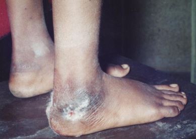
Figure 1: Clinical Picture Right Heel

C:\Users\cse\Desktop\2.jpg

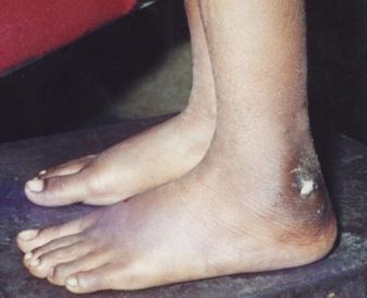
Figure 2: Clinical Picture Left Heel

Sinuses were of size about ½ x ½ cm. with no active discharge but a minimal soakage. The sinuses were present about 1 cm. below and behind the lateral malleolus on left side while 2 cm. below and behind lateral malleolus on right side (Figure 1 & 2). On the right side it was present over the distal most and of an oblique healed incision of previous operation. Sinus had their edges undermined, some granulation tissue over right side. The skin around the sinuses was excoriated and showed hyperpigmentation.Movements of both ankles were free while inversion was restricted and eversion absent.

Radiographs revealed central osteolytic lesions in both calcanei of size about 1 and ½ cm. x 1 cm. (Figure 3), with marginal sclerosis and osteoporosis of whole calcanei.


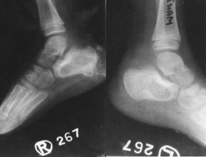
Figure 3: X ray of Both Heel

Laboratory data included, haemoglobin 10 gm%, total leukocyte count 9,600/mm3 with 62% polymorphonuclear cells, 36% lymphocytes and 2 % eosinophils. His ESR was 5mm at one hour and his urinalysis was normal.Biopsy taken through both sinus tracts with the help of a thin curette revealed tuberculous lesion.

Treatment consisted of chemotherapy with Rifampicin 10 mg/kg. body weight once daily empty stomach, Isoniazid 5 mg/kg body weight once daily, Ethambutol 25 mg/kg body weight once daily and Pyrazinamide 35 mg/kg body weight in two divided doses. A regular vitamin “B” complex supplementation too was prescribed. Patient was discharged with advice for bed rest and follow up every month to see his progress, clinically and radiologically.

Over a period of three months, his sinuses healed and the patient could stand and walk without pain and showed no relapse after a 18 months follow-up.

Discussion

Osteoarticular tuberculosis is a leading cause of morbidity and mortality in many developing countries and actually in the entire world due to the increased incidence of TB in HIV-positive patients. Osteoarticular tuberculosis represents 1.7-2% of all forms of TB. It is the third amongst extrapulmonary TB after the peritoneal and lymph node forms of TB. It is a disease that must be diagnosed quickly before the disease progresses and injures or destroys the bone and joint.

In osteoarticular tuberculosis, the spine and the hip joint are the most commonly involved areas, with the ankle and foot being rarely involved and represent only 1% of all infections of TB and about 10% of osteoarticular involvements. Clinical presentation of calcaneal tuberculosis is non-specific and thus diagnosis is often delayed. The tubercular sinus can also be infected and this may lead to misdiagnosis [1,2].

Tuli (1991) reports 1% to 3% incidence of osteoarticular involvement of all the tuberculosis cases. In his series of 980 patients, he found 3.26% cases with involvement of calcaneum, however, he has not distinguished how many of them were involving calcaneum alone and how many had other bones and joints involved too[3].

                Wilkinson (1964) states that tuberculosis does not distinguish different joints and bones forming the foot and usually affects several of them. He further states that in infants it mainly involves metatarsals, in children tarsal bones; while ankle becomes more susceptible as the ossification advances[4].

                Bosworth (1959) states that in round bones the pathology of tuberculous affection is same as that in diaphysis of long bones i.e. expansive central lesion due to slow development of the disease and relative softness of cortical structures in children [5].


Shannon (1990) stated that in adults skeletal tuberculosis involves spine in 50% cases, hip in 15% and ankle, wrist, elbow and others in 20% cases. He also quotes report of American Academy of Paediatrics which says that resistance to Isoniazid is 15% in south east Asia. A third drug is therefore recommended in first two months [6].

Tuli (1991) advocates at least one month of drug therapy and general treatment before any major surgery is done. Antituberculous drugs in their doses according to body weight of the patient could be followed by appropriate surgery if it seems necessary.

In this institution, we use four drugs up to 2 months, two drugs for next 10 months. Dosages are calculated according to the body weight of the patient and in child have to be reviewed in every follow-up with weight measurement, complete hemogram & ESR. An X-ray after every 2 to 3 months is also required. Surgery can be planned after one month of anti-tuberculous treatment and after assessing the patient for any clinical or radiological improvement [7].


Conclusion

The location of tuberculosis at the calcaneum is extremely rare. The diagnosis and treatment of calcaneal tuberculosis are often delayed because of the unawareness of the surgeon and less dramatic signs and symptoms of calcaneal osteomyelitis than osteomyelitis of long bones. Thus a high index of suspicion is mandatory for early diagnosis, prompt treatment and better clinical outcome. The clinical observations of this case highlight the unusual bilateral skeletal involvement of tuberculosis and should be considered to prevent delay in diagnosis and therapeutic management.


References

1.                Anil Agarwal, Kumar Shashi Kant, Tarun Suri, Neeraj Gupta, IndreshwarVerma, Abbas Shaharyar. Tuberculosis of the calcaneus in children. Journal of Orthopaedic Surgery 2015;23(1):84-9.

2.                Lamiae Chater, MounirArroud, MoulayAbderrahmaneAfifi. Tuberculosis of the calcaneus in children. International Journal of Mycobacteriology 3 (2014),57-59.

3.                Tuli SM. In: Tuberculosis of the Skeletal System (Bones, Joints, Spine and Bursal Sheaths). 2ndEdn, Jaypee Brothers Medical Publishers (P) Ltd., New Delhi, 1997; 115.

4.                Wilkinson M. C. The treatment of Bone and Joint Tuberculosis, Ann. R. Coll. Surg. Engl. 37:19, 1965.

5.                Bosworth D. N. Treatment of bone and joint tuberculosis in children. J. Bone and Joint Surgery, 41-A, No. 7, 1959.

6.                Shannon F. B. et al. Multifocal Cystic Tuberculosis of Bone. J. Bone and Joint Surgery, 72-A, 1089, 1990.

7.                B. Swain, S. Mishra, K. Pattnaik, P. Dutta. Tuberculosis of Calcaneum: A case report. Ind. J. Tub. 2001, 48, 209-10.

1.