Varma H S. et al. : Double Bone Graft in Hemiarthroplasty for Fracture Neck of Femur.

Use of Double Bone Graft to Stabilize Prosthesis in Uncemented Partial Hip Replacement in Elderly Patients with Fracture Neck of Femur


Varma H S, Pandey K K, Peepra D, Rajpoot Manish S.


Investigation performed at Department of Orthopaedics, NSCB Medical College, Jabalpur, Madhya Pradesh, India


Abstract

Background: Partial hip replacement (cemented or uncemented) is the frequently done surgery in elderly patients with fracture neck of femur. Problems with cementing techniques are reported like increased infection rate and cardiac problems particularly in elderly patients with compromised cardiopulmonary reserve. Austin Moore prosthesis has its own complication like femoral loosening and frequent revision.
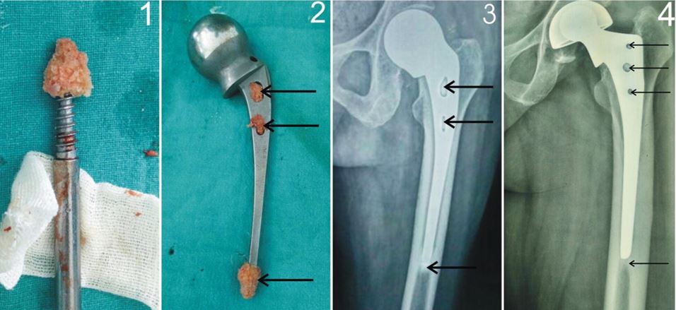
Material& Methods: We analysed results of 30 patients of fracture neck femur with Dorr type A and B proximal femoral morphology, treated by uncemented partial hip replacement (20 patients with  Austin Moore prosthesis,10 patients with  bipolar prosthesis) in elderly age group ( range 60 to 75 years) where double bone graft were used  for additional stabilization of the stem of prosthesis in femoral canal which included transverse peg grafts in fenestrations and conical graft over the tip of the stem of prosthesis to centralise the stem.

Results: The mean follow up was 9.3 years (range from 2 to 15 years).  According to Harris Hip score, final outcome was excellent in 21 hips, good in 7 hips and poor in 2 hips.Prosthesis subsidence was found in uncemented partial hip replacement with mean of 2.5 mm (range from 0 mm to 3.5 mm ) which was well tolerated  due to bone plugs formed   around the tip and fenestrations of prosthesis except in one patient who had complained of thigh pain which required revision with cemented modular prosthesis subsequently. Groin pain was reported in another one patient due to acetabular erosion which required cemented total hip replacement subsequently. There was no incidence of hip dislocation.

Conclusions: Our study shows that in addition to the transverse bar by fenestration grafts, bone block formed around the tip of the stem by conical graft provided strong pillar support and once it is integrated, it prevents excessive subsidence of the prosthesis with excellent result.

Key-words: Uncemented Bipolar prosthesis, uncemented Austin Moore Prosthesis, Double Bone graft.

Address for Correspondence:Dr Krishna Kumar Pandey HIG 27 Dhanwantrinagar , Jabalpur, M. P. 482003. India.

Email:drpandeykk@yahoo.com


How to site this article:Varma H S, Pandey K K, Peepra D, Rajpoot Manish S.Use of Double Bone Graft to Stabilize Prosthesis in Uncemented Partial Hip Replacement in Elderly Patients with Fracture Neck of Femur. OrthopJMPC 2016;22(2):37-41.


Introduction

Femoral neck fracture is the common hip fracture in elderly age group of patients.


Prosthetic replacement can be partial or total depending upon the case [1]. Uncemented Partial hip replacement with  unipolar Austin Moore Prosthesis is the treatment of choice in elderly patients with limited mobility but it is controversial in otherwise healthy patients of 60 years or older. For them reduction and internal fixation, cemented or uncemented bipolar partial hip replacement and total hip replacement are usually recommended.There is evidence in the literature supporting better functional outcome and less need for re-operation with hip arthroplasty compared to internal fixation in the treatment of displaced femoral neck fractures in the elderly. [2,3,4]

Austin Moore prosthesis is still commonly done in developing countries in elderly patients with low functional demand and poor candidate for revision surgery [5], having no failure history of about 20 years in few case reports. The responsible factor attributed for early failure is the technical error in implantation of the prosthesis [6].  Few studies suggest high incidence of femoral loosening and acetabular erosion with unipolar prosthesis. [7] Although Cemented prosthesis are claimed to avoid thigh pain but it has its own complication like transient hypotension, increasedinfection and cardiac problems particularly in patients with compromised cardiopulmonary reserve. [8, 9, 10]

In view of complication of cementing with cemented prosthesis and femoral loosening with unipolar (Austin Moore) prosthesis we have used double bone graft to fix the stem of prosthesis transversely by fenestration peg grafts and prepared conical graft at the tip of stem vertically to stabilise the prosthesis.

Material and Method

A total of 40 patients of the fracture neck of femur were operated by a senior orthopaedic surgeon in elderly age group (range 60 to 75 years), with uncemented bipolaror uncemented unipolar (Austin Moore) prosthesis. 30 patients met the required criteria for the study.10 patients were lost in the follow-up. Among 30 patients, 20 patients, comparatively older with less functional demand were treated with Austin Moore prosthesis and rest 10 patients, comparatively younger with  community ambulatory activity  with uncemented bipolar prosthesis. We included patients with Dorr type A and B proximal femoral morphology. We excluded patients with wide femoral canal (Dorr Type C femur) and also those patients having intra-operatively loose fitting of the stem that was felt during rasping of the proximal femur. Patients with loose fitting stem were excluded from the study and their surgery was completed with cemented prosthesis.

In our study, each patient was operated in lateral decubitus position with affected hip above and about 8 to 10 cm incision was taken with Moore approach. Skin, subcutaneous tissue and fascia were cut in the line of the incision. Gluteus maximus muscle fibres were split from anterior to posterior direction and retracted with hand held retractors. External rotators were cut in the single line of incision and secured.Capsule was cut in T shape manner, then head was extracted out and size of the prosthesis was measured.Bone graft was taken from the removed head of the femur, Conical graft was prepared with the help of femoral head extractor and its size was measured with K nail gauge. Fenestrations of the prosthesis were packed with bone graft and conical graft was fixed to the tip of the stem. Neck was prepared and rasping of the proximal femur up to two third of the length of stem was done with keeping proper ante version and lateralisation in mind. If the femoral canal was found press-fit for the stem, the prepared stem was implanted. If the femoral canal was felt wide for the stem, the patient was excluded from the study and cemented prosthesis was. The capsule and external rotators were repaired. Wound was closed in layers. Patients were mobilised to partial weight-bearing for six weeks and then allowed full weight-bearing without support. Active hip abduction exercises and quadriceps exercise were initiated postoperatively. Follow up was done at 6 weeks, 3 month, 6 month and then once a year for clinical and radiological evaluation. Among clinical criteria absence of pain and limp and ability to do daily living activities independently were assessed. Among radiological criteria sign of acetabular erosion and femoral stem subsidence (distance from the calcar to the prosthesis tip) were assessed.

Result

The mean follow up was 9.3 years (range from 2 to 15 years). There were excellent clinical and radiological results in all patients except in three patients. According to Harris Hip score, final outcome was excellent in 21 hips, good in 7 hips and poor in 2 hips. Prosthesis subsidence was found in uncemented partial hip replacement with mean of 2.5 mm (range from 0 mm to 3.5 mm) which was well tolerated due to bone plug formed around the tip of the stem and in the fenestrations of prosthesis except in one patient who had complaints of thigh pain due to femoral loosening which required revision with cemented modular bipolar prosthesis subsequently. Groin pain was reported in another one patient due to acetabular erosion which required cemented total hip replacement subsequently. There was no incidence of hip dislocation.


Figure 1 -Preparation of conical graft with the help of femoral head extractor

Figure 2 -Austin Moore prosthesis with fenestrations peg grafts and conical tip graft  

Figure 3- Arrows show the bone plug formed in fenestrations and around the tip of stem of Austin Moore Prosthesis in a patient with 10 year follow up

Figure 4 -Arrows show the bone plug formed in fenestrations and around tip of stem of bipolar prosthesis in a patient with 10 years follow-up


Discussion

Primary partial prosthetic replacement for recent fracture neck of femur is recommended in elderly ambulatory patients because of complications associated with internal fixation like nonunion and osteonecrosis. [11, 12]  

During procedure adequacy of calcar seating, residual femoral neck length and metaphyseal fill were kept in mind and errors in sizing the prosthesis were also avoided to prevent prosthesis failure. More than 12 mm length of the neck remnant, measured from the superior margin of the lesser trochanter to the resection margin at the calcar of femur ; Less than 1 mm for Calcar seating, measured from the medial prosthetic collar to calcar  and the relative metaphyseal fill greater than 70% of the stem of prosthesis in the medullary canal of femur at the level of tip of lesser trochanter on anteroposterior radiograph are  considered adequate. Equal prosthetic head size measured from femoral head gauge is considered satisfactory [13]

 Dorr’s classification [14] was used to assess anatomy of proximal femur with three types: - Type A - Good cortical bone stock with narrow medullary canal. We did rasping and sometimes reaming also.

Type B - Relatively good cortical support with wide medullary canal with no change of morphology. We prepared ourself for Bone graft impaction for such case and kept bone cement standby for those patients where stem fitting was felt loose. Once we did cementing, we excluded the case from our study.

Type C- Loss of much medial and posterior cortex with very wide medullary canal. We excluded these cases from beginning and did cemented prosthesis.

Hudson et al. noted significantly higher mortality rate with internal fixation than partial hip replacement in their study of 367 femoral neck fracture in patients with 65 to 80 years of age group, although revision rate in both method of treatment was similar. They also noted similar complications and revision rates in unipolar and bipolar prosthesis. [15]

Lu-Yao et al found 20% to 36% re-operation rate within 2 years after internal fixation and 6% to 18% re-operation rate in partial hip replacement group in their meta-analysis of 106 reports of displaced femoral neck fractures.[16] Bhandari et al in his meta-analysis in 1162 patients reported significantly less revision rate with patients with arthroplasty than patients with internal fixation group, although in arthroplasty group blood loss, infection rate , operative time and mortality were higher. [17] Rogmark et al. reported reported higher failure rate in internal fixation group and higher complication in arthroplasty group in their 2 year of prospective study of 409 ambulatory patients with 70 years or older having Garden stage III or IV fractures. [18] A Cochrane database survey with 17 trials involving 1920 patients showed no significant complication with cemented prostheses patients compared with cement less prostheses patients, at a mean follow-up of 1 year. Similarly, no significant differences were found between unipolar and bipolar hemiarthroplasty (seven trials, 857 participants, 863 fractures). [19] That’s why we used bipolar prosthesis and unipolar Austin Moore prosthesis in our study.

Although prosthetic replacement is reserved for patients 70 years old or older patients with a life expectancy of about 10 to 15 years but in our study reason for inclusion of patients younger than 70 years were old neck fracture due to long waiting list for operation theatre and inability to accept second revision surgery that is significantly common in patients with internal fixation. [15,16,17].

Conclusion

Our study showed that in addition to the transverse bar by fenestration grafts, bone block formed around the tip of the stem by conical graft provided strong pillar support and once it was integrated, it prevented subsidence of the prosthesis with excellent result.


References

1.                MaryaSKS,Thukral,SChandeep. Prosthetic replacement in femoral neck fracture in the elderly: Results and review of the literature.Indian J Orthop,2008 Jan-March;42(1):61-67.

2.                Davison JN, Calder SJ, Anderson GH, Ward G, Jagger C, Harper WM, et al. Treatment for displaced intracapsular fracture of the proximal femur: A prospective, randomized trial in patients aged 65 to 79 years. J Bone Joint Surg Br. 2001;83:206–12.  

3.                Parker MJ, Pryor GA. Internal fixation or arthroplasty for displaced cervical hip fractures in the elderly: A randomized controlled trial of 208 patients. Acta Orthop Scand. 2000;71:440–6.

4.                Roden M, Schon M, Fredin H. Treatment of displaced femoral neck fractures: A randomized minimum 5-year follow-up study of screws and bipolar hemi prostheses in 100 patients. Acta Orthop Scand. 2003;74:42–4.

5.                Nather A, Seow C, Lau P, Chan A. Mortality and morbidity for elderly patients with fracture neck of femur treated by hemiarthroplasty. Injury . 1995;26:187–190.

6.                MUE DD,  Yongu WT,  Mohammad H,  Kortor JN,  Elachi IC, and  Donwa JO. Intra –operative implantation errors during hemiarthoplasty. J West AfrColl Surg. 2012 Oct-Dec; 2(4): 79–94.

7.                Drinker H,MurryWR.The universal proximal femoral endoprosthesis. J Bone Joint Surg Am.1979;61(8):1167-74).

8.                Lo WH, Chen WM, Huang CK, Chen TH, Chiu FY, Chen CM. Bateman bipolar hemiarthroplasty for displaced intracapsular femoral neck fractures: Uncemented versus cemented. ClinOrthopRelat Res. 1994;302:75–82

9.                Parvizi J, Holiday AD, Ereth MH, Lewallen DG. Sudden death during primary hip arthroplasty. ClinOrthopRelat Res. 1999;369:39–48.

10.                Byrick RJ. Cement implantation syndrome: A time limited embolic phenomenon. Can J Anaesth. 1997;44:107–11.


11.                Leighton RK, Schmidt AH, Collier P, Trask K. Advances in the treatment of intracapsular hip fractures in the elderly. Injury. 2007;38:S24–34.

12.                Nikolopoulos KE, PapadakisSA,KaterosKT,Themistocleous GS, VlamisJA,Papagelopoupos PJ, Nikiforidid PA. Long-term outcome of patients with avascular necrosis, after internal fixation of femoral neck fractures.Injury 2003 Jul;34(7):525-8.

13.                MUE DD,  Yongu WT,  Mohammad H,  Kortor JN,  Elachi IC, and  Donwa JO. Intra –operative implantation errors during hemiarthoplasty. J West AfrColl Surg. 2012 Oct-Dec; 2(4): 79–94.

14.                Dorr L, Faugere M, Mackel A. Structural and cellular assessment of bone quality of proximal femur. Bone.1993;14:231–242. Majority of our patients were of Dorr type B morphology.

15.                Hudson JI, Kenzora JE, Hebel JR, Gardner JF, Scherlis L, Epstein RS, et al. Eight-year outcome associated with clinical options in the management of femoral neck fractures. ClinOrthopRelat Res. 1998;348:59–66.

16.                Lu-Yao GL, Keller RB, Littenberg B, Wennberg JE. Outcomes after displaced fractures of the femoral neck: A meta-analysis of one hundred and six published reports. J Bone Joint Surg Am. 1994;76:15–25.

17.                Bhandari M, Devereaux PJ, Swiontkowski MF, Tornetta P, 3rd, Obremskey W, Koval KJ, et al. Internal fixation compared with arthroplasty for displaced fractures of the femoral neck: A meta-analysis. J Bone Joint Surg Am. 2003;85:1673–81.

18.                Rogmark C, Carlsson Å, Johnell O, Sernbo I. A prospective randomized trial of internal fixation versus arthroplasty for displaced fractures of the neck of the femur: Functional outcome for 450 patients at two years. J Bone Joint Surg Br. 2002;84:183–8.

19.                Parker MJ, Gurusamy K. Arthroplasties (with and without bone cement) for proximal femoral fractures in adults. Cochrane Database Syst Rev. 2006;3:CD001706.