Jati S. et al. : Comparative Study between Interlock Nailing And Anterior Bridge Plating for humerus fracture.


Comparison of Outcome of Comminuted Fractures of Shaft Humerus Treated with Interlock Nailing and Anterior Bridge Plating


JatiS, Singh P, Jain N


Investigation performed at Department of Orthopaedics, Sri Aurobindo Medical College and Post Graduate Institute, Indore, Madhya Pradesh, India


Abstract

Background: Humeral shaft fractures are uncommon injuries constituting about 3-6 % of all fractures and are most commonly caused by high energy trauma and usually occurs in middle third of the shaft. Operative treatment for fracture of shaft humerus has always been controversial and subject of debate among the orthopaedic surgeon particularly after bridge plating was introduced few years back. [1,2]

Objectives: In this study, we have compared the two system of surgical treatment i.e. Interlocking nail and bridge plating by minimal invasive percutaneous plate osteosynthesis. MIPPO both in terms of intra-operative and post-operative outcome.

Methods: This was randomized control trial study. All patients with comminuted fractures of shaft of humerus that met the criteria for operative interventions presenting to the Department of Orthopaedics, SAIMS in the study period and giving informed consent were included in the study. Sample size was taken 15 patients in each group.

Results:The usual mode of injury in both the groups was fall or assault and road traffic accident.  The operating time, Hospital stay, dosage of antibiotic, Early mobilization, Amount of blood loss, Time taken in the procedure for both the groups were comparable with no significant difference. ASES shoulder scoring was similar in both the groups at final follow up.

Keywords: ILN- interlocking nail, anterior bridge plating, MIPPO

Address for Correspondence:DrSaketJati, Dept. of Orthopaedics, SAIMS, Indore-452003, India.

Email:saket555@gmail.com


How to site this article:Jati S, Singh P, Jain N. Comparison Of Outcome Of Comminuted Fractures Of Shaft Humerus Treated With Interlock Nailing And Anterior Bridge Plating. OrthopJMPC 2016;22(2):22-30.


Introduction

Fractures of the shaft of humerus have been treated conservatively since ages.

Sarmiento et al[3] since 1977 have published good results with functional cast bracing for fractures shaft humerus with residual angular deformities usually functionally and aesthetically acceptable.However conservative treatment cannot be recommended in every case.

Chapman et al [4] in their study to compare the clinical and radiographic results for locked intramedullary nail and plate reported follow-up of average thirteen months. 42 fractures (93 percent) in the Plate group were healed by 16 weeks versus 33 fractures (87 percent) in the nail group (p = 0.70). Shoulder pain and a decrement in shoulder range of motion were significant associations with nail (p = 0.007 for both variables) but not with Plate. A decrement in elbow Range of motion was significantly associated with Plate (p = 0.03), especially for fractures of the distal third of the diaphysis (p = 0.123)4.

In 1996 , the concept of MIPPO was introduced by Krettek and Tscherne[5] for fractures of distal femur.

Bridge plating is used to stabilize comminuteddiaphyseal fracture of long bones by an elastic construct, without an attempt to anatomically reduce the comminuted fragments,with an aim only to restore length , alignment and rotation of distal fragment in relation to proximal fragment. The elasticity of the construct allows fracture healing by callus formation,unlike rigid compression plating.A long elastic plate is able to withstand bending stresses better, as load is distributed over a longer area , so that load per unit area is less and chances of implant failure reduces considerably.

In 2005 Apivatthakakul T et al[6] explored the possibility of anterior bridge plating for shaft of humerus fractures in a cadaveric study.Since then several papers on anterior bridge plating for fractures shaft of humerus have been published in literature.

Yu, Liu et al[7] in their meta-analysis compared Bridge plating using MIPPO with conventional plate osteosynthesis, found that radial nerve injury which is as common as 6.5 % in conventional plate osteosynthesis was very rare in Bridge plating. The incidence of non-union and delayed union was also less in bridge plating.

Singisetti et al[8] compared nailing with conventional plate osteosynthesis and found no difference in outcome. There are several other studies comparing interlock nailing with conventional plate osteosynthesis but very few with Bridge plating.

KejianLian et al[9]in their study in 2013 had compared MIPPO with interlock nailing of shaft humerus fractures and found the outcome comparable in two groups.

Changulani et.al [10]in their study compared the results of the humerus intramedullary nail and dynamic compression plate in 47 patients. This study proves that nail can be considered a better surgical option for the management of diaphyseal fractures of the humerus.

Early restoration of joint motion to normal physiologic function and minimal morbidity is now regarded as ideal fracture treatment. Though plate fixation has given high rates of union, it requires extensive surgery, increase chances of infection or nerve damages. Conventional nail of Kuntscher, Rush Pins, and Enders nail have been used with varied results. These devices do not provide rotational stability; locked intramedullary nailing technically avoids these problems by transverse locking screws at each end, thus allowing early mobilization. Specially the shaft fractures with severe comminution and bone loss can be effectively treated by this method.

The treatment ofcomminuted humeral shaft fractures, the choice of bridge plating or Intramedullary nailing remains controversial.

Aims and Objective

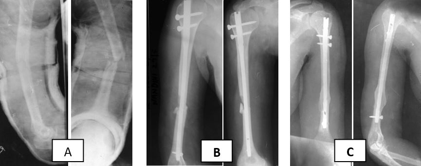
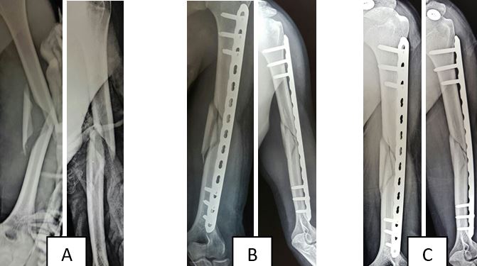
To compare and analyse outcome of treatment of comminuteddiaphyseal fracture humerus by interlocking nail with bridge plating in terms of –Early mobilization, Range of Movement, Size of incision, Hospital stay, Number of antibiotic dosage, Blood loss, Fracture healing, Complications.

Material and Methods

The study was conducted in the Department of Orthopedics, Traumatology and Reconstructive surgery Sri  Aurobindo  Medical College and Post Graduate Institute, Indore on total 30 patients, 15 for nail and 15 for bridge plate, from September 2012 to September 2014.

Inclusion criteria:

Age: >18 yrs, Both sex, Humeral shaft fractures treated within 1week by nailing or plating, Patients with grades 1 and 2 open fractures, Polytrauma, Unstable fractures

Exclusion criteria:

Epiphyseal plate open, Fracture line extending into the metaphysis, Patients with grade 3 compound fractures, Patients with pathological fractures, Patients with neglected fractures of the humerus.

The patients presenting to the hospital with humerus diaphyseal fracture and fulfilling inclusion criteria were randomised by computer generated table and treated surgically by bridge plating or interlocking nailing.

Only ante-grade nailing was done because of greater familiarity with the method among the


surgeons. From the second day, isometric exercises were carried out.

For bridge plating only anterior surface of humerus was used. In all cases minimally invasive approach was used. Reduction was achieved by manual traction.

Immediate active mobilization was done in all cases.

All patients were followed up in outpatient for Clinical and radiological assessment at 6weeks and at 3, 6 months.

The outcomes were assessed in terms of functional outcome, ability to return to previous jobs after 6 months, union time, union rate and the incidence of complications. Functional outcome was assessed using the American Shoulder and Elbow Surgeons Score.


Results


Blood loss during procedure:

BLOOD LOSS(ml)

0-50ml

50-100ml

100-150ml

150-200ml

200-250ml

250-300ml

TOTAL

NAILING

2

(6.67%)

4

(13.33%)

6

(20%)

3

(10%)

0

(0%)

0

(0%)

15 (50%)

PLATING

3

(10%)

6

(20%)

3

(10%)

2

(6.67%)

1

(3.34%)

0

(0%)

15 (50%)

TOTAL

5

(16.67%)

10

(33.34%)

9

(10%)

5

(16.67%)

1

(3.34%)

0

(0%)

30 (100%)


Mean

SD

Nailing

108.34 ml

182.57

Plating

98.34 ml

222.11

Table 1: Table showing Blood loss in the plating and nailing group


Two-sample T for Blood loss in Nailvs Blood loss in Plate

P value =  0.8938 This difference is considerednot to be statistically significant.

 t = 0.1347, df = 28


Duration of Procedure:

Duration

30mins -60 mins

Number

%

60 mins-90 mins

Number

%

90mins – 120mins

Number

%

120 mins -150mins

Number

%

150 mins – 180 mins

Number

%

Total

Number

%

Nailing

2

6.67%

8

26.67%

3

10%

2

6.67%

0

0%

15

50%

Bridge Plating

0

0%

8

26.67%

5

16.67%

1

3.34%

1

3.34%

15

50%

Total

2

6.67%

16

53.34%

8

26.67%

3

10%

1

3.34%

30

100%


Mean

SD

Nailing

85

100.995


Bridge Plating

95

100.995


Table 2: Table showing Duration of Surgery in the plating and nailing group

Two-sample T for Duration of procedure  Nail vs Bridge Plate

P Value:0.7883. This difference is considered not to be statistically significant.

t = 0.2712 , df = 28


Number of dose of Antibiotic –

No of doses of antibiotics

4 doses

%

5-8 doses

%

8 -12 doses

%

12-16 doses

Total

Nailing

4

13.34%

9

30%

1

3.34%

1

3.34%

15

50%

Bridge Plating

3

10%

9

30%

2

6.67%

1

3.34%

15

50%

Total

7

23.34%

18

60%

3

10%

2

6.67%

30

100%


Mean

SD

Nailing

6.23

11.95

Bridge Plating

6.76

11.95

Table 3 : Table showing Antibiotic dose in the plating and nailing group

Two-sample T for  No. of antibiotic doses  Nail vs  Bridge Plate

P Value :   0.9042 This difference is considered not to be statistically significant.

t = 0.1215 , df = 28


Number of days of hospital stay –

No of days of hospital stay

1-3 days

%

4-6 days

%

7-9 days

10-12 days

Total

%

Nailing

4

13.33%

9

30%

2

6.67%

0

0%

15

50%

Bridge Plating

5

16.67%

7

23.34%

2

6.67%

1

3.34%

15

50%

Total

9

30%

16

53.34%

4

13.33%

1

3.34%

30

100%


Mean

SD

Nailing

4.6

7.18

Bridge Plating

4.8

9.91

Table 4 : Table showing Hospital Stay in the plating and nailing group

Two-sample T for  No. of days of hospital stay Nail vs  Bridge Plate

P value :  0.9500  This difference is considered not to be statistically significant.

t = 0.0633, df = 28

ASES Scoring:

ASES Score

Excellent

%

Good

%

Fair

%

Poor

%

Total

%

Nailing

12

(40%)

1

3.33%

1

3.33%

1

3.33%

15

50%

Bridge Plating

12

(40%)

2

6.67%

0

0%

1

3.33%

15

50%

Total

24

(80%)

3

10%

1

3.33%

2

6.67%

30

100%


Table 5 : Table showing ASES Scoring in the plating and nailing group


Chi square : 0.667., DF : 3, P value : 0.8809


Figure 1 : 28 year old female fracture left humerus A-O type 12.B2 treated with Interlock nailing. (A) Pre-operative x-ray. (B) immediate post-operative x-ray. (C) 4 month post-operative x-ray.


Figure 2 : 28 year old female fracture left humerus A-O type 12.B2 treated with Interlock nailing. Functional Result at Final Follow up.


Figure 3:24 year male fracture left humerus A-O type 12 B.3  Bridge plating with Locking Compression plate. (A) Pre-operative x-ray. (B) immediate post-operative x-ray. (C) 3 month post-operative x-ray.


Discussion

The nailing and bridge plating groups were similar with respect to age, sex, dominant limb, injured limb, mode of injury, immediate treatment, injury surgery interval which indicated that the randomization had been effective. A total of 53% were male and 46.67% female in both nailing and plating group. In the study of Changulani M et al. [10] 86.9% were males and 13% females in nailing group, while in plating group 79.2% were males and 20.8% were females. The


mean age of the patients in our study 37.6 years in nailing and 42.73 years in plating group which compared to 39 years in nailing and 35 years for plating group in their study.In a study of Kejain Lain et al.[9]comparing intramedullary nailing with bridge plating, he found that mean operating time was more in Intramedullary nailing (126 mins). In present study we found that mean operating time was almost equal for both the groups (nailing- 85 mins, bridge plating -95 mins).  Similarly mean blood loss was statistically bit more in Nailing group as compared with bridge plating group in the study of kejain et.al.[9].It was 147ml in bridge plating and 195 ml in nailing group, while in our study blood loss difference between two groups was statistically in significant and was about 108 ml in nailing group and 98 ml in plating group. In our study ASES score in both groups was comparable, while in kejain et al.[9] study ASES score was better in plating group. In our study, one patient ( 22 years old) in bridge plating group had poor results in ASES score due implant failure. Which is comparable to the  study  done by Bell et.al and Dabezies E. J. et.al [11,12] in which they found 2-5% osteosynthesis failure rate in plating group. Bridge plating does not cause shoulder dysfunction which may occur in intramedullary nailing. In our study one patient from nailing group had shoulder dysfunction as revealed by poor score. Post-operative hospital stay are comparable in the study done by RaghvendraS et.al.[13].

Conclusion

In our study outcome of bridge plating and Interlock nailing for treatment of comminuted fracture shaft humerus was comparable with respect to time taken for surgery (p = 0.78), Amount of blood loss( p = 0.8938), duration of hospital stay( p = 0.95), antibiotic dosage( p = 0.904) and ASES Score. In none of the cases we encountered radial nerve palsy. In nailing group the poor result was due to shoulder stiffness while in bridge plating group poor result was due to implant failure.We found no significant difference in terms of radiological union time and functional outcome. Further study with large sample size is recommended to validate these results.


References

1.                Rockwood, C. A., Green, D. P., Heckman, J. D., &Bucholz, R. W. (2001). Rockwood and Green's fractures in adults. Philadelphia: Lippincott Williams & Wilkins.

2.                Canale, S. T., Beaty, J. H., & Campbell, W. C. (2013). Campbell's operative orthopaedics. Philadelphia, PA: Elsevier/Mosby.

3.                Sarmiento A, Kinman PB, Galvin EG, Schmitt RH, Phillips JG. Functional bracing of fractures of the shaft of the humerus. J Bone Joint Surg Am. 1977;59:596–601.

4.                Chapman J R, Henley M B, Agel J, et al. Randomized prospective study of humeral shaft fracture fixation: Intramedullary nails versus plates. J Orthop Trauma; 14(3): 162–6.

5.                Farouk O, Krettek C, Miclau T, Schandelmaier P, Guy P, Tscherne H. Minimally invasive plate osteosynthesis: does percutaneous plating disrupt femoral blood supply less than the traditional technique? J Orthop Trauma. 1999;13:401–406. doi: 10.1097/00005131-199908000-00002.

6.                Apivatthakakul T, Arpornchayanon O. Minimally invasive plate osteosynthesis (MIPO) combined with distraction osteogenesis in the treatment of bone defects. A new technique of bone transport: a report of two cases. Injury. 2005;36(4):530–538. doi: 10.1016/j.injury.2004.05.036

7.                Yu, Bin-fendgMD; Liu, Liang-Le-MD,Yang, Guo-Jing MD;Zhang, Lei MD, Lin,Xi-peng MD. Comparison of minimally invasive plate osteosynthesis and conventional plate osteosynthesis for humeral shaft fracture: A meta- analysis. Medicine: September 2016 – volume 95 – issue 39 – p e4955.

8.                Singisetti K, Ambedkar M. Nailing versus plating in humerus shaft fractures: A prospective comparative study. IntOrthop 2010;34:571-6.

9.                Lian K, Wang L, Lin D, Chen Z. Minimally Invasive Plating Osteosynthesis for Mid-distal Third Humeral Shaft Fractures. Orthopedics. 2013; 36: e1025-e1032. doi: 10.3928/01477447-20130724-18 

10.                M. Changulani, U. K. Jain, and TulsiKeswaniIntOrthop. Comparison of the use of the humerus intramedullary nail and dynamic compression plate for the management of diaphyseal fractures of the humerus. A randomised controlled study. IntOrthop. Jun 2007; 31(3): 391–395.

11.                Dabezies EJ, Banta CJ, Murphy CP, d’Ambrosia RD. Plate fixation of the humeral shaft for acute fractures with and without radial nerve injuries. J Orthop Trauma 1992;6:10-3.

12.                Bell MJ, Beauchamp CG, Kellam JK, McMurty RY. The results of plating humeral shaft fractures in patients with multiple injuries: the Sunnybrook experience. J Bone Joint Surg [Br] 1985;67-B:293-6.

13.                Radhavendra S, Bhalodiya HP. Internal Þ xation of fractures of the shaft of the humerus by dynamic compression plate or intramedullary nail: A prospective study. Indian J Orthop( serial online) 2007( cited 2009 Jul 1)41:214-8.

14.                fixation nails versus plates journal of orthopaedic trauma march 2000  volume14 (3):162-166.

15.                Lin, 1998. Lin J:  Treatment of humeral shaft fractures with humeral locked nail and comparison with plate fixation. J Trauma   1998; 44:859.

16.                Lin J, Inoue N, Valdevit A, et al. Biomechanical comparison of antegrade and retrograde nailing of humeral shaft fracture. ClinOrthopRelat Res. jun1998;(351):203-13.