Vidyarthi A, Turkar R.: Pelvic Fracture – Results of early External Fixation


Pelvic Fracture - Results of Early External Fixation


Vidyarthi A, Turkar R.


Investigation performed at Department of Orthopaedics, NSCB Medical College, Jabalpur, Madhya Pradesh, India


Abstract

Background: The behavior of pelvic ring fracture following surgical fixation has been very sparsely studied. Pelvic ring injuries, depending on their severity had been treated by variety of closed method. The purpose of the study is to assess the short-term functional outcome of pelvic ring fracture following surgical fixation.

Method: Present prospective study consists of 62 patients of pelvic fracture admitted in department of orthopedics, NSCB medical college and hospital Jabalpur (M.P) during the period of Oct. 2010 to Sep. 2011. All patient of type A (stable) fracture treated by conservative method. Out of 40 patients of unstable pelvic fracture (involving pelvic ring) 14 were treated by surgical method. Out of 14 patients 4 were treated by external fixation, 8 by internal fixation, and 2 by combined external and internal fixation. Functional outcome assessment was done by Majeed Scoring System [1].

Results:  Functional outcome score in surgically treated patient is excellent in 7 patients, good in 4, fair in 2, and poor in 1 patient at 3 month follow-up. Pain is the most common complaint after 3 month of surgical fixation, it is seen in 4 patients (28.57%) but only 1 patient changed his job because of this pain.

Conclusion:Unstable Pelvic ring injury has poor outcome, it should be treated by operative methods for optimal functional outcome.

Keywords: Pelvic Ring injury, external Fixation


Address for Correspondence:Dr. Ashok Vidyarthi, Professor Bungalow P-3 Doctors Colony N.S.C.B. Medical College, Jabalpur (M.P.)482003, India.

Email:vidyarthi_ashok@rediffmail.com


How to site this article:Vidyarthi A, Turkar R.Pelvic Fracture - Results of Early External Fixation. OrthopJMPC 2016;22(2): 3-6.


Introduction

Pelvic fracture constitute 3% of all skeletal injuries [2]. Among poly trauma patients the incidence is about 25%. Henderson (1989) followed 26 patients with major pelvic disruption and found low backache in 50%, limp in 32%, and work disability in 43% of patients [3]. Tornetta et al (1996) achieved a high long-term success rate with operative management of pelvic fractures while Mirada et al found no difference in the outcome among operatively and non-operatively managed cases of pelvic fractures [2,4]. The actual outcome is likely to be much more adversely affected with the cultural practices of sitting and squatting. So pilot study was planned to assess the short term functional outcome following surgical fixation in pelvic ring injury, in Indian scenario.

Materials and Methods

Present prospective study consists of 62 patientsof pelvic fracture admitted in Department of Orthopedics, NSCB medical college and hospital Jabalpur (M.P) during the period of Oct. 2010 to Sep. 2011. An anteroposterior (AP) x-ray of the pelvis was included in the initial radiographic examination of all patients with blunt trauma. The pelvis has a remarkable ability to recoil to a near-normal alignment after trauma. This elastic recoil can mask severe instability. Fractures noted on the AP x-ray should prompt further investigation of the pelvis with inlet and outlet views.

All patients with unstable type of pelvic injuries were planned for surgery. 14 patients were treated by surgical method in this study. There were 7 patients with type B and 7 with type C pelvic injury. Out of 14 patient 4 were treated by external fixation, 8 by internal fixation, and 2 by combined external and internal fixation. All unstable pelvic fracture were managed conservatively initially Conservative management includes bed rest, pelvic belt, skeletal traction etc.

Results

Higher incidence was observed in young active person in 3rd 4th and 5th decades (74.19%). The incidence was more in males than female (M:F= 1.8:1). Fall was the commonest (56.45%) mode of injury in patient with pelvic fracture followed by RTA (road traffic accident). Out of 62 patients of pelvic fracture, 40 (64.51%) had unstable type of injury. Anteroposterior compression injuries (53.23%) were the commonest type of injuries. Out of 14 patients, 1 (7.14%) got infection at operated site in the form of pin tract infection. Persistent pain seen in 4 (28.57%) operated patients. Limb length discrepancy seen in 1 (7.14%) patient. This discrepancy was 10 mm. Average time to start mobilization after surgical fixation was 19 days (range 1-62 days) for all operated patients, 10 days(range 1-24 days) for patient with type B pelvic injury, and 28 days (range 3-62 days) for patients with type c pelvic injury. Follow up duration for all patients ranged from 3 months to 8 months.


Pre op x-ray

Post op x-ray shows good compression at fracture site.

At 1 and half after fixate application pt start doing his all work with fixator insitu. [ Functional Majeed score 62].

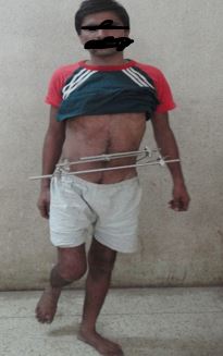
At 6 month follow-up patient can walk, sit and do all his routine work. [ Functional Majeed score 86].


Majeed functional outcome score in surgically treated patient is excellent in 7 patients, good in 4, fair in 2, and poor in 1 patient at 3 month follow-up. Pain is the most common complaint after surgical fixation, it is seen in 4 patients (28.57%) but only 1 patient required change inhis job because of this pain.

Discussion

Historically, pelvic ring injuries, depending on their severity had been treated by a variety of closed methods. Unstable pelvic injuries treated by these conventional measures often result in significant disability, moreover the mortality can reach up to 21.8%.[5,6,7,8] There was a growing body of evidence that the application of an external skeletal frame will reduce venous and bony bleeding and improve tamponade by reducing and maintaining the pelvic volume to the extent that other interventions are rarely required.[4,7,9,10]. Recently, biomechanical studies showed that external frame could not ensure sufficient stability to allow mobilization without the risk of re-displacement of the fragments particularly those with vertical instability. External fixation can be used temporarily in vertically unstable injury as a part of emergency treatment to allow the patient to be placed with trunk in the upright position to improve ventilation [12,13]. External fixation used as definitive fixation in Tile’s type B (partially unstable) pelvic injury. Our results agree with other studies stating that anterior or posterior fixation, or both could restore excellent stability and adequate consolidation of the unstable (Type-C) pelvic injuries with subsequent decrease in morbidity and mortality [6,13,14]. They were mobilized relatively earlier without significant risk of re-displacement of the fragments. Early mobilization minimized the complications associated with prolonged recumbency. It has been emphasized that surgical treatment should be carried out 5-7 days post trauma when the patient’s general condition allows [13,14] It is the author’s opinion to perform internal fixation for unstable pelvic injuries as soon as the general condition stabilized even up to 6weeks after the injury. The functional results are often affected by the associated skeletal or extra skeletal injuries as well as other variables [5,8,12,13].

Conclusion

Unstable pelvic injury should be treated operatively as soon as general condition of the patient allows. Surgical treatment gives superior results even when performed as late as 6 weeks. External fixation of Tile’s type B  unstable fracture allows early mobilization of the patient and avoids  early and late complication associated with conservative management.Tile’s type C vertically unstable (>10mm displacement) injury should be treated by internal fixation because an external fixator does not correct the malalignment and is not rigid enough to provide stability in vertical plane.


References

1.                Majeed S.A. .; grading the outcome of pelvic fracture.J.  bone and joint. Surg. 71B (2) 304-306, 1989

2.                TornettaP,Dickson K, Matta JM. Outcome of rotationally unstable pelvic ring injuries treated operatively. ClinOrthopRelat Res 1996;329:147-51.

3.                Henderson RC. The long term results of non-operatively treated major pelvic disruption. J Ortho Trauma 1989;3:41-7.

4.                Miranda MA, Rieman BL, Butterfield SL, Burk CJ. Pelvic ring injuries: A long term functional outcome study. ClinOrthopRelat Res 1996;329:152-9.

5.                Goldstein A, Phillips T, Sclafani SJ, Scalea T, Duncan A, Goldstein J et al. Early open reduction and internal fixation of the disrupted pelvic ring. J Trauma 1986; 26: 325-333.

6.                Tile M. Pelvic ring fractures: should they be fixed? J Bone Joint Surg 1988; 70-B: 1-12.

7.                Van den Bosch EW, Van der Kleyn R, Hogervorst M, Van Vugt AB. Functional outcome of internal fixation for pelvic ring fractures. J Trauma 1999; 47: 365-371.


8.                Gruen GS, Leit ME, Gruen RJ, Reitzman AB. The acute management of haemodynamically unstable multiple trauma patients with pelvic ring fractures. J Trauma 1994;36: 706-711.

9.                Matta JM, Saucedo T. Internal fixation of pelvic ring fractures. ClinOrthop 1989; 242: 83-97.

10.                Lindahl J, Hirvensalo E, Böstman O, Santavirta S. Failure of reduction with an external fixator in the management of injuries of the pelvic ring. J Bone Joint Surg 1999; 81-B(6): 955-962.

11.                Kregor PJ, Chip Routt ML. Unstable pelvic ring disruptions in unstable patients. Injury 1999; 30: B19-28.

12.                Pohlemann T, Gansslen A, Schellwald O, CulemannU,Tscherne H. Outcome after pelvic ring injuries. Injury 1996; 27 (Suppl 2): B31-38.

13.                Webb LX, Gristina AG, Wilson JR, Rhyne AL, Meredith JH, Hansen ST Jr. Two-hole plate fixation for traumatic symphysis pubis diastasis. J Trauma 1988; 28: 813-817.

14.                Hirvensalo E, Lindahl J, Böstman O. A new approach to the internal fixation of unstable pelvic fractures. ClinOrthop 1993; 297: 28-32.  

15.