Introduction
In 1957, Kaplan[1], brought attention to the patho-anatomy of the complex dorsal dislocation of the metacarpophalangeal joint (MCP Joint ) of the index finger and its management. Since then various authors reported this phenomenon in other digits like middle [2], ring, little fingers either as isolated or in combination. Dislocation can be either in volar or dorsal directions. 50% cases are complete and complex dislocations for which treatment of choice is open reduction.
Complexity of dislocation is due to its unique anatomical event i.e. entrapment of fibro cartilaginous volar plate (detached from weakest point i.e. metacarpal neck) between metacarpal head and proximal phalangeal base. Other supporting anatomical structures which help in entrapment are lumbricals on radial side, flexor tendon and pretendinous band on ulnar side, natatary ligament with volar plate are on distal and dorsal to metacarpal head, superficial transverse ligament across metacarpal neck volar and proximally [1] Besides this lateral collateral ligaments, which are now abnormally displaced, locks the proximal phalanx in dorsal displaced position.
Case report
A 18 year old male, farmer, presented with mild pain and swelling over both hands around index finger’s MCP joint with 1 week old history of fall on outstretched volar aspect of index finger.
On examination there was tenderness, swelling and mild extension at 2nd metacarpophalangeal joint of both index fingers and these were slightly ulnar deviated. Bilaterally interphalangeal joints of index finger were flexed (Figure 1). Volarly there were bony bulge formed by the head of 2nd metacarpal head with puckering of skin dorsally bilaterally. Neurovascular status of both digits was normal.
X-ray of both hand AP and lateral was obtained and shows dorsal dislocation of proximal phalanx of index finger bilaterally.(Figure 2)
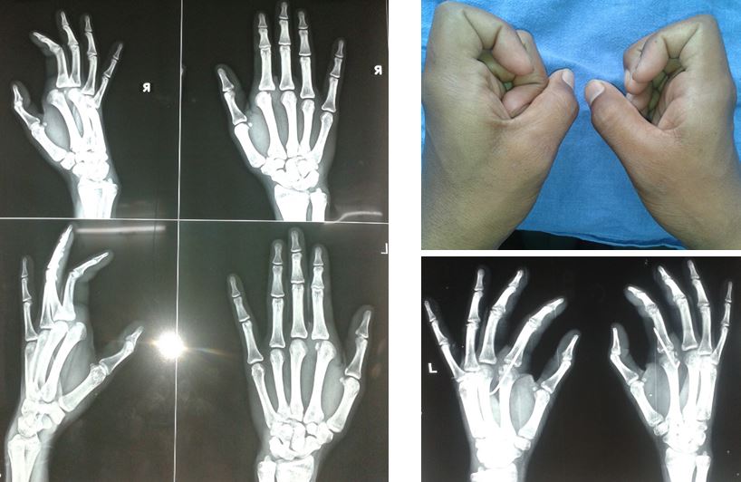
Figure 1: Clinical Photograph showing B/L Dislocated MCP Joint. Figure 2: Pre Op X ray both Hands. Figure 3: Post op X ray after reduction and K wire fixation.
After unsuccessful attempt of closed reduction, we planned for open reduction by dorsal approach.
Under all aseptic condition a 3 cm long straight skin incision was made on dorsum of 2nd metacarpophalangeal joint. . Extensor tendon with dorsal capsule was split longitudinally. With traction joint was distracted and volar plate identified carefully, which was main hurdle in reduction. It was longitudinally cut.
With the division of volar plate the proximal phalanx was restored to its normal position and there was freedom of movement at 2 nd MCP joint. Under C-arm guidance the position of joint was confirmed and a k- wire placed in 30-degree flexion in MCP joint (Figure 3). There was avulsion of minute chip fragment from dorsal cortex with dorsal capsule, which was excised and dorsal capsule was repaired. Tourniquet was released and homeostasis achieved and the wound was closed in layers. .
The patient was given cock-up slab for 2 weeks, after which k- wire removed and active early range of motion exercise was begun using a removable 10 degree dorsal extension block splint. After 4 weeks, the splint was used only for protection and discontinued at 6 weeks. At 6 weeks follow up range of movement at metacarpophalangeal joint and both interphalangeal joints were normal bilaterally. There was decreased grip strength i.e. 18kg on right and 16kg on left. Neurovascular evaluation was with in normal limits. X-rays confirmed maintenance of reduction. Patient return to full activity after 12 weeks.
Discussion
Kaplan et al [1] and Barry K et al [3] described a volar approach to complex MCP joint dislocations. This approach has certain disadvantages like risk of damaging the radial digital nerve of index finger during exposure[4], limited view of entrapped volar plate, extensive and unnecessary release of other supporting volar structures. Because of these demerits some authors like Becton et al [5] advocated dorsal approach, which has certain definitive advantages over volar approach. Like it is simple and easy, with less chance of damaging the radial nerve [3,4], accurate management of fractured osteochondral fragment of metacarpal head, there is complete exposure of fibro cartilaginous volar plate that is main structure blocking reduction.
There are few demerits of this approach like it is not possible to repair the volar plate which results delay in recovery and instability to the joint. Other offending structures like natatory ligament, lumbrical muscle, and superficial transverse ligament cannot be addressed with this approach.
Conclusion
The complex dorsal dislocation of MCP joint can be managed with dorsal as well as volar approach. The dorsal approach has advantage over the other as discussed above. Though, further clinical evaluation is to be done to assess the effectiveness of both methods.


