Introduction
Low-back pain is the leading cause of disability worldwide. It is the second most common symptom-related reason for seeking care from a primary care physician.1 While low back pain rarely indicates a serious disorder, it is a major cause of pain, disability, and social cost. The lifetime prevalence is over 60%. The costs associated with low back pain include the direct cost of medical care and the indirect costs of time lost from work, disability payments, and diminished productivity.2 The extent of chronic low back pain among Indian population is alarmingly high, with approximately 79% of women between 20 to 50 years suffering from chronic pain. Lower back pain alone affects around 80% of women compared to 59% of men.3
Intervertebral disc (IVD) degeneration is a major contributing factor for discogenic low back pain (LBP), causing a significant global disability.4 It is a common joint disease of all orthopedic diseases. It is mainly caused by degenerative changes of the lumbar intervertebral disc; external forces; or nerves, horsetails and other nerves.5 The PIVD consists of an inner core proteoglycan-rich nucleus pulposus (NP) and outer lamellae collagen-rich annulus fibrosus (AF) and is confined by a cartilage end plate (CEP), providing structural support and shock absorption against mechanical loads. Thus, changes to degenerative cascades in the PIVD cause dysfunction and instability in the lumbar spine.6
Patients exhibit back pain, lower limb radiation neuralgia and neurological dysfunction.5 The relationship between lumbar disc prolapse and radicular pain was first described by Mixter and Barr. Mixter and Barr in 1932, described lumbar discectomy by which an L2 to S1 exploratory laminectomy led to removal of a “mass one centimeter in diameter” that was “pressing on the left fifth nerve root and displacing the cauda equina to the right”. In 1934, they first published the surgical treatment of lumbar disc herniation (LDH). 6 However, first discectomy was done by Oppenheim and Fedre Krause in 1906 though the first publication was done by Mixter and Bar.7
Surgical treatment is well known to be beneficial for patients with LDH who fail to respond to conservative care.8 Surgery is offered to patients with persistent leg pain that is refractory to conservative treatment. The open surgical technique has been described since the early 20thcentury. Since its introduction, alternative methods for operating disc pathologies have been developed.9 With the continuous progress of microsurgery, the surgical techniques of LDH treatment have been developed rapidly. Later in 1977, Caspar and Yasargil first applied the conventional microdiscectomy (CMD) to the surgical treatment of LDH. 10, 11
Newer techniques were developed with the objective of achieving less tissue trauma in a fast and efficient way. 9 The minimally invasive technique of transmuscular tubular discectomy (TD) was introduced in 1997 by Foley and Smith which is a procedure that combines spinal endoscopy and the techniques used in microdiscectomy.12 Hence, with the introduction of the microscope, the original laminectomy was refined into microdiscectomy (MD).9
Material and method
The study was conducted at the Department of Orthopaedics at R.D. Gardi Medical College, Ujjain. This study was completed within two years after receiving approval from the ethics committee. This is a prospective observational study.
Written informed consent was obtained from all patients before enrolling them for the study. The patients admitted in the department of orthopaedics coming with a complain of lower back pain with radicular symptoms. were enrolled for this study as per the following exclusion and inclusion criteria.
Inclusion criteria was patients with unilateral back pain with radicular symptoms (pain, paresthesia weakness), lumbar or lumbosacral single level prolapsed intervertebral disc patients, patient not responding to conservative treatment for 6weeks and patients above 20years of age and of both genders.
Exclusion criteria was age less than 20 years, revision surgery, infection and bleeding disorders, more than one level involvement or bilateral symptoms, patients who are not fit for surgery, patient with dynamic instability and patients with congenital narrow canal, multilevel disc herniations, cauda equina syndrome, spondylolisthesis, central canal stenosis, pregnancy, and severe somatic or psychiatric diseases As per study criteria 32 patients with lower back pain with radicular symtoms was included in this study.
After admission of patients a detailed, careful history was taken. Patient was assessed clinically to evaluate general condition; vitals were recorded and detailed spine examination was done.
Radiological assessment was done to identify the level of herniation and preoperative routine investigation was done. By chit system 16 patients were placed in group A underwent microscopic tubular discectomy and remain 16 into group B underwent open discectomy.
Clinical outcomes were evaluated by Oswestry disability index (ODI) scores and visual analog scale (VAS) scores for leg and back pain. Back and leg VAS and ODI scores were assessed before surgery (preoperative), at the 6 weeks from surgery (postoperative), and subsequently at 1year.

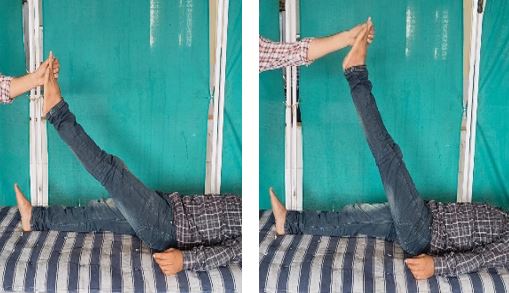
Figure 1: A and B, pre op and post op SLRT


