Orthopaedic Journal of MP Chapter

Publisher: Madhya Pradesh Orthopaedic Association www.mpioa.com
E-ISSN:2582-7243, P-ISSN:2320-6993
2024 Volume 30 Number 1 Jan-Jun

A case series on floating knee injuries with ipsilateral femur and tibia fracture

Singh V1*, Gupta N2, Nagdev S3, Dwivedi VM4

1* Vivek Singh, Professor, Department of Orthopaedics, R D Gardi Medical College, Ujjain, Mp, India.

2 N Gupta, Department of Orthopaedics, R D Gardi Medical College, Ujjain, Mp, India.

3 S Nagdev, Department of Orthopaedics, R D Gardi Medical College, Ujjain, Mp, India.

4 V M Dwivedi, Department of Orthopaedics, R D Gardi Medical College, Ujjain, Mp, India.

Background: The floating injuries occur due to high velocity trauma following motor vehicle accidents. Management of such complex injuries is a challenging task even in experienced hands as there are no standard treatment guidelines. All the patient were first stabilized initially by external fixator and subsequently taken up for definitive surgery. Early fixation and aggressive mobilization ensure fracture union and leads to fewer complications.

Material and method: In this study, 10 cases of fracture ipsilateral femur and tibia were operated in R.D. Gardi Medical College, Ujjain between February 2022 to February 2024 with the sequence of early fixation by application of external fixator on the day of admission, followed by delayed definitive surgery through ORIF with plate osteosynthesis or CRIF with intermedullary nail as needed according to the fracture. This study incorporates all fractures associated with floating knee viz, fracture shaft femur, fracture distal femur (intra/extra articular), fracture proximal tibia with diaphyseal extension and fracture shaft tibia, also includes compound, comminuted fractures.

Results: With the surgical management protocol followed by us, union was achieved in all the 10 cases. Post operative infections was observed in 1 patient out of 10 and knee stiffness was noted in about 3 patients. The present study had a minimum follow up of 6 months.

Conclusion: The surgical management protocol implemented yielded outstanding functional and radiological outcomes across all cases. Patients demonstrated a remarkable ability to resume their pre-injury functional activities and achieved full weight-bearing walking without assistance within six months post-surgery.

Keywords: floating knee injuries, ipsilateral femur and tibia fracture, polytrauma

Corresponding Author How to Cite this Article To Browse
Vivek Singh, Professor, Department of Orthopaedics, R D Gardi Medical College, Ujjain, Mp, India.
Email:
Singh V, Gupta N, Nagdev S, Dwivedi VM, A case series on floating knee injuries with ipsilateral femur and tibia fracture. ojmpc. 2024;30(1):21-28.
Available From
https://ojmpc.com/index.php/ojmpc/article/view/185
Manuscript Received Review Round 1 Review Round 2 Review Round 3 Accepted
2024-06-06 2024-06-12 2024-06-18 2024-06-24 2024-06-30
Conflict of Interest Funding Ethical Approval Plagiarism X-checker Note
Authors state no conflict of interest. Non Funded. The conducted research is not related to either human or animals use. 13.21 All authors have accepted responsibility for the entire content of this manuscript and approved its submission.

© 2024by Singh V, Gupta N, Nagdev S, Dwivedi VMand Published by Madhya Pradesh Orthopaedic Association. This is an Open Access article licensed under a Creative Commons Attribution 4.0 International License https://creativecommons.org/licenses/by-nc/4.0/ unported [CC BY NC 4.0].

Introduction

In 1975, Blake and McBryde introduced the term 'floating knee' to describe ipsilateral fractures of the femur and tibia. This combination of injuries transcends mere bone lesions, typically resulting from high-energy trauma in patients with multiple injuries and a multitude of other lesions. Following initial evaluation, patients should be stratified, with only stable individuals undergoing immediate reduction and internal fixation, while others may require external fixation. Definitive internal fixation of both bones consistently produces optimal outcomes across various series. When both fractures (femoral and tibial) are extra-articular, nailing of both bones represents the preferred fixation method. In cases involving articular fractures, plates emerge as the standard of care. A substantial portion, approximately 40%, of floating knees necessitate a combination of implants. Although associated ligamentous and meniscal lesions are common, they may be inconsequential in cases of intraarticular fractures, which typically carry the poorest prognosis for this type of injury. (1) Major lower limb injuries involve many or all components of lower limb architecture namely skin, soft tissue, osseous, vascular and neural element which makes for prompt and precise evaluation and management for optimizing functional outcome. In major lower limb trauma, often in polytrauma setting the important question is whether to salvage or amputate the injured limb. Unfortunately, data on this issue is often conflicting and confusing. For both cultural and practical reasons, patient prefers to retain their own limb even though deformed, provided it is painless and retains its function. Clinical examination is of paramount importance, such injuries are often associated with head injuries, chest or abdominal trauma, and other significant musculoskeletal trauma. Detailed examination is necessary to exclude associated injuries. While assessing the patient, aggressive resuscitation should be initiated preferably in operation theatre. As described by Blake, McBryde and Fraser classification, we have included all fracture patterns associated with floating knee with variable degree of comminution and compounding.

Fraser et al.(3) initially categorized floating knee injuries into three types: Type I, involving fractures of the femur and tibia shaft without involvement of the knee joint articular surface; Type II, where fractures extend into the knee joint. Type II was further divided into subtypes: Type IIa, involving tibial plateau; Type IIb, featuring an intercondylar fracture of the distal femur; and Type IIc, encompassing both tibial plateau and distal femur articular surface. This classification was recently revised by Ran to incorporate disruption of the extensor apparatus. According to Ran, Type I fractures are extra-articular, Type II are intra-articular, and Type III are accompanied by patella fractures (4)

ojmpc_185_01.JPG
Figure 1: Fraser classification of floating knee11 Type I, IIA ,IIB, IIC

Material and method

This prospective interventional study includes patients admitted in Orthopedics dept, RDGMC, Ujjain, MP with a diagnosis of ipsilateral fracture of the femur and tibia, who were managed in a single first-level trauma centre between February 2022 and February 2024. 10 cases of fracture ipsilateral femur and tibia were taken into account. Inclusion criteria was patients diagnosed with a floating knee injury, patients with lower limb trauma, regardless of the mechanism or nature of the injury, patients with skeletal maturity, defined as those aged 15 years and above and patients managed surgically with a minimum follow-up period of 6 months. Exclusion criteria was patient with neurovascular injury, patients with any history of malignancy or pathologic fractures, patients whose follow-up was discontinued, patients with a history of metabolic or other types of bone diseases and patients with periprosthetic fractures.

All admitted patients underwent initial stabilization in the emergency unit. In cases of polytrauma, management followed the Advanced Trauma Life Support (ATLS) protocol. Comprehensive evaluations were conducted to rule out neurovascular and additional injuries. Initial injury stabilization was tailored to each patient's specific and general physiological condition. Perioperative planning relied on standard radiographic images and 3D CT scans. These imaging modalities were utilized for further fracture classification and surgical strategizing. Patients with open injuries classified according to the Gustilo-Anderson classification system underwent emergency debridement, sterile coverage, and external fixation of the fracture as part of damage control surgery/orthopedics (DCS) on the day of admission.

Patients with clean wounds were administered a first-generation cephalosporin (1 g ceftriaxone as the initial choice). For patients with contaminated wounds, an additional aminoglycoside (amikacin 500 mg) and metronidazole 500 mg were added. This antibiotic regimen was maintained for Gustilo type III open fractures. Prophylactic tetanus therapy was also administered to all patients presenting with open injuries. Patients in an unstable general condition and with closed fractures were either admitted to the trauma ward or the surgical intensive care unit (SICU) for intensive management. Those with suspected haemothorax or pneumothorax received chest drains for appropriate management.

Additionally, colour doppler was conducted before the procedure for every suspected patient of vascular injury. All patients eligible for surgical stabilization underwent either general or spinal anesthesia. The surgical approach, reduction technique, and implant selection were determined according to the fracture pattern and classification based on Fraser’s classification (2), as well as the patient’s physiological state upon presentation and the condition of the soft tissue. All surgical procedures were conducted when patients were hemodynamically stable. Patient with shaft femur fracture preferably treated with antegrade intermedullary nail whereas those with fracture distal femur (intra and extra articular) with plate osteosynthesis by distal femur locking plate.


In distal femur fracture associated with Hoffa’s fracture, anteroposterior CC screw was additionally used along with plate. In majority of cases, anterolateral approach (swash buckler) was preferred and in some patient with medial condyle comminution, medial buttress plate was also used by medial approach. For tibia fracture, proximal tibia with diaphyseal extension were treated with plate osteosynthesis by locking hockey plate and majority cases were operated by anterolateral approach. Medial plate was also used in case of severe comminution. However, all diaphyseal tibial fractures were managed by intermedullary nail. In instances of severe soft tissue compromise, external fixation was the preferred method for definitive fixation. The postoperative protocol encompassed temporary immobilization, infection prophylaxis, and meticulous wound dressing to facilitate proper wound and soft tissue healing. Patients with significant soft tissue defects underwent skin grafts or flap surgery as deemed necessary.

Rehabilitation, including physiotherapy, commenced once proper wound healing was attained, typically between 10 to 21 days following surgery. Patients were encouraged to achieve full range of motion (ROM) in both hip and knee joints, incorporating isometric exercises for quadriceps and isotonic exercises for hamstrings, as tolerated. Non-weight-bearing ambulation using crutches was permitted for six weeks, provided the contralateral limb was unaffected, followed by a transition to partial weight-bearing. Full weight-bearing was only permitted after clinical and radiological union had been confirmed. Active ROM exercises were delayed for some patients who underwent knee-spanning external fixation.

On average, patients attained full weight bearing after four weeks for extra-articular fractures and eight to ten weeks for intra-articular fractures. The discharge of patients from the hospital was primarily based on the patient’s general clinical condition, while the hospital length of stay (LOS) was influenced by the severity of the initial trauma.

Upon discharge, patients were scheduled for postoperative follow-up appointments at the outpatient orthopedic clinic at two weeks, one month, and once every three months for one year, followed by yearly appointments thereafter. Routine follow-up examinations included radiographic assessments (anteroposterior, lateral, and oblique views) of the injured segments and evaluations of functional outcomes using the Karlstrom and Olerud grading system (fig. 2)

CriterionExcellentGoodAcceptablePoor
Subjective symptoms from thigh or legNoneIntermittent slight symptomsMore severe symptom, impairing functionConsiderable function impairment: pain at rest
Subjective symptoms from knee or ankle jointNoneSame as aboveSame as aboveSame as above
Walking abilityUnimpairedSame as aboveWalking distance restrictedUses cane, crutch or other support
Work and sportsSame as before the accidentGiven up some sport; work same as before accidentChange to less strenuous workPermanent disability
Angulation, rotational deformity or both0<10°10-20°>20°
Shortening0<1 cm1-3 cm>3 cm
Restricted joint mobility0<10° at ankle; <20°10 -20° - at ankle; 20-40°>20° at ankle; >40°

Figure 2: the Karlstrom and Olerud grading system

Postoperative complications were documented and classified as either early, including wound infection and septic shock, or late, such as knee stiffness, nonunion and nerve palsy. All clinical and functional outcomes were assessed on 6 months follow up.

Case 1

ojmpc_185_03 (2).JPG
Figure 3: Images A, B and C - Preoperative radiographs (A , B) showing AP and Lateral view comminuted fracture distal femur with intercondyle extension . ( C ) showing fracture distal 1/3rd shaft tibia & fibula.

ojmpc_185_04.jpg
Figure 4: Post-operative radiographs showing Knee spanning external fixator which is extended upto distal tibia.

ojmpc_185_05.jpg
Figure 5: (A) and (B) – intraoperative pictures of distal femur fixation by anterloateral approach (Swash buckler)


Figure 6: A, B, C and D– Showing immediate postoperative radiographs (A,B) – Distal femur plate osteosynthesis with distal femur locking plate and anteroposterior screw for hoffas fracture. (C, D)- Tibia interlock nailing and fibula rush nail.

ojmpc_185_07.jpg
Figure 7: A, B, C and D – 3 months post operative radiographs of a case showing significant callus formation at distal femur fracture.

ojmpc_185_08.jpg
Figure 8: A and B. Three months follow up showing active and passive knee range of motion.

Case 2:

ojmpc_185_09.JPG
Figure 9: Images- A &B– preoperative radiographs showing fracture distal femur with intercondylar extension, communited patella fracture and segmental communited fracture of proximal 1/3 rd and distal 1/3rd shaft tibia with intraarticular extension to ankle joint and distal 1/3rd fibula fracture.

Patients demographics (Masterchart)

Data compilation of 10 cases included in the study . Abbreviations used :

MOI- Mechanism of injury , SOI- Site of injury, OA/OTA- Arbeitsgemeinschaft für Osteosynthesefragen/Orthopedic Trauma Association, FE- Femur, TI- Tibia, LOS – Length of stay, IM- Intramedullary, NA- Not applicable, RTA- Road Traffic Accident, FFH-Fall from hight, HO- Heavy object injury


Table 1: Patients demography

CASEAGEGENDERMOISOIFRASER CLASSOA /OTA CLASS FEOA /OTA CLASS TIGUSTILO CLASS TIASSOCIATED INJURY TIMANAGEMENT DONE FEMANAGEMENT DONE TICOMPLICATIONS TIKARLSTORM OLERUD SCORELOS (days )SCORE
1.17MALERTALeftII B33C42C2+NAComminuted fracturePlatePlategood25
3.143C2patella ,medial
malleolus fracture .
2.30MALERTALeftII B33C42B2FE -INONEPlateNailgood22
2.2TI -II
3.42MALEFFHRightI32C242C2TI -INONENailNailExcellant18
4.46MALERTALEFTIIA33C242C2FE -IIIpsilateral patellaAntegradePlateExcellant23
fracture .nailing
5.51MALEHORightIIC33C341C2FE -IIMedial malleolarPlatePlateKnee stiffnessPoor17
TI -IIfracture (ipsilateral )
6.25MALERTALEFTIIA32C241C1NANonePlatePlateKnee stiffnessAcceptable22
7.67FEMALEFFHRightI32A242A1FE -IIpsilateral multipleAntegradeAntegradeNoneexcellant25
TI -Irib fractureIM nailIM nail
8.33MALERTARightI32B242A1TI -III AIpsilateral shaftPlateIM NailWound infectiongood39
radius fracture
9.58FEMALEFFHLeftII A3241C1NAIpsilateral calcaneumAntegradePlateDVT smallExcellent31
Alfracturenailsephanous vein
10.19MALERTARightI32B241A3NABilateral clavicle and multiple rib fracturenail AntegradeAntegrade nailNoneExcellent24

ojmpc_185_10.jpg
Figure 10: (A,B) showing immediate post external fixation, uniplanar fixator applied for tibial fracture seen in above radiograph.(C,D) intraoperative pictures showing distal femur plating and patellar TBW with encirclage.

ojmpc_185_11.jpg
Figure 11: showing immediate postoperative radiographs. (A,B,C) – showing distal femur fracture fixation with distal femur locking plate with patellar TBW and encirclage. (D,E) showing tibial fracture fixation with long hockey plate done by minimally invasive percutaneous plate osteosynthesis (Mippo technique) and medial malleolus fracture fixed with CC screw , fibula fracture fixation done by 2.5 mm rush nail.

ojmpc_185_12.jpg
Figure 12: A, B, C and D- Three months follow up- xray

ojmpc_185_13.jpg
Figure 13: A, B and C- Follow up pictures of patient


Results

In the present study, a comprehensive analysis of the available data yielded several key findings. The mean age of the cases was 38 years, with a range from 17 to 67 years. Out of the 10 cases examined, 8 were males and 2 were females. Fractures were distributed evenly between the left and right lower extremities, with 5 cases on each side. The majority of cases (60%) resulted from road traffic accidents, while 30% were attributed to falls from altitude, and 10% were caused by heavy objects.

Regarding treatment, both intramedullary nails and plates were utilized for femoral and tibial fractures, with each method employed in 5 cases. Concomitant injuries were observed in 7 cases, adding complexity to the management approach. Notably, 50% of tibial fractures and 40% of femoral fractures were classified as open, with Gustilo type II being the most common subtype in both cases. Evaluation at the 6-month follow-up revealed promising outcomes, with 80% of cases achieving excellent to good results according to the Karlström–Olerud scoring system. However, 10% of cases demonstrated acceptable results, while 10% exhibited fair to poor outcomes, underscoring the challenges inherent in managing these complex injuries.

Discussion

The prognostic indicators in floating knee cases encompass associated injuries and the type of fracture, whether it's open, intra-articular, or comminuted. Achieving a favourable final outcome necessitates the proper management of associated injuries, employment of external fixation, intramedullary nailing for both fractures, plate osteosynthesis and combination of implants. Notably, optimal results were attained when both fractures underwent intramedullary nailing, facilitating an earlier return to normal activity levels compared to alternative treatment approaches. Many surgeons regard the type of fracture (open, intra-articular, comminuted) and the severity grade of soft tissue and associated injuries as crucial prognostic indicators for both initial and final outcomes in patients with floating knee injuries. These factors represent significant risks for unfavourable outcomes in such cases. Through multivariate analysis, certain studies have identified intra-articular involvement of the knee joint as a significant contributing factor affecting the final outcome of floating knee injuries. Within these variables, the Fraser type plays a pivotal role in determining the ultimate result, with knee involvement considered the most influential factor. Some studies have shown poorer outcomes when one or both fractures involve the joint compared to cases where both fractures are diaphyseal. According to the Blake and McBryde classification, type I cases tend to have better outcomes, whereas type II cases typically exhibit comparatively poorer outcomes, likely due to articular injury and knee stiffness following prolonged protection.

In a study by Rethnam et al., 29 patients with floating knee injuries were managed over a 3-year period. The mechanism of injury was RTA in 27 patients. There were 38 associated injuries.

Twenty patients had intramedullary (IM) nailing for both fractures. The complications were knee stiffness, foot drop, delayed union of tibia, and superficial infection. The mean age of the study group was 28 years (18-56). The right side was involved in 19 and left side in 10 patients. There were 20 Type 1, 3 Type 2A, and 6 Type 2B floating knee injuries according to the Fraser classification. Results of this study showed that the frequency of injured men was higher than in females and the most common cause of injury was vehicle accidents. It was also shown that almost half of the patients were in the age group of 20-29. (8)

Dwyer showed that the middle third of the shaft of both femur and tibia was most commonly (75%) involved, as in other reports. Concomitant injuries were common and were observed in 40 of the 60 patients, and delayed mobilization in all of them. A higher percentage (27%) of patients underwent amputations compared with the maximum of 25% in other studies (5). The reported incidence of open fractures is consistently high, 57% to as high as 81%. The most common pattern is an open tibial and closed femoral fracture. The obviously deformed and bruised limb is easily distracting. Associated trauma to the head, chest, abdomen, pelvis, and long bones of contralateral extremity is common. The reported rate of such injuries may be as high as 89%, highlighting violence involved. (6) In largest series reported till date by Kao et al., 110 (26%) patients had head injury, 37 (8.8%) had pelvis fracture, and 230 (55%) had contralateral serious extremity injury. Surprisingly, incidence of vascular injury is reportedly low (7). Paul et al. reported (29%) vascular injuries in their series of 21 patients (9). This high rate was, however, not shared in larger studies. Kao et al. in fact, did not comment on vascular insult in their report on 419 patients. (7) Most of injuries to head, chest, and abdomen were life threatening. Adamson et al., in their study, encountered 71% major associated injuries with 21% vascular injuries. The reported mortality rate ranged from 5% to 15%, reflecting seriousness of associated injuries. (10)

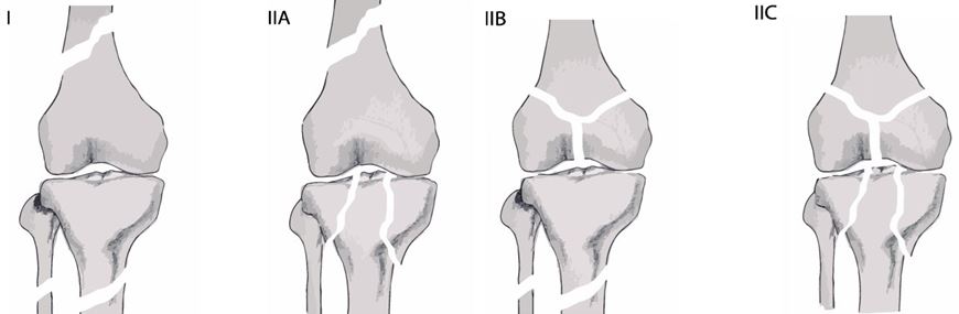
Piétu et al., in a retrospective analysis of 172 cases according to the Fraser classification, reported that 71.5% of the cases were type I and the repartition of the type II in the three subgroups was as follow: II A 8.2%, II B 11.6%, and II C 8.7%. At least one of the fractures was open in 69.2% of the patients. The average Injury Severity Score (ISS) was 19.5. The polytraumatized patients (ISS over 18) represented 37.7% of cases. The surgical procedure started within 6 first hours in 62% of injured people. The Intramedullary nailing was preferred method at femur site (73%, 126 cases), tibia Intramedullary nailing was performed in 54.4% of cases, and external fixation was used in one in every four patients. (11)

In a study by Abalo et al. on 43 patients with floating knee injuries, results showed that there were 32 males and 11 females with a mean age of 37 years. All patients had sustained their injuries in motor vehicle accidents. According to Fraser's classification, there were 21 type I, 10 type IIa, 7 type IIb, and 5 type IIc. The complications encountered were 1 case of fat embolism, 8 cases of knee stiffness, 10 cases of delayed union, 9 cases of infections, and 7 cases of nonunion. (12)


In our study, an in-depth examination of the data revealed significant findings. The average age of the individuals under review was 38 years, ranging from 17 to 67 years. Among the 10 cases analyzed, 8 were male and 2 were female. Fractures were equally distributed between the left and right lower extremities, with 5 cases observed on each side. Predominantly, road traffic accidents accounted for 60% of the cases, followed by falls from altitude contributing to 30%, and heavy object-related incidents representing 10% of the cases. Among the 10 patients, 4 (40%) were classified as Fraser type I, while 3 (30%) were categorized as type IIA. Additionally, 2 patients (20%) fell into the type IIb category, and 1 patient (10%) was identified as type IIC. (13)

In terms of treatment, both intramedullary nails and plates were employed for addressing femoral and tibial fractures, with an equal distribution of each method observed across 5 cases. The presence of concomitant injuries complicated the management strategy in 7 cases. Noteworthy findings include 50% of tibial fractures and 40% of femoral fractures being classified as open, with Gustilo type II being the predominant subtype in both instances. Our study observations revealed that fractures treated with interlock nails demonstrated favorable postoperative knee range of motion and early weight-bearing capability. Conversely, fractures managed with plate osteosynthesis were associated with prolonged pain, knee stiffness, delayed weight bearing, and an elevated risk of infection due to prolonged intraoperative durations. Patients treated with a combination of implants exhibited superior functional outcomes compared to those managed solely with plate osteosynthesis for both fractures.

Evaluation conducted at the 6-month follow-up indicated promising outcomes, with 80% of cases achieving excellent to good results based on the Karlström–Olerud scoring system. However, 10% of cases demonstrated acceptable results, while another 10% exhibited fair to poor outcomes, highlighting the inherent challenges associated with managing such complex injuries.

Conclusion

The floating knee presents as a complex injury extending beyond mere ipsilateral fractures of the femur and tibia. Prognostic indicators for both initial assessment and final outcomes include associated injuries and the type of fracture, Our experience underscores the persistent high rate of complications associated with the floating knee, regardless of the treatment modality employed. Surgeons should prioritize complication reduction in managing this injury. We advocate for an initial assessment of patients to identify potential life-threatening injuries, followed by prompt surgical fixation of both fractures, preferably utilizing external fixation in emergency settings. Additionally, attention should be given to addressing soft tissue injuries. Internal fixation, thorough evaluation of knee ligaments for associated injuries, and rigorous postoperative rehabilitation are essential components for achieving favorable functional outcomes. Adherence to meticulous management protocols is paramount throughout the treatment process.

References

1. Blake R, McBryde A. , Jr The floating knee: ipsilateral fractures of the tibia and femur. South Med J 1975;68:13-16. [Crossref][PubMed][Google Scholar]

2. Muñoz Vives J, Bel JC, Capel Agundez A, Chana Rodríguez F, Palomo Traver J, Schultz-Larsen M, Tosounidis T. The floating knee: a review on ipsilateral femoral and tibial fractures. EFORT Open Rev. 2017 Mar 13;1(11):375-382. doi: 10.1302/2058-5241.1.000042. PMID: 28461916; PMCID: PMC5367526 [Crossref][PubMed][Google Scholar]

3. Fraser RD, Hunter GA, Waddell JP. Ipsilateral fracture of the femur and tibia. J Bone Jt Surg Br Vol. (1978) 60-b(4):510–5. doi: 10.1302/0301-620X.60B4.711798 CrossRef Full Text | [Crossref][PubMed][Google Scholar]

4. Ran T, Hua X, Zhenyu Z, Yue L, Youhua W, Yi C, et al. Floating knee: a modified Fraser'classification and the results of a series of 28 cases. Injury. (2013) 44(8):1033–42. doi: 10.1016/j.injury.2012.12.012 PubMed Abstract | CrossRef Full Text | [Crossref][PubMed][Google Scholar]

5. Dwyer AJ, Paul R, Mam MK, Kumar A, Gosselin RA. Floating knee injuries: Long-term results of four treatment methods. Int Orthop. 2005;29:314–8 [Crossref][PubMed][Google Scholar]

6. Kumar R. The floating knee injury. J Clin Orthop Trauma. 2011;2:69–76 [Crossref][PubMed][Google Scholar]

7. Kao FC, Tu YK, Hsu KY, Su JY, Yen CY, Chou MC. Floating knee injuries: A high complication rate. Orthopedics. 2010;33:14 [Crossref][PubMed][Google Scholar]

8. Ulfin Rethnam,corresponding author1,4 Rajam S Yesupalan,2 and Rajagopalan Nair3J Trauma Manag Outcomes. 2007; 1: 2,Published online 2007 Nov 26. The floating knee: epidemiology, prognostic indicators & outcome following surgical management Paul GR, Sawka MW, Whitelaw GP. Fracture of the ipsilateral femur and tibia: Emphasis on intraarticular and soft tissue injury. J Orthop Trauma. 1990;4:309–14 [Crossref][PubMed][Google Scholar]

9. Adamson GJ, Wiss DA, Lowery GL, Peters CL. Type II floating knee: Ipsilateral femoral and tibial fractures with intraarticular extension into the knee joint. J Orthop Trauma. 1992;6:333–9 [Crossref][PubMed][Google Scholar]

10. Piétu G, Jacquot F, Féron JM. The floating knee: A retrospective analysis of 172 cases. Rev Chir Orthop Reparatrice Appar Mot. 2007;93:627–34 [Crossref][PubMed][Google Scholar]

11. Abalo A, Randolph S, Ayouba G, Walla A, Dossim A. Floating knee: Epidemiology and results of treatment. Niger J Orthop Trauma. 2011;10:23–7 [Crossref][PubMed][Google Scholar]


12. Management of the Floating Knee in Polytrauma Patients - Scientific Figure on ResearchGate. Available from: https://www. researchgate. net/figure/Fig-1-Frasers-classification-of-floatingknee_fig2_281099004 [accessed 25 Apr, 2024 [Crossref][PubMed][Google Scholar]

Disclaimer / Publisher's Note

The statements, opinions and data contained in all publications are solely those of the individual author(s) and contributor(s) and not of Journals and/or the editor(s). Journals and/or the editor(s) disclaim responsibility for any injury to people or property resulting from any ideas, methods, instructions or products referred to in the content.