Orthopaedic Journal of MP Chapter

Publisher: Madhya Pradesh Orthopaedic Association www.mpioa.com
E-ISSN:2582-7243, P-ISSN:2320-6993
2024 Volume 30 Number 1 Jan-Jun

Functional outcome of elastic nail fixation for Intertrochanteric fracture in medically high-risk elderly

Tirkey R1, Vidyarthi A2, Rajak CB3*

1 R Tirkey, Netaji Subhash Chandra Bose Medical College, Jabalpur, Madhya Pradesh, India.

2 A Vidyarthi, Netaji Subhash Chandra Bose Medical College, Jabalpur, Madhya Pradesh, India.

3* Chandra Babu Rajak, Department of Orthopaedics, Netaji Subhash Chandra Bose Medical College, Jabalpur, Madhya Pradesh, India.

Introduction: Intertrochanteric fractures in elderly population are major health problem. There are so many implants available to treat these fractures. We used enders nail for intertrochanteric fractures in medically compromised and high-risk patients.

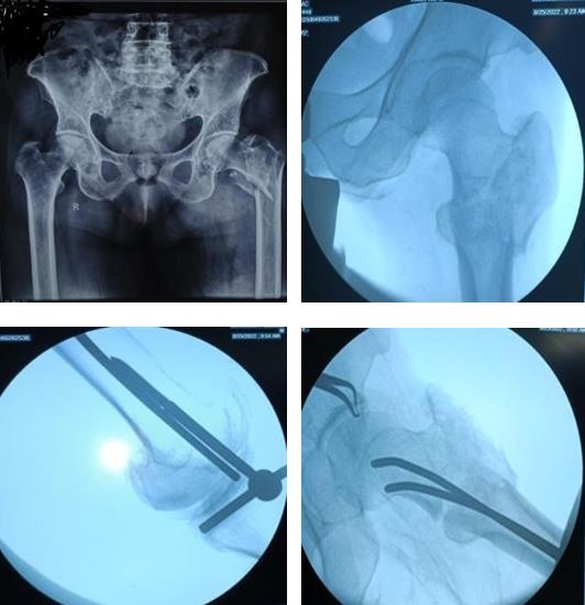
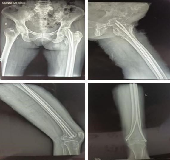
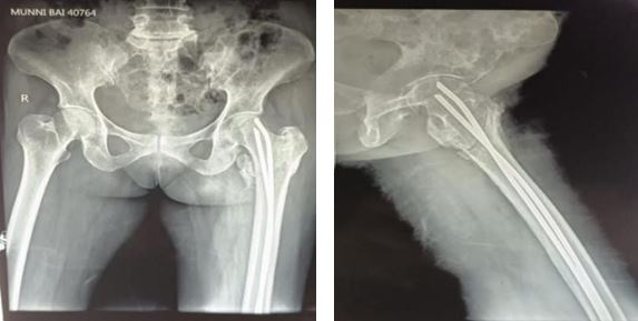
Material and method: Twenty-seven patients of intertrochanteric fractures of femur were operated by condylocephalic ender’s nail. We included elderly patient with age more than 60 year with high risk and medically compromised conditions. The clinical and radiographically assessment was done in all cases at 4-week, 6-week, 12 week and 6 months.

Results: Fracture healing was achieved in average 12.5 week (ranging from 10 week to 24 week). The mean Harris score was 82.

Conclusion: Our experience suggests that the chief indication of enders’ nail fixation is in the treatment of intertrochanteric femur fracture in critically ill patient who cannot tolerate anaesthesia for an hour and more. Ender’s nailing appears the least traumatic form of internal fixation.

Keywords: Intertrochanteric femur fractures, enders nailing, old patients

Corresponding Author How to Cite this Article To Browse
Chandra Babu Rajak, , Department of Orthopaedics, Netaji Subhash Chandra Bose Medical College, Jabalpur, Madhya Pradesh, India.
Email:
Tirkey R, Vidyarthi A, Rajak CB, Functional outcome of elastic nail fixation for Intertrochanteric fracture in medically high-risk elderly. ojmpc. 2024;30(1):17-20.
Available From
https://ojmpc.com/index.php/ojmpc/article/view/184
Manuscript Received Review Round 1 Review Round 2 Review Round 3 Accepted
2024-06-06 2024-06-12 2024-06-18 2024-06-24 2024-06-30
Conflict of Interest Funding Ethical Approval Plagiarism X-checker Note
Authors state no conflict of interest. Non Funded. The conducted research is not related to either human or animals use. 12.32 All authors have accepted responsibility for the entire content of this manuscript and approved its submission.

© 2024by Tirkey R, Vidyarthi A, Rajak CBand Published by Madhya Pradesh Orthopaedic Association. This is an Open Access article licensed under a Creative Commons Attribution 4.0 International License https://creativecommons.org/licenses/by-nc/4.0/ unported [CC BY NC 4.0].

Introduction

Intertrochanteric femur fractures are major public health problem because their frequency and the associated complications in older patient. In this already fragile population, the aim of treatment is to achieve prior functional level with low rate of complications and mortality. [1], [2] Various devices have been used which usually require an incision in the upper thigh involving considerable soft tissue dissection and significant blood loss. [3],[4] In 1970, Enders described a method of treatment for intertrochanteric femur fracture using flexible condylocephalic intramedullary nail. Advantage claimed in this operation is less technical, minimal blood loss and low morbidity and mortality. [8] [15] [10] In this article we described our experience with ender’s nail in intertrochanteric fracture in medically compromised and high-risk patient in the Neta ji Subhash Chandra Bose medical College Jabalpur.

Materials and methods

This prospective study includes 27 intertrochanteric fractures in medically compromised high risk elderly patient. Out of 27, 18 were stable and 9 were unstable fractures as per Evans classification. We included elderly patient with age more than 60 year with high risk and medically compromised. Presence of co-morbidities like 4 patients were having both diabetes mellitus and hypertension both. 9 patients were having ischemic heart disease with hypertension and Diabetes. 7 patients were having Chronic obstructive pulmonary disease with hypertension out of 3 also having diabetes mellitus.4 patients were having chronic renal failure with liver cirrhosis. Out of 3 having hypertension and 1 were having diabetes mellitus. 2 patients were having cerebrovascular disease. and 5 patients were having bedsore. Out of 27, 11 were female and 16 were male. All patient were walking except one hemiparetic patient who needed support to walk. The clinical and radiographically assessment was done in all cases at 4-week, 6-week, 12 week and 6 months.

Operative procedure – Under effect of anaesthesia the patient is put on fracture table in supine position with legs well abducted to allow the medial aspect of the knee. After draping reduction was achieved under c- arm. A nail of size was assessed pre operatively in normal limb from mid inguinal point to grater trochanter up-to adductor tubercle. Nail width was preoperatively assessed with more than 80 percent of isthmus under zero magnification x-ray. At diaphysio-metaphysial junction medial and lateral aspect of distal femur 1 cm skin incision was taken followed by entry was made with curved awl and enders nail was inserted firstly medial side enders nail was hammered up to cancellous bone of head followed by lateral side enders nail was also hammered in cancellous bone of femoral head and enders nail eye were locked using enders locking pins. And limb was kept in boot and bar brace to prevent rotation. Quadriceps strengthening exercises, knee mobilization and ankle pump were encouraged from the post operative day 1 with back care and chest physiotherapy was also encouraged.

Non wight bearing was advised for 6 weeks (non wight bearing toe touch with walker was permitted according to patient self-confident in post operative 4 week). Partial weight bearing was initiated after post op 6 week. it was gradually progressive to full weight bearing as per tolerance and absence of radiological evidence of collapse. Successive reviews were done at 12 week and 6 months during which rotation in flexion and extension, limb length discrepancy and knee range of motion were assessed.

Results

Patients were assessed at 4 week, 12 weeks and 6 months, out of 27 all were left the hospital in 12 – 15 days. The mean Harris score was 82. Out of 4 lost to follow up. bone healing was achieved in average 12.5 week ( ranging from 10 week to 24 week) .


Figure 1: A, B, C and D, Pre op x-ray and intra operative image intensifier pictures


Figure 2: A, B, C and D, 4 week follow up X-ray showing healing of fracture without displacement.


Figure 3: A and B At 6-month follow up, bone union achieved without displacement and shortening

Complications

Complications of enders nailing in 27 patients

ComplicationsNoPercentage
External rotation (15%)417.3 %
Shortening (avg 1.5 cm)521%
Varus deformity28.6%
Migration proximal14.3 %
Migration Distal14.3%
InfectionNil0 %
Total with complication1256.3%

Table 1: Complications of the surgery

Discussion

The series of cases emphasised again the advantage of enders nailing in intertrochanteric fracture, the operative procedure is simple, short and associated with minimal soft tissue and blood loss, as a results morbidity and mortality are lower than the other methods. Biomechanically placement of enders’ nails more medial than other devices that’s why fatigue failure was rare. Due to flexibility of nail lead to micromovement at fracture site accounts for the absence of delayed union and non-union. [11], [13] [19] Knee pain accounts for 1/3rd of patient but it resolves spontaneously or after removal of implant. [16]

Conclusion

Our experience suggests that the chief indication of enders’ nail fixation is in the treatment of critically ill patient who cannot tolerate anaesthesia for an hour and more. Even of proximal femoral nail and Dynamic hip screw with small incision causes bleeding and stress on physiology. enders nailing is the least traumatic form of internal fixation.

References

1. Veronese N, Maggi S. Epidemiology and social costs of hip fracture. Injury 2018:49:(8). 1458- 1460 [Crossref][PubMed][Google Scholar]

2. Brauer CA, CoCa-Perralion M, Cutler DM, Rosen AB. Incidence and mortality of hip fracture in the united states JAMA. 2009;302 (14):1573:302. [Crossref][PubMed][Google Scholar]

3. Roberts KC,BroxWT, Jevesevar DS, Sevarino K. Management of hip fracture in the elderly. J Am AcadOrthop Surg. 2015:23(2):131-137 [Crossref][PubMed][Google Scholar]

4. Braithawaite Rs , Col NF. Wong JB. Estimating hip fracture morbidity, mortality and costs. J Am Geriator soc 2003;35(9) :753 -765 [Crossref][PubMed][Google Scholar]

5. Silva Mj. Biomechanics of osteoporosis fractures Injury. 2007;38. [Crossref][PubMed][Google Scholar]

6. Bijhler J. (1975) Percutaneous intramedullary nailing of intertrochanteric fractures. In: The Hip, Proceedings of the Hip Society. Mosby, St Louis, pp. 170-179 [Crossref][PubMed][Google Scholar]

7. Einoder B. J. (1978) Ender nailing for proximal fractures. J. Bone Joint Surg. 618,239, (Abstract) [Crossref][PubMed][Google Scholar]

8. Ender H. G. (1973) Fixation Trochanterer Mit Fedemageln Nach Ender und Simon-Weidner. Lagenbecks. Arch. Chir. 332,935 [Crossref][PubMed][Google Scholar]

9. Kuderna H. , Bohler N. and Collon D. J. (1976) Treatment of intertrochanteric and subtrochanteric fractures of the hip by the Ender method. J. Bone Joint Surg. 58A, 604 [Crossref][PubMed][Google Scholar]

10. Iwegbu C. G. (1980) The place of Ender’s nails in the evolution of methods of treatment of trochanteric fractures of the femur. M.Ch.Orth. Thesis, University of Liverpool [Crossref][PubMed][Google Scholar]

11. Wynn Jones C. , Morris J. , Hirschowitz D. et al. (i977) A comparison of the treatment of trochanteric fractures ofthe femur by internal fixation with a nail plate and the Ender technique. Injury9,35 [Crossref][PubMed][Google Scholar]

12. . Nordin S, Zulkifil O, Faisham WI. Mechanical failure of DHS fixation in intertochanteric fracture femur. Med J Malaysia 2001;56:12-7 [Crossref][PubMed][Google Scholar]

13. Moon MS, Woo YK, Kim ST. A clinical study of trochanteric fractures of the femur: Outcome of the treatment in regard to osteoporosis and type of the treatment. I Korean Orthop Assoc 1991;26:1693702. [Crossref][PubMed][Google Scholar]

14. Hall LG. Comparison of nail-plate fixation Ender’s nailing for intertrochanteric fractures. J Bone Joint Surg 1981;63-B:24-8. [Crossref][PubMed][Google Scholar]

15. Russian LA, Sonni A. Treatment of intertrochanteric and subtrochanteric fractures with Ender intramedullary rods. Clin Orthop 1980;148:203-12. [Crossref][PubMed][Google Scholar]

16. Emami M, Manafi A, Hashemi B, Nemati A, Safari S (2013). Comparison of intertrochanteric fracture fixation with dynamic hip screw and bipolar hemiarthroplasty techniques. Arch Bone Joint Surg 1:14– 17. [Crossref][PubMed][Google Scholar]


17. an KP, Mohan R, Newman RJ (2002). The Gotfried percutaneous compression plate compared with the conventional classic hip screw for the fixation of intertrochanteric fractures of the hip. J Bone Joint Surg Br 84:1922. [Crossref][PubMed][Google Scholar]

18. M. ROOFMAN , L. SOLOMAN. Enders nail fixation for intertrochanteric fracture, department of orthopaedics surgery , university of Witwatersrand and johannesberg Hospital , johannesberg [Crossref][PubMed][Google Scholar]

19. Garg B, Marimuthu K, Kumar V, Malhotra R, Kotwal PP (2011) Outcome of short proximal femoral nail antirotation and dynamic hip screw for fixation of unstable trochanteric fractures. A randomised prospective comparative trial. Hip Int 21:531–536. [Crossref][PubMed][Google Scholar]

20. Adams CI, Robinson CM, Court-Brown CM, McQueen MM (2001). Prospective randomized controlled trial of an intramedullary nail versus dynamic screw and plate for intertrochanteric fractures of the femur. J Orthop Trauma 15:394–400. [Crossref][PubMed][Google Scholar]

21. Saudan M, Lubbeke A, Sadowski C, Riand N, Stern R, Hoffmeyer P (2002) Pertrochanteric fractures. is there an advantage to an intramedullary nail?. a randomized, prospective study of 206 patients comparing the dynamic hip screw and proximal femoral nail. J Orthop Trauma 16:386393 [Crossref][PubMed][Google Scholar]

22. Zeng C, Wang YR, Wei J, Gao SG, Zhang FJ, Sun ZQ, Lei GH (2012) Treatment of trochanteric fractures with proximal femoral nail antirotation or dynamic hip screw systems: a metaanalysis. J Int Med Res 40:839–851 ayali C, Agus H, Ozluk S (2006) Treatment for unstable intertrochanteric fractures in elderly patients: internal fixation versus cone hemiarthroplasty. J Orthop Surg 14:240–244. [Crossref][PubMed][Google Scholar]

Disclaimer / Publisher's Note

The statements, opinions and data contained in all publications are solely those of the individual author(s) and contributor(s) and not of Journals and/or the editor(s). Journals and/or the editor(s) disclaim responsibility for any injury to people or property resulting from any ideas, methods, instructions or products referred to in the content.