Orthopaedic Journal of MP Chapter

Publisher: Madhya Pradesh Orthopaedic Association www.mpioa.com
E-ISSN:2582-7243, P-ISSN:2320-6993
2023 Volume 30 Number 2 Jul-Dec

Pauwels’ osteotomy in fracture neck of femur in type II and type III

Tirkey R1, Barua VK2*, Vidhyarthi A3

1 R Tirkey, Netaji Subhash Chandra Bose Medical College, Jabalpur, M P, India.

2* Vinay Kumar Barua, Department of Orthopaedics, Netaji Subhash Chandra Bose Medical College, Jabalpur, M P, India.

3 A Vidhyarthi, Netaji Subhash Chandra Bose Medical College, Jabalpur, M P, India.

Background: Pauwels’ osteotomy is a promising procedure to treat non-union in fracture neck of femur of type II and type III, with good success rate.

Material and methods: This study is conducted in department of orthopaedics, Netaji Subhash Chandra Bose Medical College & Hospital, Jabalpur (M.P.) India from 1st December 2022 to 31st December 2023. This prospective study and functional analysis of Pauwel’s Osteotomy in fracture neck of femur in type II and type III was done on 10 patients.

Results: Out of 10, union was achieved in 8 patients and 2 patients were lost to follow up. Average time of union of fracture was 15 weeks. All the patients were able to squat, sit cross-legged and stand up on one leg.

Conclusion: Valgus osteotomy and fixation with dynamic hip screw has high success rate in young patients with neglected and ununited intracapsular fracture neck of femur as far as the union of fracture is concerned.

Keywords: Pauwels osteotomy, fracture neck of femur

Corresponding Author How to Cite this Article To Browse
Vinay Kumar Barua, , Department of Orthopaedics, Netaji Subhash Chandra Bose Medical College, Jabalpur, M P, India.
Email:
Tirkey R, Barua VK, Vidhyarthi A, Pauwels’ osteotomy in fracture neck of femur in type II and type III. ojmpc. 2023;30(2):31-36.
Available From
https://ojmpc.com/index.php/ojmpc/article/view/175
Manuscript Received Review Round 1 Review Round 2 Review Round 3 Accepted
2023-12-07 2023-12-13 2023-12-19 2023-12-25 2023-12-31
Conflict of Interest Funding Ethical Approval Plagiarism X-checker Note
Authors state no conflict of interest. Non Funded. The conducted research is not related to either human or animals use. 14.32 All authors have accepted responsibility for the entire content of this manuscript and approved its submission.

© 2023by Tirkey R, Barua VK, Vidhyarthi Aand Published by Madhya Pradesh Orthopaedic Association. This is an Open Access article licensed under a Creative Commons Attribution 4.0 International License https://creativecommons.org/licenses/by-nc/4.0/ unported [CC BY NC 4.0].

Introduction

Fracture neck of femur is aptly called as “the unsolved fracture”. This is because even with so much of advances in orthopedic field, there is no simple method of treatment which can give consistently successful results for this fracture. Management of this fracture especially in younger patients is a really demanding and challenging task for any orthopaedic surgeon (1). Fracture neck of femur is common in old people as many of them are osteoporotic. With improvement in quality of life leading to increased life expectancy, the incidence is even more common nowadays. Due to the congested vehicular traffic, it is also commonly seen in young patients after road traffic accidents and many a times they are polytraumatized. Many of these fractures are unstable. Because of its peculiar blood supply, a fracture neck of femur may cause circulatory disturbance leading to avascular necrosis and non-union. So, every fracture neck of femur should be treated as an emergency (2). It should be reduced accurately anatomically and fixed stably by one of the many implants available now. Usually, undisplaced stable fractures have a good prognosis and displaced unstable fractures a poor prognosis.

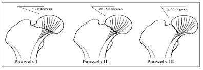
In Madhya Pradesh, because many patients go to native bone setters for treatment of fractures, these patients present with non-union of fracture neck of femur. Another factor leading to non-union is the angle of inclination of fracture. Usually, horizontal fractures with less than 30° of angle unite well and those with more than 30° may result in non-union even when treated expertly (3). This is because in fractures with more than 30° of inclination the resulting forces will act as shearing forces leading to displacement of fragments and non-union. The Pauwels’ principle which was described in 1927 is used, even today successfully. Pseudoarthrosis of femoral neck will unite, if inclination of pseudoarthrosis is changed in such a way that the shearing forces are converted into compression forces and converting unstable fracture into stable one. This leads to endochondral ossification of the fibrocartilage at pseudoarthrosis making the fracture to unite (4). Since, our patients require squatting for their routine daily activities, it is important to preserve the natural femoral head by making the fracture unite. One should not think of prosthetic replacement for every patient with fracture neck of femur. The best end result after fracture neck of femur treatment is the patient’s own healed femoral head and neck and every attempt must be made to achieve that goal (5).

Non-union after femoral neck fracture is defined as lack of radiographic evidence of union 6 months after fracture. King [6], in 1939, in his comprehensive review of both recent and old case of fracture neck femur, emphasized that 3 weeks old can be arbitrarily called old and ununited, as it can be assumed that the head of the femur is devoid of its blood supply, has less chances of osseous union and that secondary changes are more probable, than if operation were performed earlier. If the fracture neck of femur remains untreated for more than 3 weeks, internal fixation alone is likely to have high rate of non-union, as reported by Barnes et al. [1]; in their series on fractures of more than 1-week duration they had non-union rate as high as 50%.

The femoral neck fracture is probably the fracture, for which there exists the larger number of methods of osteosynthesis. Internal fixation of femoral neck fracture is followed by certain incidence of fixation failure. Nonunion may occur in one-third of patients, with higher rate in vertical or displaced fractures [7]. Number of methods of internal fixation when used alone has failed to achieve the desired end result. Osteoporosis directly influences degree of displacement and quality of internal fixation. The deficient bone stock and posterior comminution play a significant role in biological failure to unite the fracture. Age and sex of patient, osteoporosis, degree of displacement and quality of fracture reduction are the factors that have been found to affect the end result [8, 9]. The appropriate treatment for non-union of a femoral neck fracture depends on the age of the patient, his or her medical status, viability of the femoral head, size of the remnant femoral neck, osteoporosis, duration of the disease and finally the state of joint space. In patients younger than 55 years who are in good general medical condition, with no osteoporosis and with a reasonable size of femoral neck and normal joint space, it is desirable to preserve the femoral head, particularly if the patient’s lifestyle and social and religious customs require squatting and sitting in a cross-legged position [10]. Treatment options for femoral neck non-union with preservation of the femoral head include refixation of the fracture, refixation and bone grafting, a pedicle graft to provide blood supply, or a valgus osteotomy with fixation. Cortical bone grafts have been associated with dis-impaction or angulations of the head leading to failure [11]. The initially reported success of Meyer’s procedure has not been reproduced in a large series and the procedure has been considered unreliable [12]. Arthrodesis has a high failure rate but, when successful, it leads to a functional but immobile hip. Different techniques of vascularized bone grafting have been introduced, often with excellent results [13, 14], but the usefulness of these techniques is limited because of donor-site morbidity and limb length discrepancy with residual varus deformity.

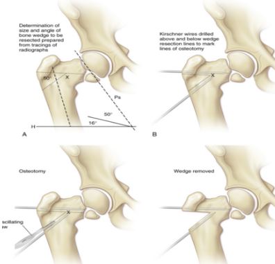
We believe, that valgus osteotomy acts as a biological stimulus for healing of these fractures, promoting osteogenesis as a result of conversion of shearing forces to compressive forces across the fracture site. The osteotomy is relatively easy to perform, cost effective, provides stability and often is definitive one-time surgery. Valgus osteotomy and internal fixation with dynamic hip screw and angle barrel plate plays a twofold role, it converts the shearing forces into the compression forces by placing the fracture site perpendicular to the resultant of body weight forces; and it buttresses the head of femur from below to improve stability provided by the internal fixation. Osteosynthesis with dynamic hip screw in its optimum position in the femoral head, supplemented by the buttressing effect of the distal osteotomy fragment, maintains coaptation and immobilization of the fragments and provides a high degree of stability. Following osteotomy, valgus orientation of the proximal femur decreases the lever arm and therefore increases contact pressure on the head. keeping in mind the facts of above-mentioned studies, it seems that performing valgus osteotomy in a hip with changes of AVN may lead to progression of disease and a painful hip later on, and therefore we excluded these patients from our study.


Many authors have reported that preoperative presence of osteonecrosis is not a contraindication for osteotomy [17].

There is a criticism about the difficulty in performing THR in these patients, when required at a later stage. However, Marti et al. [17] reported no such problems. Kirby [18] stated that the standard prosthesis could be used for THRs in majority of the cases.

Material and method

This Study is conducted in Department of Orthopaedics, Netaji Subhash Chandra Bose Medical College & Hospital, Jabalpur (M.P.) India from 1st December 2022 to 31st December 2023. This prospective study and functional analysis of Pauwels’ osteotomy in fracture neck of femur in type II and type III, was done on 10 subjects. Patients with fracture neck of femur, non-union fracture neck of femur under age group 25 to 70 years were included in the study. Patient not included in the study were patients not willing to be operated, pathological fracture of neck of femur, fracture neck femur with secondary osteoarthritis of hip joint, patients with advanced Avascular Necrosis (AVN) changes on plain radiography, patients with significant resorption of femoral neck with proximal fragment less than 2.5 cm.

Pauwels osteotomy: This procedure makes use of the Pauwels principle which states that, if the fracture inclination is reduced to less than 30°, the forces acting on the fracture are converted into compression forces making the fracture unite. Here, a laterally based wedge is removed at the level of lesser trochanter and when the osteotomy is closed, the fracture line will become more horizontal. The fracture and osteotomy are fixed stably using AO 145°, AO 150° and AO 155°, angled blade plate or DHS. The preoperative Pauwels’ angle will be calculated for each fracture and the osteotomy will be planned in such a manner so as to achieve a Pauwels’ angle of 30° or less. The goal of osteotomy is to achieve compression at the fracture site. The site of osteotomy is at the level of lesser trochanter and desired wedge will be resected depending on Pauwels’ angle as described by Pauwels F and later modified by Mueller ME.

The patient is taken on fracture table and manual reduction achieved under C-ARM guidance. Lateral incision given from 2cm above the greater trochanter to 6-7 cm distally. Superficial and deep dissection done and greater trochanter exposed.

K wire inserted to fix the reduction temporarily followed by Richards screw insertion and then k wires are used to mark the osteotomy site and angle with the help of pre-operative angle calculations and under C-ARM guidance and the wedge is resected and then abduction of the leg done and traction is released and using AO 145°, AO 150° and AO 155°, angled blade plate or DHS plate is fixed with cortical screws. Surgical site washed with normal saline and betadine 5% solution and suturing done in layers. post operative 2nd and 5th day check dress was done. post-operative physiotherapy was started. Suture removal done at post operative 14th day. Patient was advised non weight bearing for 6 weeks.

175_01.JPG
Figure 1: Pauwels classification for femoral neck fractures

175_02.JPG
Figure 2: Steps of Pauwels Osteotomy

175_03.JPG
Figure 3: A, B, C, D Intra-operative c-arm pics


175_04.JPG
Figure 4: A, B, C, D, E, Case-1: 45 year old male, DOI- 12/06/2023, Operated on-28/08/2023

Observations

Patients presented with pain over affected hip and inability to walk being the most common complaints. Radiographic images confirmed the diagnosis and MRI was done for evaluation of the femoral head vascularity.

Table 1: Age of the patients

AgeNo of casesPercent
25-35220
35-45440
45-55110
55-65330
65-7500
Total10100

Table 2: Sex of patients

SexNo of casespercent
Female220
Male880

175_05.JPG
Figure 5: A,B,C,D,E, Case-2: 38 year old male, DOI- 26/04/2023, Operated on 18/06/2023


Table 3: Harris hip score at final follows up

CaseHHS
Case-196%
Case-282%
Case-387%
Case-475%
Case-559%
Case-685%
Case-783%
Case-871%
Case-951%
Case-1064%

Results

There were 10 patients in our study, 20% cases in our study belongs to age group 25-35years, 40% belongs to 35-45years, 10% belongs to 45-55years and rest 30% belongs to age 55-65 (mean 45.3 years).


Eight were males and two females. The majority of patients were in the age group of 35-45 years (Four patients). Mean duration of injury was 9.8 weeks with a range of 3—20 weeks at the time of osteotomy and fixation. There were 7 type 2 and 3 type 3 fractures according to Pauwels’ type. Out of 10, union was achieved in 8 patients and 2 patients were lost to follow. None of the fractures was complicated by intra-articular penetration of blade. The average time of union following procedure among different Pauwels’ types are shown in Table below. Average time of union of fracture was 15 weeks. There was no statistically significant difference in the time taken in weeks to unite following procedure between type 2 and type 3. There was no relation to the type of fracture, degrees of wedge resection for the osteotomy and the abduction restriction. All patients who had lengthening had wedge resection of 20-30 degree. However, all the patients were able to squat, sit cross-legged and stand up on one leg.

Table 4: Average time of union of fracture

No. of casesUNION TIME(WEEKS)
114
214
315
417

Discussion

Pauwels’ recognized that non-union of femoral neck fracture would consolidate within few months, if shearing force acting on non-union fracture site were transformed into compression forces [15]. Good results had been reported in femoral neck fracture treated with primary osteosynthesis and valgus intertrochanteric osteotomy [16, 17]. Marti et al. [17] reported 86% union in 50 patients at an average of 3.6 months, treated by this method alone. Sameer Gupta et al (19) operated 60 patients (mean age, 35 years) by valgus subtrochanteric osteotomy and repositioning of the osteotomy and fixation with a dynamic hip screw and a 135° single-angled barrel plate for closed un-united femoral neck fractures after failed internal fixation (n=27) or neglected (>3 weeks) fractures (n=33). Bone union was achieved in 56 patients after a mean of 3.9 (range, 3-5.5) months. The mean Harris hip score improved from 65 to 87.5. Outcome was excellent in 30 patients, good in 24, and poor in 6. Four of the patients developed avascular necrosis; 2 of whom nonetheless achieved a good outcome (19). In our study, follow-up did not reveal any avascular necrosis and subchondral collapse. Postoperatively we achieved an average fracture plane of 30 degree (25–40 degree), as we aimed for fracture plane of less than 30 degrees. This technique resulted in union in 8 of 10 patients as the use of dynamic hip screw added compression at the fracture site. In dynamic hip screw fixation, powered instruments could be used, which saves time and reduces blood loss. In our present study, because of the delay in intervention and the young age of patients, Pauwels’ osteotomy was performed as a head-salvaging procedure. Union was achieved in 80% of our patients, which is quite good for any surgical intervention as 2 patients were lost to follow up.

Among the united fractures, all were able to sit cross-legged, squat and stand on affected leg, which are needed in day-to-day life. This is also another advantage over the head-replacing procedure. Follow-up period of 1.5 year in our study was a limitation in this regard and longer follow-up is required for evaluation of collapse in femoral head.

Conclusion

We conclude by stating that valgus osteotomy and fixation with dynamic hip screw and angle barrel plate has high success rate in young patients with neglected and ununited intracapsular fracture neck of femur as far as the union of fracture is concerned, and internal fixation with dynamic hip screw and angle barrel plate is technically simple, with additional advantage of compression at fracture site by hip screw.

References

1. Barnes JT, Brown JT, Garden RS, Nicoll EA (1976). Subcapital fractures of the femur. a prospective review. J Bone Joint Surg 58B:2–24. [Crossref][PubMed][Google Scholar]

2. Ballmer FT, Ballmer PM. Pauwels' osteotomy for non-unions of femoral neck fractures. OrthopedicClinics of North America 21 (4): 759-767,1990. . [Crossref][PubMed][Google Scholar]

3. Bombelli R: The role of osteotomy as a consequent therapy. In: Osteoarthritis of the Hip. 2nd ed. , Berlin: Springer-Verlag, pp. 49-119, 1983 [Crossref][PubMed][Google Scholar]

4. Braune W, Fischer 0: Rotations of the hip line: experiments on man, loaded and unloaged. In The Human Gait. Berlin: Springer-Verlag, pp. 94-103,1987. [Crossref][PubMed][Google Scholar]

5. Marti RK, Schüller HM, Raaymakers EL. Intertrochanteric osteotomy for non-union of the femoral neck. J Bone Joint Surg Br. 1989;71:782-7. [Crossref][PubMed][Google Scholar]

6. King T (1939). Closed operation for intracapsular fracture of the neck of the femur. Br J Surg. 26:721. [Crossref][PubMed][Google Scholar]

7. Lu-Yoa GL, Keller RB, Littenberg B, Wenliberg JE (1994). Outcomes after displaced fractures of the femoral neck. A metaanalysis of one hundred and six published reports. J Bone Joint Surg Am 76:15–23. [Crossref][PubMed][Google Scholar]

8. Garden RS (1964). Stability and union in subcapital fractures of the femur. J Bone Joint Surg Br. 46:630–647. [Crossref][PubMed][Google Scholar]

9. Dalen N, Jacobson B (1986). Rarefied femoral neck trebecular patterns. fractures displacement and femoral head vitality in femoral neck fractures. Clin Orthop 207:97–98. [Crossref][PubMed][Google Scholar]

10. Hammer AJ (1992). Nonunion of subcapital femoral neck fractures. J Orthop Trauma. 6:73–7. [Crossref][PubMed][Google Scholar]


11. Baksi DP (1986). Internal fixation of ununited femoral neck fracture combined with muscle pedicle bone grafting. J Bone Joint Surg Br. 68:239–245. [Crossref][PubMed][Google Scholar]

12. Zukerman JD, Koval KJ (1993) Hip trauma. In. Frimoyer JW (ed) Orthopaedic knowledge update 4. American Academy of Orthopaedic Surgeons. Rosemont. pp 525–538 [Crossref][PubMed][Google Scholar]

13. LeCroy CM, Rizzo M. Gunneson EE et al (2002). Free vascularised fibular bone grafting in the management of femoral neck non union in patients younger than fifty years. J Orthop Trauma 16:464–472. [Crossref][PubMed][Google Scholar]

14. Nagi ON, Dhillon MS. Goni VG (1998) Open reduction. internal fixation and fibular autografting for neglected fracture of the femoral neck. J Bone Joint Surg Br 80-B:798–804. [Crossref][PubMed][Google Scholar]

15. Pauwels F (1935). Der Schenkelhalsbruch ein mechanisches Problem. Grundlagen des Heilungsvorganges, Prognose and kausale Therapie. Ferdinand Enke Verlag, Stuttgart. [Crossref][PubMed][Google Scholar]

16. Rinaldi E, Marengh P, Negri V (1984). Osteosynthesis with valgus osteotomy in the primary treatment of subcapital fractures of the neck of the femur. Ital J Orthop Traumatol. 10(3):313–320. [Crossref][PubMed][Google Scholar]

17. Marti RK, Schuller HM. Raaymakers EL (1989). Intertrochanteric osteotomy for nonunion of the femoral neck. J Bone Joint Surg Br 71:782–787. [Crossref][PubMed][Google Scholar]

18. Kirby H (2002). Femoral neck non-union. osteotomy or arthroplasty. Tech Orthop. [Crossref][PubMed][Google Scholar]

19. Sameer Gupta, Sunil Kukreja, Vivek Singh. Valgus osteotomy and repositioning and fixation with a dynamic hip screw and a 135º. single-angled barrel plate for un-united and neglected femoral neck fractures. Journal of Orthopaedic Surgery 2014;22(1):13-7. [Crossref][PubMed][Google Scholar]

Disclaimer / Publisher's Note

The statements, opinions and data contained in all publications are solely those of the individual author(s) and contributor(s) and not of Journals and/or the editor(s). Journals and/or the editor(s) disclaim responsibility for any injury to people or property resulting from any ideas, methods, instructions or products referred to in the content.