Orthopaedic Journal of MP Chapter

Publisher: Madhya Pradesh Orthopaedic Association www.mpioa.com
E-ISSN:2582-7243, P-ISSN:2320-6993
2022 Volume 28 Number 2 Jul-Dec

Functional and radiological outcome of locking compression plate for distal end radius fracture

Patidar A1, Jadhav A2*, Jain A3, Bhinde S4, Singh V5, Rathore S6, Sharma S7

1 A Patidar, Department of Orthopaedics, RD Gardi Medical College, Ujjain, Madhya Pradesh, India.

2* Aditya Jadhav, Senior Resident, Department of Orthopaedics, RD Gardi Medical College, Ujjain, Madhya Pradesh, India.

3 A Jain, Department of Orthopaedics, RD Gardi Medical College, Ujjain, Madhya Pradesh, India.

4 S Bhinde, Department of Orthopaedics, RD Gardi Medical College, Ujjain, Madhya Pradesh, India.

5 V Singh, Department of Orthopaedics, RD Gardi Medical College, Ujjain, Madhya Pradesh, India.

6 SS Rathore, Department of Orthopaedics, RD Gardi Medical College, Ujjain, Madhya Pradesh, India.

7 SK Sharma, Department of Orthopaedics, RD Gardi Medical College, Ujjain, Madhya Pradesh, India.

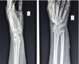
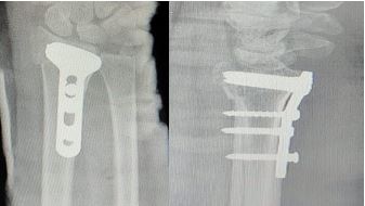
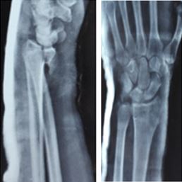
Background: Fracture distal end of radius are one of the most common skeletal injuries encountered in orthopaedic departments. Various modalities for treatment like close reduction and cast application, external fixators, ORIF with plate etc. Close reduction and cast application or K-wire fixation, external fixation is associated with immobilisation, wrist stiffness, loss of reduction postoperatively. So, we performed volar plating for distal end radius fractures and assessed the results.

Study design: Prospective case series of 48 patients who underwent volar plating for distal end radius fracture at CRGH associated with R D Gardi Medical College, Ujjain.

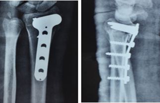
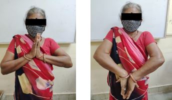
Methods: We performed ORIF with plating (Volar approach) in 48 patients (32 male and 16 female) of distal end radius fractures, depending on inclusion and exclusion criteria. Fractures were classified according to Frykman classification. All patients were operated under tourniquet and anaesthesia. Results were analysed using Green and o'brien scores for functional and Lidstrom classification for radiological outcome.

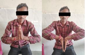
Results: Out of 48 patients, as per Frykman classification 1 had type III, 30 had type IV, 7 had type V,8 had type VI and 2 had type VII fractures. RTA as a (52.1%) mode of trauma was higher as compared to Fall (47.9%). Average time to clinico-radiological union was 8 weeks with the longest follow up time 6 months. Average time to wrist mobilisation was 8 days. According to Green and o'brien scores, 22 patients had excellent,20 good and 6 fair results. As per Lidstrom classification 26 patients had excellent,18 good and 4 fair results.

Conclusion: Volar plating for fracture distal end radius reduced the chances of wrist joint stiffness, loss of reduction and good results increased the patient compliance.

Keywords: distal end radius fracture, Volar plating, locking compression plate

Corresponding Author How to Cite this Article To Browse
Aditya Jadhav, Senior Resident, Department of Orthopaedics, RD Gardi Medical College, Ujjain, Madhya Pradesh, India.
Email:
Patidar A, Jadhav A, Jain A, Bhinde S, Singh V, Rathore S, Sharma S, Functional and radiological outcome of locking compression plate for distal end radius fracture. ojmpc. 2022;28(2):75-82.
Available From
https://ojmpc.com/index.php/ojmpc/article/view/162
Manuscript Received Review Round 1 Review Round 2 Review Round 3 Accepted
2024-08-03 2024-08-10 2022-12-17 2022-12-24 2022-12-31
Conflict of Interest Funding Ethical Approval Plagiarism X-checker Note
Authors state no conflict of interest. Non Funded. The conducted research is not related to either human or animals use. 12.54 All authors have accepted responsibility for the entire content of this manuscript and approved its submission.

© 2022by Patidar A, Jadhav A, Jain A, Bhinde S, Singh V, Rathore S, Sharma Sand Published by Madhya Pradesh Orthopaedic Association. This is an Open Access article licensed under a Creative Commons Attribution 4.0 International License https://creativecommons.org/licenses/by-nc/4.0/ unported [CC BY NC 4.0].

Introduction

Fractures of the distal end of the radius accounts around 15% of all fractures diagnosed and treated in the emergency rooms. In the words of Dr. Abraham Colles “this fracture takes place about an inch and a half above the carpal extremity of the radius and there is a 17% lifetime probability for distal radius fractures”. Most of these injuries are ‘closed’, with no evidence of breach of the surrounding skin.

Fractures of distal end of radius are one of the most common skeletal injuries. Though the management of these fractures remained a controversial issue.1 These were often treated with closed reduction and longer immobilisation but the difficulty here is the possibility that displacement may persist even in the least complex fractures. Other problem with this method is immobilisation of wrist and forearm for at least 6 weeks and the further time required to regain the functions of forearm wrist and hand by physiotherapy. During this entire time duration, patient's ability to carry out day to day activities is hampered. The need of the hour is the treatment modality that restores and maintains anatomy and allows early functional mobility which allows patient to carry out his activities of day-to-day life with minimal discomfort.

A bimodal age distribution is seen, with differing trends in males and females. In females, the probability of these fractures rises with age, especially from the age of 40 years and onwards; in people below the age of 40 years, the incidence is more in males. In young people, these fractures usually result secondary to high velocity injury such as a motor vehicle accident or fall on an outstretched hand. While in the elderly, the fracture commonly occurs from low-energy or moderate trauma, like a fall from standing height. This is due to the fragile bone in older adults because of osteoporosis.

Management for distal radius fractures has evolved significantly over the years. Closed Manipulative Reduction (CMR) and below elbow cast application has been the main treatment. However, the outcomes are often less than satisfactory with loss of reduction, limitation of function and disabilities. Several studies have shown that after a distal radius fracture, patients function more effectively when the anatomy is restored. Despite this fracture being very common, we lack evidence to support a single reparative technique as against the use of others. It is difficult to compare “fractures of the distal radius” because of the many different specific patterns of lesions. In addition, different plates are used for fixation, different outcome tools are used, and the fractures also vary in their complexity.

Numerous other methods of treating injuries of this nature like closed percutaneous pinning, external fixation, buttress plating have enjoyed recognition from time to time, testifying the fact that there is no ideal modality of treatment. The anti-glide effect of Buttress plates helps to reduce and stabilise intra-articular fractures.2 However, the need for protection of fracture, till it consolidates and the chances of loss of reduction on mobilisation are still areas of concern with Buttress plating.

Treatment of comminuted, displaced intra-articular or potentially unstable fractures of the distal radius with open reduction and internal fixation with locking compressions plates (LCP) and screws has increasingly been found to be the better alternative.3

The functional outcome of treatment of fracture of the distal aspect of the radius is influenced by the anatomical reduction of the articular surface and the extraarticular alignment of the distal part of the radius.4 By directly restoring the anatomy, plating allows secure internal fixation with resultant early return of wrist function. Furthermore, the increase in the incidence of sympathetic dystrophy with immobilisation over long duration is circumvented by this novel method of fixation.

This new fixation technique of using LCP for treating distal end radius fracture shows promise in terms of stable intraoperative fixation and restoration of acceptable anatomy, resulting in early mobilisation and good recovery of function. 5,6,7 This holds true even for osteopenic bones.7,8 Stable internal fixation with minimal complications can be achieved using distal radius LCP. 7,9 We report a series of 48 distal radius fractures treated by this method of internal fixation with satisfactory results in all.

Material and method

Inclusion Criteria was patient with distal end radius close fracture, aged between 18 to 60 years. Also, patient with complete clinical records and medically and surgically fit for surgery. Exclusion Criteria was patient with associated ipsilateral fracture, compound fracture, pathological fracture, age group <18 and > 60.

Protocol was followed once patient was admitted and planned for surgery. First, fracture was stabilized with slab immobilization and limb elevation given then surgery delayed till soft tissue becomes favourable for surgery. Patients were admitted and physical fitness for surgery was obtained. Fractures were classified using Frykman classification and randomization was done based on day of admission and mode of treatment was decided. patient operated with volar distal radius plating application.


Post operatively intravenous antibiotic regimen given, along with IV fluids and pain control medications. Joints were mobilized after 48 hrs. Patients were encouraged for active finger and elbow movements.At Follow-up Patients were regularly followed 6,12 and 24 weeks, and every 4 weeks thereafter until radiographic healing and full functions are achieved.

During follow-up we evaluated any possible loss of reduction. Also, assessment and analysis of any complication. Functional outcome was assessed according to the Green and O'Brien scoring system.

Observation and Result

The study comprised a total of 48 patients of fractures of the distal radius according to inclusion and exclusion criteria for the final evaluation of results. The study was performed between January 2019 to june 2020.

The mean age of the patients taken up for the study was 33.35 years. There were 16 female patients (33.3%) and 32 male patients (66.7%). Mode of trauma due to RTA 25(52.1%) is more as compare to Fall 23 (47.9%). Left side 28 (58.30%) is involved more as compared to right 20 (41.70%).

Out of 48 patients, we had Frykman type-III-01(2.1%) type-IV-30(62.5%), type-V-07(14.6%), type-VI-08(16.7%), type-VII-02(4.2%) fracture. As per Green and O’Brien scores, 22 patients have excellent, 20 have good and 6 have fair results. And as per Lidstrom classification 26 patients have excellent, 18 have good and 4 have fair results.

Table 1: Functional outcome   

FUNCTIONAL SCOREFREQUENCYPERCENT
EXCELLENT2245.8
GOOD2041.7
FAIR612.5
Total48100.0

Table 2: Radial angle

RADIAL ANGLEFREQUENCYPERCENT
EXCELLENT2245.8
GOOD1939.6
FAIR714.6
Total48100.0

Table 3: Volar tilt presentation.   

RADIAL LENGTH(MM)FREQUENCYPERCENT
EXCELLENT3164.6
GOOD1225.0
FAIR510.4
Total48100.0

Table 4: Radial length shortening.

VOLAR TILTFREQUENCYPERCENT
EXCELLENT510.4
GOOD4185.4
FAIR24.2
Total48100.0

Table 5: Lidstrom Classification Outcome

OUT COMEFREQUENCYPERCENT
EXCELLENT2654.2
GOOD1837.5
FAIR48.3
Total48100.0

Table 6: Final Result

RESULTFREQUENCYPERCENT
EXCELLENT2041.7
FAIR612.5
GOOD2245.8
Total48100.0

Case 1:

ojmpc_162_01.jpg
Figure 1: Pre op x-Ray

ojmpc_162_02.jpg
Figure 2: Post op x-ray


ojmpc_162_03.jpg
Figure 3 and 4: Follow up functional results

Case 2:

ojmpc_162_04.jpg
Figure 5: Preop Xray

ojmpc_162_06.jpg
Figure 6: Post op x-ray

ojmpc_162_07.jpg
Figure 7 and 8: Follow up of patient with functional outcome

Discussion

Over the last decade, several studies have been directed towards clarifying which surgical treatment method would be best for fracture of the distal extremity of the radius. In this context, Osada et al recently documented the increasing popularity of open reduction and internal fixation, especially since the introduction of locked volar plates in 2001.13 They demonstrated that locked volar plates are well tolerated, allow early mobilization, and provide good support for muscle forces after the surgical reduction, even in intra-articular fractures. The majority of the studies have used subjective tools for measuring quality of life, such as the Gartland and Werley calculation and the DASH calculation while others have given greater emphasis to the radiographic parameters obtained after surgical reduction of fractures of the distal extremity of the radius.12-15

In present study patients evaluated functionally by Green and O'Brien score and radiologicaly by Lidstrom classification. The average age of the patients in our study was 33.35 years. The eldest patient in the study was 65 years of the age and the youngest patient was 22 years old (mature skeleton) as compare with other studies like Kevin C, Chung et al had average age of the patients was 48.9 years with minimum age 18 years and maximum years 83 years.16 In Rohit et al average age of the patients was 57 years with minimum age 17 years and maximum years 79.17 In Kilic et al average age of the patients was 45 years with minimum age 18 years and maximum years 77.18 In Anakwe et al average age of the patients was 48 years with minimum age 22 years and maximum years 67.19

In present study, most of the patients were males 32 (66.7%) as compared with females 16 (33.3%). In Margaret Fok WM et al study there were 56 (57.7%) male and 41 (42.3%) female while Tank Gyaneshwar study had female 52 (65%) and male was 28 (35%) in number.20,21


In the present study, the mode of injury was road traffic accident in 25 patients (52.1%), fall in 15 patients (47.9%). In the study of Ayhan Kilic et al also found road traffic accident in 13 patients (48.1%) and fall in 14 patients (51.9%) while Chung KC et al found 42 patients (48.3%) had road traffic accident and 45 patients (51.7%) had trauma due to fall.16,18 Arora Rohit et al in his study got 40 patients with road traffic accident and 60 patients due to fall. 17

In present study, out of 48 patients, 3 patients (6.3%) had an associated injury, similarly in Jakob M et al study out of 73 patients, 21 (28.8%) had associated injury and In Aggarwal AK et al study out of 16 patients, 10 (62.5%) had associated injuries.22,23 The percentage of associated injuries was low in our study because of exclusion criteria, while in other studies considered all the varieties of injuries.

In present study, 3.5mm size plate was used. Similarly, in Aggarwal AK et al study also 3.5 mm size plate was used. Jakob M, et al in his study, 2 mm plate used. While Santiago A et al used 2.4 mm plate size.22-24 The use of plate and screws allows more accurate treatment after reconstruction of joint under direct vision and reestablishment of radial length, both intermediate and lateral columns can be buttressed and cancellous

bone graft may be added where it is needed. The 2 screws usually give good purchase in distal fragments. A 3.5 mm T plate can be used for the intermediate column but not for the lateral column and also this plate is too big for small fragments, and it is difficult to obtain a good purchase in comminuted distal fragments.25 There is extensive work to show that locked volar plates are well tolerated, allow early movement and maintain position even for intraarticular fractures.26,27 Proposed advantages of locked volar plating include improved pull-out strength even in osteoporotic bone.28 Internal fixation using a dorsal plate, which is greatly advocated, achieves anatomical reduction with good stability.

However, a variety of complication has been documented, including irritation of subcutaneous tissue, tenosynovitis of extensor tendons, rupture of extensor tendon and even chronic pain.29 In view of this fixed angle locked volar plate for the distal end of radius have gain much space among orthopaedic surgeons, since these plates are not only provide stable fixation but also avoids the above-mentioned complication.30,31 Volar surgical approach that avoids need for an extensive dorsal dissection. The plate is positioned in well-padded area beneath pronator quadratus to avoid flexor tendon irritation and it is thought that patient tolerate volar wrist scar better than dorsal one.32,33 As they are less obvious and the blood supply to the radius is less likely to be disturbed.

Dorsal plate fixation is biomechanically effective in buttressing a dorsally displaced fracture of distal radius. Osada et al compared the biomechanical properties of dorsal and volar fracture fixation plate designs in a cadaver model.34 They reported that if, the volarily placed titanium symmetry plate was used to fix a colles – type fracture, the distal fragment of radius to develop a dorsal angulations of about 9 degrees, if early active mobilization of fingers was initiated during the postoperative period, on the other hand, Leung et al demonstrated no statistical difference between axial loading transmission though the intact radius and a distal radius fracture fix with a volar locking plate.35 In fact, the volar locking plate showed advantages over dorsal plating in the fixation of dorsally unstable distal radius fracture. In addition, volar plate fixation is a valuable method because of decreased risk of inducing dorsal soft tissue complications. The dorsal approach often needs dissection of the extensor retinaculum and sometimes, dissection of lister tubercle. Therefore, the extensor tendon generally exposed to mechanical attrition by the plates and screw.

In the volar approach, the volar anatomy of the wrist presents advantage over the dorsal aspect because there is more space between the volar cortex and the flexor tendons, and the pronator quadratus can also sometime act as a hedge to prevent soft tissue complications. The palmer cortex is relatively flat, and the plate better contoured for application from this aspect rather

than on the dorsal cortex of the distal radius.36 The volar cortex of the distal radius was very often not as communited when compared with dorsal cortex. Anatomical reduction of palmar cortex may avoid the shortening of the radius, which is important for its restoration. The volar plate system used in our study was a locking plate system and this must be one of the reasons for retaining good anatomical reduction.

The optimal placement of distal screw is important, they must be inserted at the radius styloid, beneath the lunate facet, and near the sigmoid notch. Therefore, the plate is positioned near the volar radius margin. But fixation implants placed over or distally to the watershed line can exert pressure on the flexor tendon and cause injury. The watershed line is defined as a transverse ridge that is the most volar aspect of the volar margin of the radius distally. Distal to this line, the radius slopes in a dorsodistal direction and become prominent, palmarly.

The course of flexor pollicis longus tendon is close to the palmar rim of the distal radius. The plate placed very close to the wrist joint can support the palmar aspect of the articular surface.


However, it sometimes causes flexor tendon impairment in the very distal area, it is not possible for the reattached pronator quadratus muscle to protect the flexor tendon. As a result, the tendon can abrade against the plate and sharp edges of the screw heads. It also must be emphasized that protruding screw heads can cause tendon irritation. To avoid rupture of flexor pollicis longus tendons, care must be taken especially in a very distal fracture, type C3 fractures and osteoporotic bone. Adequate image intensifier control to verify the extraarticular and subchondral position of screw and plate is also quite important. Jakob M used double plating when open reduction of dorsal displaced fracture of the radius is indicated to restore congruency and extra-articular anatomy.19,22 It Is very important to select the proper plate width to provide satisfactory subchondral support across the entire articular surface as well as to capture volar ulnar fragments with at least one threaded peg. Volar prominence of the plate is often associated with an insufficient fracture reduction and residual dorsal tilt of the dorsal fragment as well as with plate application distal to the watershed line.20. The use of newer plate may have changed outcomes in some way, however the basic principle of fracture reduction, stable fixation and respect for the soft tissues remain paramount regardless of implant. In study of Agrawala S, all the fractures were excess through volar approach and fixed with titanium LCP (2.4mm and 3.5mm distal radius LCP).11

If, any instability was encountered after volar plate fixation and additional lateral column plate was added to improve stability of construct through same incision as volar plate alone will be inadequate in fixation of fracture with complex pattern. Furthermore, the increase in incidence of sympathetic dystrophy with immobilization over long duration is circumvented by this novel method of fixation. In present study, most of the fractures were united

in the time duration of 6 to 8 weeks. The difference in union rate was due to variable factors, like there was trend towards increasing union time with higher energy fracture type like in type C fracture but this did not prove significant. While in Anakwe RE, et al average time of bony union was 12 weeks.30 Overall mean time to fracture union was 8.4 weeks (6-28 weeks) in the study of Phadnis J et al.37 Excessive distraction of the hardware to obtain satisfactory reduction can result in delayed union, nonunion, complex regional pain syndrome or digital stiffness. 38,39

Avoidance of malunion is important, since a poor anatomical result adversely affects recovery of function as was reflected in the difficulties in patients with malunion had with activities of daily living.

The study of keating et al, concluded that malunion with dorsal tilt could be because of inadequately contouring the plate resulting in excessive dorsal angulation of the distal fragments. They suggested that restoration of the normal volar tilt is the single most important determinant of functional outcome. Despite the high rate of malunion they noticed and acceptable level of function after rehabilitation.

Conclusion

Results of present study have been encouraging. The operation is technically demanding, we believe that restoration of joint and the articular anatomy led to desired results of range of movement, grip strength, pain intensity and functional status. Consequently, it seems rational to use LCP for distal radius fracture with volar approach as an effective treatment method in terms of early functional mobilisation compared to other available methods.

References

1. Kapoor H, Agarwal A, Dhaon BK. Displaced intra-articular fractures of distal radius; a comparative evaluation of results following closed reduction, external fixation,and open reduction with internal fixation. Injury. 2000;31(2):75-79. [Crossref][PubMed][Google Scholar]

2. Nana AD, Joshi A, Lichtman DM. Plating of the distal radius. J Am Acad Orthop Surg. 2005; 13(3): 159-171. [Crossref][PubMed][Google Scholar]

3. Kumar D, Breakwell L, Deshmukh SC, et al. Tangential views of the articular surface of the distal radius: Aid to open reduction and internal fixation of fractures. Injury. 2001; 32:783-786. [Crossref][PubMed][Google Scholar]

4. Koi K, Hattori Y, Otsuka K, et al. Intra-articular fractures of the distal aspect of the radius: Arthroscopically assisted Reduction Compared with Open Reduction and Internal Fixation. J Bone Joint Surg. 1999; 81:1093-110. [Crossref][PubMed][Google Scholar]

5. Konstantinidis L, Helwig P. Strohm PC et al. Clinical and radiological outcomes after stabilisation of complex intra-articular fractures of the distal radius with the volar 2. 4 mm LCP. Arch Orthop Trauma Surg. 2009; 130(6):751-757 [Crossref][PubMed][Google Scholar]

6. Kwan K, Lau TW, Leung F. Operative treatment of distal radial fractures with locking plate system - a prospectively study. J. Hand Surg. Am. 2010; 21(1):31-39 [Crossref][PubMed][Google Scholar]


7. Smith DW, Henry MH. Volar Fixed-Angle Plating of the Distal Radius. J Am Acad Orthop Surg. 2005; 13(1):28-36. [Crossref][PubMed][Google Scholar]

8. Berglund LM, Messer TM. Complications of volar plate fixation for managing distal radius fractures. J Am Acad Orthop Surg. 2009; 17(6):369-377. [Crossref][PubMed][Google Scholar]

9. Jupiter JB, Marent-Huber M. Operative of distal radial fractures with 2. 4-millimeter locking plates: A multicenter prospective case series. J Bone Joint Surg. Am. 2010; 92:96-106 [Crossref][PubMed][Google Scholar]

10. Pratap D, Deane AK, Peshin C. Radiological evaluation of distal radio-ulnar joint congruency in distal radius fractures. International Journal of Orthopaedics. 2018;4(1):787-91. [Crossref][PubMed][Google Scholar]

11. Agarwala S, Mohrir GS, Gadiya SD. Functional outcome in distal radius fractures treated with locking compression plate. Bombay Hospital J. 2012;54(2):209-19. [Crossref][PubMed][Google Scholar]

12. Van Eerten PV, Lindeboom R, Oosterkamp AE, Goslings JC. An X-ray template assessment for distal radial fractures. Arch Orthop Trauma Surg. 2008;128(2):217-21. [Crossref][PubMed][Google Scholar]

13. Osada D, Kamei S, Masuzaki K, Takai M, Kameda M, Tamai K. Prospective study of distal radius fractures treated with a volar locking plate system. J Hand Surg Am. 2008;33(5):691-700. [Crossref][PubMed][Google Scholar]

14. Gruber G, Zacherl M, Giessauf C, Glehr M, Fuerst F, Liebmann W. Quality of life after volar plate fixation of articular fractures of the distal part of the radius. J Bone Joint Surg Am. 2010;92(5):1170-8. [Crossref][PubMed][Google Scholar]

15. Lozano-Calderón S, Moore M, Liebman M, Jupiter JB. Distal radius osteotomy. In The elderly patient using angular stable implants and norian bone cement. J Hand Surg Am. 2007;32(7):976-83 [Crossref][PubMed][Google Scholar]

16. Chung KC, Watt AJ, Kotsis SV. Treatment of unstable distal radial fractures with the volar locking plating system. J Bone Joint Surg Am. 2006; 88:2687-94. [Crossref][PubMed][Google Scholar]

17. Arora R, Lutz M, Hennerbichler A, Krappinger D. Complications following internal fixation of unstable distal radius fracture with a palmar locking-plate. J Orthop Trauma. 2007;21(5):316-22. [Crossref][PubMed][Google Scholar]

18. Kilic A, Kabukcuoglu Y, Ozkaya U, Gul M, Sokusu S, Ozdogan U. Volar locking plate fixation of unstable distal radius fractures. Acta Orthop Traumatol Turc. 2009;43(4):303-8. [Crossref][PubMed][Google Scholar]

19. Minegishi H, Dohi O, An S, Sato H. Treatment of unstable distal radius fractures with volar locking plate. Ups J Med Sci. 2011;116(4):280-4. [Crossref][PubMed][Google Scholar]

20. Fok MWM, Klausmeyer MA, Fernandez DL, Orbay JL, Bergada AL. Volar plate fixation of intraarticular distal radius fractures: A retrospective Study J Wrist Surg. 2013;2(3):247-54. . [Crossref][PubMed][Google Scholar]

21. Tank G, Gupta AK, Parvez A, Singh G, Jaiswal S, Kumar A. Anatomical and functional evaluation of distal end radius fractures managed by volar plating. Pros study J evol Med dental Sci. 2013;7(2):802. [Crossref][PubMed][Google Scholar]

22. Jakob M, Rikli DA, Regazzoni P. Fractures of distal radius treated by internal fixation and early fuction. J. Bone Joint Surg. 2000;82-B;340-4 [Crossref][PubMed][Google Scholar]

23. Aggarwal AK, Nagi ON. Open reduction and internal fixation of volar Bartons fractures: A prospective study J ortho sur. 2004;12(2):230-4. . [Crossref][PubMed][Google Scholar]

24. Santiago A, Calderón L, Souer S, Mudgal C, Jupiter JB, Ring D. Wrist Mobilization Following Volar Plate Fixation of Fractures of the distal part of radius. J Bone Joint Surg Am. 2008; 90:1297-304. [Crossref][PubMed][Google Scholar]

25. Rikli DA, Regazzoni P. Fractures of the distal end of the radius treated by internal fixation and early function: A preliminary report of 20 cases. J Bone Joint Surg. 1996;78(4):588-92. [Crossref][PubMed][Google Scholar]

26. Leung F, Tu YF, Chew WY, Chow SP. Comparison of external and percutaneous pin fixation with plate fixation for intra-articular distal radial fractures: A randomized study. J. Bone Joint Surg Am. 2008;90A (1):16-22 [Crossref][PubMed][Google Scholar]

27. Rizzo M, Katt BA, Carothers JT. Comparison of locked volar plating versus pinning and external fixation in the treatment of unstable intraarticular distal radius fractures. HAND. 2008; 3:111-7. [Crossref][PubMed][Google Scholar]

28. Mudgal CS, Jupiter JB. Plate fixation of osteoporotic fractures of the distal radius. J. Orthop Trauma. 2008;22(8): S106-15 [Crossref][PubMed][Google Scholar]

29. Sobky K, Baldini T, Thomas K, Bach J, Williams A, Wolf JM. Biomechanical comparison of different volar fracture fixation plates for distal radius fractures. Hand (N Y). 2008;3(2):96-101. [Crossref][PubMed][Google Scholar]


30. Anakwe R, Khan L, Cook R, McEachan J. Locked volar plating for complex distal radius fractures: Patient reported outcomes and satisfaction. J Orthop Surg Res. 2010; 5:51. [Crossref][PubMed][Google Scholar]

31. Krukhaug Y, Gjerdet NR, Lundberg OJ, Lilleng PK, Hove LM. Different osteosynthesis for Colles’ fracture: a mechanical study in 42 cadaver bones. Acta Orthop. 2009;80(2):239-44. [Crossref][PubMed][Google Scholar]

32. Fernandez DL. Should anatomic reduction be pursued in distal radial fractures. J Hand Surg. 2000;. 25:523-7 [Crossref][PubMed][Google Scholar]

33. Baratz ME, Des Jardins JD, Anderson DD, Imbriglia JE. Displaced Intra-articular fractures of the distal radius: Effect of fracture displacement on contact stresses In a cadaver model. J Hand Surg Am. 1996; 21:183-8. [Crossref][PubMed][Google Scholar]

34. Osada D, Viegas SF, Shah MA, Morris RP, Patterson RM. Comparison of different distal radius dorsal and volar fracture fixation plates: A biomechanical study. J Hand Surg. 2003;28A:94– 104. [Crossref][PubMed][Google Scholar]

35. Leung F, Zhu L, Ho H, Lu WW, Chow SP. Palmar plate fixation of AO type C2 fracture of distal radius using a locking compression plate: A biomechanical study in a cadaveric model. J Hand Surg. 2003;28B:263-6. [Crossref][PubMed][Google Scholar]

36. Wong KK, Chan KW, Kwok TK, Mak KH. Volar fixation of dorsally displaced distal radial fracture using locking compression plate. J Orthop Surg. 2005; 13:153-7. [Crossref][PubMed][Google Scholar]

37. Phadnis J, Trompeter A, Gallagher K, Bradshaw L, Elliott DS, Newman K. Midterm fuctional outcome after the internal fixation of distal radius fractures journal of orthopaedic surgery and research. 2012; 7:4. . [Crossref][PubMed][Google Scholar]

38. Kaempfe FA, Wheeler, Peimer CA, Hvisdak KS, Ceravolo J, Senall J. Severe fractures of the distal radius: Effect of amount and duration of external fixator distraction on outcome. J Hand Surg (Am). 1993;18-A:33-41. [Crossref][PubMed][Google Scholar]

39. Papadonikolakis A, Shen J, Garrett Jp, Davis Sm, Ruch Ds. The effect of inreasing distraction on digital motion after external fixation of the wrist. J Hand Surg (Am). 2005; 30:773-9. [Crossref][PubMed][Google Scholar]

Disclaimer / Publisher's Note

The statements, opinions and data contained in all publications are solely those of the individual author(s) and contributor(s) and not of Journals and/or the editor(s). Journals and/or the editor(s) disclaim responsibility for any injury to people or property resulting from any ideas, methods, instructions or products referred to in the content.