Orthopaedic Journal of MP Chapter

Publisher: Madhya Pradesh Orthopaedic Association www.mpioa.com
E-ISSN:2582-7243, P-ISSN:2320-6993
2022 Volume 28 Number 2 Jul-Dec

Prospective study to evaluate the benefits of percutaneous K-wires system in Proximal Humeral Fractures in Elderly Population: Our Experience

Bajoria R1, Bhinde S2*, Shubhi B3

1 RS Bajoria, Department of Orthopaedics and Trauma Centre, JA Group of Hospitals, Gwalior.

2* Sandeep Bhinde, Department of Orthopaedics and Trauma Centre, JA Group of Hospitals, Gwalior, Mp, India.

3 B Shubhi, Department of Orthopaedics and Trauma Centre, JA Group of Hospitals, Gwalior.

Introduction: Proximal humeral fractures are extremely common injuries. The incidence of fracture of the proximal humerus is bimodal in geriatric due to osteoporosis and in adolescents due to high-energy trauma. Recent trends are shifting away from open reduction and massive internal fixation (by plates and screws) toward closed reduction and percutaneous fixation as this method is less invasive soft tissue damage and minimal risk of iatrogenic avascular necrosis. The purpose of this study was, to preserve the biological integrity of the humeral head and to secure anatomical reduction with multiple k wires with angular stability also improve stability of fixation osteoporotic humeral bones.

Material and methods: A prospective study was performed on all patients diagnosed with intra-articular proximal humerus fracture presenting to us from Nov 2018 to June 2022. Only patients with closed, displaced two or three part fractures as per Neer’s classification were included in the study. Fractures were managed by close reduction and percutaneous pinning with K wires and some cases which were linked by a fixator rod using clamps.

Results: Twenty patients comprising of 12 females and 08 males were followed for an average period of 12 months. Mean age of the patients was 58 years. Fall was the most common mode of injury in 15 patients followed RTA injury in 5 patients. All patients achieved full functional range of motion by the end of 4 months. Out of Twenty patients 06 (30%) patients showing excellent results, 12 (60%) patients having good results, and 02(10%) patients having fair results, no patient having poor result.

Conclusion: percutaneous K wire fixation using the external fixator mini clamps and rods is an effective and economical method allowing biological preservation with good results.

Keywords: Proximal humerus frature, K-wire fixation, external fixator, mini clamps, Neer Classification, Greater Tuberosity

Corresponding Author How to Cite this Article To Browse
Sandeep Bhinde, , Department of Orthopaedics and Trauma Centre, JA Group of Hospitals, Gwalior, Mp, India.
Email:
Bajoria R, Bhinde S, Shubhi B, Prospective study to evaluate the benefits of percutaneous K-wires system in Proximal Humeral Fractures in Elderly Population: Our Experience. ojmpc. 2022;28(2):65-69.
Available From
https://ojmpc.com/index.php/ojmpc/article/view/161
Manuscript Received Review Round 1 Review Round 2 Review Round 3 Accepted
2022-12-03 2022-12-10 2022-12-17 2022-12-24 2022-12-31
Conflict of Interest Funding Ethical Approval Plagiarism X-checker Note
Authors state no conflict of interest. Non Funded. The conducted research is not related to either human or animals use. 14.56 All authors have accepted responsibility for the entire content of this manuscript and approved its submission.

© 2022by Bajoria R, Bhinde S, Shubhi Band Published by Madhya Pradesh Orthopaedic Association. This is an Open Access article licensed under a Creative Commons Attribution 4.0 International License https://creativecommons.org/licenses/by-nc/4.0/ unported [CC BY NC 4.0].

Introduction

Fracture of the proximal humerus comprise nearly 4% of all fracture and 26% of fracture of humerus (1).They are the commonest fracture in elderly population, which ranks the third and the first and second being, hip and distal radius fracture respectively. Proximal humerus involves head, greater tuberosity, lesser tuberosity and proximal one fourth of the shaft.(2) Mostly common in elderly patients due to osteoporosis and less frequently in young adults due to high energy trauma. Usually high energy trauma associated with dislocation.(3)

These types of fractures are very challenging for treatment because of its osteoporotic quality in the elderly people and the deforming forces of the muscles attached. Most of proximal humerus fracture in younger as well as in the elderly patients, are stable & slightly or non-displaced , can be treated non operatively (2).These comprise nearly 80% of proximal humerus fracture. The rest of 20% requires surgical fixation either because they needs better shoulder mobility or because their fracture is more severe.(4)

Neer's classification distinguishes between the number of displaced fragments with displacement defined as greater than 45° of angulation or > 1 cm of separation. These types of fracture require stable fixation. There are different types of fixation for proximal humerus fracture like k-wires, screw fixation, T-buttress plate, conventional plate, locking plate and prosthetic replacement. Every fixation has its own complication. The proximal humerus with poor cancellous bone quality especially in older patients, results in high risk of failure of fixation with conventional Plating system.(5)

The multiple k-wire has been introduced to reduce these complications especially in older osteoporotic individual. Even minimally displaced fracture can be treated with transcutaneus pinning to early mobilize the fracture in elderly patient, thereby to avoid shoulder stiffness. Highly communited 3 & 4 parts fracture in elderly pt can be fixed with transcutaneus pinning and thereby enhance the functional out come. This study enlightens the functional outcome & management of the fracture in elderly patient of humerus involving the proximal part, with transcutaneus pinning.

Material and methods

A prospective study was performed at Department of Orthopaedics and Trauma Centre in J. A. Group of Hospitals, Gwalior (M.P.) on all patients diagnosed with intra-articular proximal humerus fracture presenting to us from November 2018 to June 2022. Fractures were classified according to Neer’s classification for proximal humerus fractures(6). Only closed, displaced two- or three-part fractures and patients of age more than 45 years were included in the study. A written informed consent was obtained from all the patients for inclusion in the study. Patient demographics, injury mechanism was noted and clinical assessment was done. Radiographic evaluation was done with anteroposterior and axillary radiographs of shoulder.

Fractures were managed by close reduction and percutaneous pinning with K wires. Fracture pattern included for study will be fracture of proximal Humerus. Selection based on some inclusion and exclusion criteria.

Inclusion Criteria was all skeletally mature patients presenting with displaced proximal humerus fracture according to NEER two-, three- and four-part fracture in elderly patients, closed fracture of proximal humerus, Age > 45 Years, patients who have given consent to this study, patients with complete clinical records, preoperative ambulatory patient and medically unfit patients (under short Anesthesia).

Exclusion Criteria was age group (<45yr age), pathological fracture from primary or metastatic tumours, undisplaced fracture, fracture associated with neurovascular deficits, shaft humerus fracture with proximal extension, Neer’s one part fracture, refusal to consent and preexisting Shoulder pathology technique(7).

Cases were operated in routine hours and emergency as per admission and availability of Operation Theater. Most of the cases can be operated between 2nd to 10th day of admission.

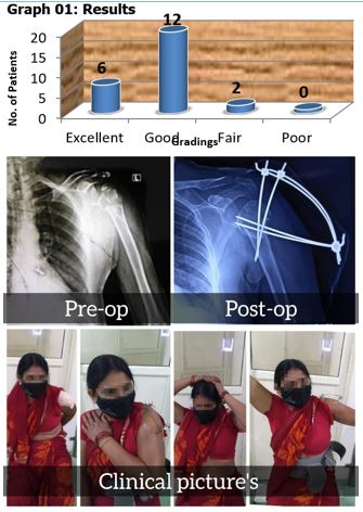
After preoperative assessment cases were posted for surgery. Under aseptic precaution and prophylactic antibiotic coverage, cases were operated with multiple k wires for proximal humerus fracture, in randomized group. Data collection procedure was included detailed study variable like preoperative and post operative clinical, radiological, surgical and functional status of involved extremity. Data collection tools had patient proforma and questioners table to show patients detail of examination, preoperative workup and surgical methods and post operative follow up. Questioners table was used for functional outcomes.

A through history was obtained including mechanism of injury and patients overall medical status, age and function and economical demands. The functional outcomes and clinical results of the patients evaluated & graded using following criteria.

In the case of a two-part surgical neck fracture, or a three-part fracture in which there is significant displacement of the shaft from under the humeral head, a trial reduction is performed to confirm the feasibility of closed reduction and percutaneous fixation. Reduction is performed by applying longitudinal traction with the arm in minimal abduction and some flexion. In case the humeral shaft is anteriorly angulated or displaced, it can be reduced by posterior pressure at fracture site lifting elbow upwards. For varus angulation a lever can be placed through a small incision into the fracture site and maneuvered to reduce it. Alternatively, two pins can be passed into head and joy sticking can be done. A 2.5-mm terminally threaded pin is held over the shoulder, and a fluoroscopic AP image is obtained. The pin is positioned over the humeral head, coming from the lateral humeral shaft into the head. A small incision is then made over the lateral arm at the level determined by the fluoroscopic image, and an artery forceps is used to spread the soft tissue down to the humeral shaft.


The tip of the artery forceps can confirm the anterior and posterior cortex of the humerus. While the assistant maintains the reduction, the surgeon drills the pin, initially horizontally to engage lateral humerus cortex and then up into the humeral head, confirming pin position with either spot radiographs or fluoroscopic control until the pin tip is just beneath the articular surface. Head is rotated internally and externally to confirm appropriate pin placement. A second pin is drilled parallel to the first pin in a similar manner. Two pins are passed from the greater tuberosity to engage into the medial cortex of humeral shaft. Additional pins can be inserted to enhance stability. Pins are connected to a fixator rod using mini clamps.

Following this shoulder immobilizer was applied for two weeks. Patients were explained pin tract care. They were instructed to daily clean the fixator and shoulder gently with chlorhexidine scrub solution followed by povidone iodine ointment at pin sites. Gentle passive shoulder mobilization was started after 2 weeks. Active mobilization of shoulder was allowed after 4 weeks. The fixator was removed when there were signs of trabecular bridging or periosteal new bone formation on radiographs and clinical improvement in the form of subsidence of pain usually by 8 weeks. Patients were evaluated for functional outcome at 4 months, 6 months and 1 year using the Constant shoulder score(8).

Follow-up is done and patient discharged on 3rd or 7th day of post op depending on wound status, regular follow up at every weekly up to 2 months and after 2 months monthly follow up to 4 months.

Results

Twenty patients comprising of 12 females and 08 males were followed for an average period of 12 months (range, 10-14 months). Mean age of patients was 58 years (range, 45- 72 years). Fall was most common mode of injury in 15 patients followed RTA injury in 5 patients. Average time between injuries to surgery was 5 days. The fracture union time ranged from 14-18 weeks with mean of 16 weeks. Out of Twenty patients 06 (30%) patients showing excellent results, 12 (60%) patients have good results, and 02(10%) patients having fair results, no patient having poor result.

Post-operative complications were noticed in total 03 patients, 02 patients had pin tract infections and one patient had mal- union. patients with pin tract infection were treated with antibiotics (oral as well as intravenous) and daily dressing, and by rigorous monitoring and were discharged after symptoms of infections had disappeared completely. patients with mal-union had acceptable level of movements, so he was not given any further surgical intervention.

Table 1: Sex Distribution

SexNo. of patientsPercentage
Male0840%
Female1260%
Total20100%

Table 2: Side Affected

SideNo. of PatientsPercentage
Right1470
Left0630
Total20100

Table 3: Mode of Injury

Mode of InjuryNo. of PatientsPercentage
RTA0525%
Fall1575%
Total20100%

Table 4: Showing Duration of Fracture Union

Duration(Weeks)Number of patientsPercentage (%)
12-140210%
14-161050%
16-180840%

Table 5: Post-Operative Complications

Non-unionMalunionPin tract Infection
NIL12

All patients achieved full functional range of motion by the end of 4 months. No nerve injury was reported.

ojmpc_161_01.jpg
Figure 1: Case 1


ojmpc_161_02.jpg
Figure 2: Case 2

ojmpc_161_03.jpg
Figure 3: Case 3

Discussion

This k-wires technique has always been providing good results with extremely low post- operative complications and has been successfully used in our study on all patients groups, osteoporotic patients with low blood loss and short post-operative stay in hospital(9). Majority of the patients with proximal humerus fractures are above 60 years old and most of these fracture in these populations is due to osteoporosis. Conservative treatment in a sling followed by functional rehabilitation under the supervision leads to satisfactory results in minimally displaced fractures whereas, displaced two and three part fractures need to be reduced and stabilized.(10)

While conservative treatment may lead to malunion and stiffness, open reduction may lead devascualrization of fragments and since most patients are elderly, chances of infection and comorbid conditions preventing extensive surgery are higher. Closed reduction and percutaneous pinning techniques, when treating the elderly patients with cardio-vascular or pulmonary diseases, in whom anesthesia is very risky or clearly contra-indicated.

In our study, all fractures were reduced conservatively, without requiring any invasion, thus making it extremely less invasive with extremely low chances of post- operative infection. Some surgeons prefer open reduction and then fixation with K-wire of such fractures, but this does not serve the original purpose. Best results of K-wire fixation of such fractures can be only achieved by conservative reduction by traction, manipulation, abduction and then fixation with K-wire. This helps in extremely low post-operative complications and provides stable fixation of such fractures. The K-wire fixation of such fracture is extremely cost effective and in the reach of the poorest candidate. Though the rehabilitation and physiotherapy exercises are being delayed in K-wire fixation, but the ultimate results are so promising that it decimates such delay in start of exercises.(9) Best part in our study was that we had used K-wires in all the 20 patients, irrespective of their ages, sex, osteoporosis, extreme displacements of fracture site, etc and achieved extremely encouraging results, intra-operatively as well as post- operatively. Thus, we can draw a clear-cut conclusion of supremacy of closed reduction and K-wire fixation such fractures. and also, some big comparative studies be carried out to clearly make conclusions in favour of K-wire fixation of these fractures.

Conclusion

Percutaneous K-wire fixation is a useful technique in select patients with proximal humerus fractures. It has extremely low post-operative complications, and at an extremely low cost, being affordable by poorest candidates. This preserves the biological integrity of the humeral head and to secure anatomical reduction with multiple k wires with angular stability. Also improves stability of fixation in osteoporotic humeral bones. K wires linked with clamps and fixator rod is an economical way to prevent K wire related complications often seen in post-operative period. So, we can conclude that K-wire fixation of humerus fractures provide extremely good post- operative results.

References

1. Pencle FJ, Varacallo M. Proximal Humerus Fracture [Internet]. StatPearls [Internet]. StatPearls Publishing; 2022 [cited 2022 Nov 7]. Available from: [Article][Crossref][PubMed][Google Scholar]

2. Proximal Humerus Fractures: Evaluation and Management in the Elderly Patient - PMC [Internet]. [cited 2022 Nov 7]. Available from: https://www. ncbi. nlm.nih.gov/pmc/articles/PMC5788098/ [Crossref][PubMed][Google Scholar]


3. Clinical and functional outcome of proximal humerus fractures treated with locking compression plate (LCP) in adults – A prospective study - IJOS [Internet]. [cited 2022 Nov 7]. Available from: https://www. ijos. co.in/html-article/12962 [Crossref][PubMed][Google Scholar]

4. Schumaier A, Grawe B. Proximal Humerus Fractures: Evaluation and Management in the Elderly Patient. Geriatr Orthop Surg Rehabil. 2018 Jan 25;9:2151458517750516. [Crossref][PubMed][Google Scholar]

5. Carofino BC, Leopold SS. Classifications in Brief: The Neer Classification for Proximal Humerus Fractures. Clin Orthop. 2013 Jan;471(1):39–43. [Crossref][PubMed][Google Scholar]

6. Neer classification of proximal humeral fractures | Radiology Reference Article | Radiopaedia. org [Internet]. [cited 2022 Nov 7]. Available from: https://radiopaedia. org/articles/neer-classification-of-proximal-humeral-fractures-1 [Crossref][PubMed][Google Scholar]

7. Evaluation of functional outcomes of a modified technique for percutaneous pinning of proximal humeus fracture [Internet]. [cited 2022 Nov 7]. Available from: https://www. researchgate. net/publication/324838667_Evaluation_of_functional_outcomes_of_a_modified_technique_for_percutaneous_pinning_of_proximal_humeus_fracture [Crossref][PubMed][Google Scholar]

8. Evaluation of the Constant score: which is the method to assess the objective strength? | BMC Musculoskeletal Disorders | Full Text [Internet]. [cited 2022 Nov 8]. Available from: https://bmcmusculoskeletdisord. biomedcentral. com/articles/10.1186/s12891-019-2795-6 [Crossref][PubMed][Google Scholar]

9. (PDF) K-Wire Fixation of Proximal Three Part Humerus Fractures - An Everlasting Result Oriented Technique [Internet]. [cited 2022 Nov 7]. Available from: https://www. researchgate. net/publication/349742589_K-Wire_Fixation_of_Proximal_Three_Part_Humerus_Fractures_-_An_Everlasting_Result_Oriented_Technique [Crossref][PubMed][Google Scholar]

10. Schumaier A, Grawe B. Proximal Humerus Fractures: Evaluation and Management in the Elderly Patient. Geriatr Orthop Surg Rehabil. 2018 Jan 25;9:2151458517750516. [Crossref][PubMed][Google Scholar]

Disclaimer / Publisher's Note

The statements, opinions and data contained in all publications are solely those of the individual author(s) and contributor(s) and not of Journals and/or the editor(s). Journals and/or the editor(s) disclaim responsibility for any injury to people or property resulting from any ideas, methods, instructions or products referred to in the content.