Orthopaedic Journal of MP Chapter

Publisher: Madhya Pradesh Orthopaedic Association www.mpioa.com
E-ISSN:2582-7243, P-ISSN:2320-6993
2022 Volume 28 Number 2 Jul-Dec

Functional outcome of patients of intertrochanteric femur fracture treated with trochanteric fixation nail in elderly patients

Shukla R1*, Rajput R2, Soni A3

1* Rajeev Shukla, Professor, Department of Orthopaedics, Sri Aurobindo Medical College, and PG Institute, Indore, Mp, India.

2 R Rajput, Department of Orthopaedics, Sri Aurobindo Medical College, and PG Institute, Indore, Mp, India.

3 AK Soni, Department of Orthopaedics, Sri Aurobindo Medical College, and PG Institute, Indore, Mp, India.

Background: Intertrochanteric fractures are disabling injuries in geriatric population and they are the most frequently operated fracture type which has the highest postoperative fatality rate of all surgically treated fractures. The objective of the study was to evaluate the functional outcomes of intertrochanteric fractures treated with trochanteric fixation nail (TFN) in elderly patients.

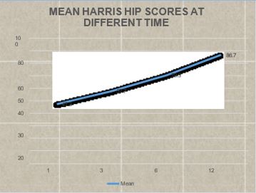
Method: This is a prospective and retrospective study with 30 patients of Intertrochanteric femur fractures carried out in the Department of Orthopaedics, Sri Aurobindo Medical College, and P.G. Institute, Indore from Jan 2015 to Aug 2020. Data collected according to Evan’s classification. Patients were followed up at intervals of 6 weeks,12 weeks,6 months and 12 months and final results were evaluated using Harris hip score at the end of 1 year.

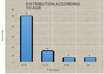
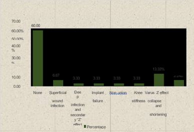
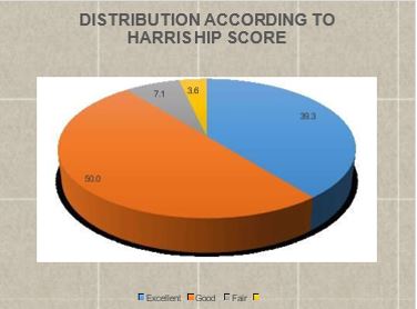
Results: The study included 30 patients, 18 males, 12 females with mean age of 70.27 years (range: 65-85 years). The fracture union rate was 96% and average union time was 12.9 weeks. Our results were excellent (39.3), good (50%), fair (7.1%) and poor (3.6%). The most common complication encountered was Varus collapse and shortening seen in 4 (13.33%) patients, superficial wound infection in 2 patients (6.67%); Varus collapse and shortening and Z effect and screw revision was seen in 2 (6.67%) while Deep infection and secondary ‗Z‘ effect, Implant failure, Non-union, Knee stiffness in 1 patient each.

Conclusion: The treatment of intertrochanteric fractures with TFN had a more favourable outcome and it is the ideal implant of choice for intertrochanteric fractures at present.

Keywords: intertrochanteric fracture, trochanteric fixation nail (TFN), Z effect

Corresponding Author How to Cite this Article To Browse
Rajeev Shukla, Professor, Department of Orthopaedics, Sri Aurobindo Medical College, and PG Institute, Indore, Mp, India.
Email:
Shukla R, Rajput R, Soni A, Functional outcome of patients of intertrochanteric femur fracture treated with trochanteric fixation nail in elderly patients. ojmpc. 2022;28(2):70-74.
Available From
https://ojmpc.com/index.php/ojmpc/article/view/158
Manuscript Received Review Round 1 Review Round 2 Review Round 3 Accepted
2022-12-03 2022-12-10 2022-12-17 2022-12-24 2022-12-31
Conflict of Interest Funding Ethical Approval Plagiarism X-checker Note
Authors state no conflict of interest. Non Funded. The conducted research is not related to either human or animals use. 13.65 All authors have accepted responsibility for the entire content of this manuscript and approved its submission.

© 2022by Shukla R, Rajput R, Soni Aand Published by Madhya Pradesh Orthopaedic Association. This is an Open Access article licensed under a Creative Commons Attribution 4.0 International License https://creativecommons.org/licenses/by-nc/4.0/ unported [CC BY NC 4.0].

Introduction

Intertrochanteric fractures are one of the most frequent fractures of the proximal femur and occur predominantly in geriatric patients and are among the most devastating injuries in the elderly. Incidence of the fractures of the proximal femur is increasing since the general life expectancy of the population has increased significantly during the last few decades. (1) Intertrochanteric fractures involve from the extra-capsular basilar neck region to the region along the lesser trochanter proximal to the development of the medullary canal. Most of the proximal femoral fractures occur in elderly individuals as a result of only moderate or minimal trauma. In younger patients, these fractures usually occur as a result of high energy trauma. Intertrochanteric fractures mostly occur due to a simple self-fall. The chances of self-fall increase with age, which is further increased by decreased muscle power, decreased reflexes, poor vision, and labile blood pressure. (1,2, 3)

Cummings et al. attributed four factors in determining whether a fall in an elderly is significant to cause fracture, the fall must be oriented in such a way that the person lands on or near the hip, the protective reflexes must be inadequate to reduce the energy of fall below the critical threshold, muscles and fat acting as local shock absorbers around the hip must be insufficient and the bone density at the hip must be inadequate to withstand the fall. (3) Prophylactic interventions to decrease the risk of falls and aggressive screening and treatment of osteoporotic patients with risk of fragility fractures are very important. (4) Unstable intertrochanteric fracture includes 3-fragment fracture with posteromedial comminution, fracture >2 intermediate fragments (lateral wall blows out), reverse oblique fracture, transverse oblique fracture and Intertrochanteric fracture with subtrochanteric extension. (5,6)

The surgical stabilization of unstable intertrochanteric fractures remains a persistent challenge. Before the introduction of appropriate fixation devices, treatment of intertrochanteric fracture was non-operative (9), consisting of prolonged bed rest in traction until fracture union (10 – 12 weeks). This is followed by a lengthy program of walking training. In elderly people, this was associated with high complication rates and to prevent these complications, the treatment of intertrochanteric fracture by reduction and internal fixation has become the standard management. (10) TFN preserve the fracture hematoma and associated with less blood loss and short operating time. The intramedullary position of the TFN prevents the excessive collapse of the proximal fragment & medialization of the distal fragment. Being an intramedullary load-sharing device, TFN aids in early postoperative mobilization, weight-bearing, and ultimately the early fracture union.

Materials and methods

This is a prospective and retrospective study with 30 patients of Intertrochanteric femur fractures carried out in the Department of Orthopaedics, Sri Aurobindo Medical College, and P.G. Institute, Indore from Jan. 2015 to Aug.

2020. Duration of the study was 2 years (one and half years for data collection and 6 months for analysis and writing) from September 2019 to August 2021. Method of collection of data and selection of cases was patients of Intertrochanteric fractures presented to the Emergency Department or OPD were treated surgically are included in the study after prior informed written consent. The patients were operated with standard methods of fixation. Patients coming for follow up were studied regularly. Inclusion criteria was all Intertrochanteric femur fractures and patient more than 65 years of age. Exclusion criteria was patient having any other injury, open fractures and patients with previous history of fracture in the same limb treated either conservatively or surgically. 30 patients were included in the study, out of which 6 were retrospective cases and 24 were prospective cases managed surgically. Patients admitted with Intertrochanteric fracture were examined and investigated with an X-ray pelvis with both hips AP and Lateral view (whenever possible).

Results

There were 21 (70%) patients in the age group 65-70 years, 5 (16.7%) patients were in the age group 71-75 years, 2 (6.7%) patients were in the age group 76-80 years and 2 (6.7%) patients were in the age group more than 80 years. Majority of the patients were in the age group 65-70 years, followed by 71-75 years. The mean age of the patients was 70.27 ± 5.82 years with a range from 65 to 85 years. There were 12 (40.0%) females and 18 (60.0%) males in the present study. There was a male predominance in the study. The male: female ratio was 1.5: 1.

22 (73.3%) patients sustained this injury due to fall and 8 (26.7%) patients sustained injury due to road traffic accidents. Fall was the most common mode of trauma. In 17 (56.7%) patient’s left side was involved and in 13 (43.3%) patients’ right side was involved. In majority of the patients left side involvement was seen in our study. All the fractures were classified as per the Evan’s Classification. In which Type 1 was considered stable fractures. Type 2 was unstable fractures. 28 (93.3%) patients had fractures of Evan’s Classification type 1 and 2 (6.7%) patients had type 2 intertrochanteric fracture.

ojmpc_158_01.jpg
Graph 1: Bar diagram showing distribution of patients according to age


One (3.3%) patient was suffering from chronic kidney disease, 4 (13.3%) patients were suffering from diabetes mellitus type-2, 4 (13.3%) patients were suffering from hypertension, 1 (3.3%) had anaemia and 1 (3.3%) patient had pulmonary tuberculosis, of the 30 patients, 19 (63.3%) patients were not having any comorbidity.

22 (73.4%) patients underwent surgical intervention within 3 days, 7 (23.3%) patients in 4-7 days, and only 1 (3.3%) patient was operated after 7 days of the injury. The mean duration between injury and day of surgery is 3.47 ± 1.85 days. It was subjected to the day of admission after trauma and availability of OT and surgical fitness of the patient.

The average operating time was 67mins (45min-110min) after incision. The fracture was reduced anatomically by closed means. Blood loss was counted intraoperatively by the number of mops used during the surgery. One mop equal to 50 ml blood loss and 1gauge piece equal to 10 ml approximately. The average blood loss was 1.4 mops, so 71ml (50-150ml).

5 patients required intraoperative blood transfusion as their pre-operative haemoglobin was less. 3 required blood transfusion postoperatively. The fracture was reduced anatomically by closed means. If that was not achieved then it was achieved by limited open reduction during surgery. 29 (96.7%) patients were managed with closed reduction and 1 (3.3%) patient, it was achieved through open reduction.

20 (66.7%) patients had a hospital stay less than or equal to 5 days, 6 (20.0%) patients had hospital stay between 6-7 days and 4 (13.3%) patients had stay of more than 7 days.

ojmpc_158_02.jpg
Graph 2: Bar diagram showing distribution according to complications

The mean hospital stay was 6.03 ± 3.13 days, with a range from 2 days to 13 days. The majority of the patients were discharged within 5 days of hospitalization. Union time was calculated radiologically on serial follow up x-rays of the patient. The follow-up x-rays were taken at 6 weeks, 3 months, 6 months and 1 year. 4 (13.3%) patients achieved fracture union in <10 weeks, 22 (73.4%) patients in 10-15 weeks, and rest 4 (13.3%) patients achieved in more than 15 weeks.

The most common complication encountered was Varus collapse and shortening seen in 4 (13.33%) patients, superficial wound infection in 2 patients (6.67%); Varus collapse and shortening and Z effect and screw revision was seen in 2 (6.67%) while deep infection and secondary ‗Z‘ effect, Implant failure, Non-union, Knee stiffness in 1 patient each. The vast majority of the patients were not having any sort of complications.Of the 30 patients included in the study, 2 (6.7%) patients died and 28 (93.3%) patients survived. Of the two patients who expired, 1 died due to chronic illness and other died due to Covid-19 infection. Both the deaths were not related to the trauma. The further analysis was carried out on the patients who survived (N=28).

2 patients had expired during the study period, so were excluded from the analysis. Of the 28 patients who followed-up till 12 months, 11 (39.3%) patients had excellent outcome at 12 months, 14 (50.0%) patients had good outcome, 2 (7.1%) patients had fair outcome and 1 (3.6%) patient had poor outcome. Majority of the patients i.e. 25 (89.3%) patients had good to excellent outcome according to Harris Hip Score. The mean Harris Hip Score at 1 month was 47.32 ± 8.06 and at 3 months it was 57.75 ± 7.74. The difference was found to be statistically significant (p=0.001), showing a significantly improved Harris Hip Score at 3 months in comparison to 1-month score. The mean Harris Hip Score at 3 months was 57.75 ± 7.74 and at 6 months it was 70.39 ± 7.95. The difference was found to be statistically significant (p=0.001), showing a significantly improved Harris Hip Score at 6 months in comparison to 3 months score. The mean Harris Hip Score at 6 months was 70.39 ± 7.95 and at 12 months it was 86.75 ± 4.45. The difference was found to be statistically significant (p=0.001), showing a significantly improved Harris Hip Score at 12 months in comparison to 6 months score.

ojmpc_158_03.jpg
Graph 3: Pie diagram showing distribution of patients according to Harris Hip Score at 12 months

There was a persistent statistically significant improvement in the mean Harris Hip Score at 3 months, 6 months and 12 months (p<0.05). Unpaired ‘t’ test applied. P-value < 0.05 was taken as statistically significant. The mean Harris Hip Score in females was 86.67 ± 4.68 and in males was 81.78 ± 18.59.


The difference was found to be statistically not significant (p=0.383), showing that the mean Harris Hip Score at 12 months is comparable between males and females. We can conclude that the Harris Hip Score is independent of the sex of the patients.

ojmpc_158_04.jpg
Graph 4: Line diagram showing mean Harris Hip Scores at different time intervals

Unpaired ‘t’ test applied. P-value < 0.05 was taken as statistically significant

Conclusion

Intramedullary nailing with the TFN has distinct advantages over other management options like shorter operating time and lesser blood loss for intertrochanteric fractures.

Early mobilization and weight-bearing are allowed in patients treated with TFN thereby decreasing the incidence of bedsores, uraemia and hypostatic pneumonia. For favourable outcomes, good preoperative planning with intraoperative reduction and sound surgical technique is vital and cannot be emphasized more. TFN is a momentous advancement in the treatment of intertrochanteric fractures which have the unique advantage of closed reduction, preservation of fracture hematoma, less tissue damage during surgery, early rehabilitation, and early return to work.

Osteosynthesis using a TFN, used in trochanteric fractures, resulted in low rates of clinical complications, excellent stabilization, few mechanical complications, and adequate functional results. Thus, the treatment of intertrochanteric fractures with TFN had a more favourable outcome and it is the ideal implant of choice for intertrochanteric fractures at present.

References

1. Abrahamsen B, van Staa T, Ariely R, et al. Excess mortality following hip fracture: Asystematic epidemiological review. Osteoporos Int. 2009;20(10):1633–1650. [Crossref][PubMed][Google Scholar]

2. Ahrengart L, Törnkvist H, Fornander P, et al. A randomized study of the compressionhip screw and Gamma nail in 426 fractures. ClinOrthopRelat Res. 2002;401:209–222. [Crossref][PubMed][Google Scholar]

3. Cummings SR, Nevitt MC. A Hypothesis. The Causes of Hip Fractures‖. J Gerontol 1989; 44:107-111. [Crossref][PubMed][Google Scholar]

4. T. Lindner,N K Kanakaris,BMarx,Acockbain. ―Fracture of the hip and osteoporosis‖ Journal of bone and joint surgery,2009;91- B:294-303. . [Crossref][PubMed][Google Scholar]

5. Dhiraj VS. Evans – classification of intertrochanteric fractures and their clinicalimportance. Trauma International 2015;1(1):7-11. . [Crossref][PubMed][Google Scholar]

6. Tronzo RG. Use of extramedullary guide pin for fractures of the upper end of the femur. Orthop Clin North Am 1974;5(3):525-7. . [Crossref][PubMed][Google Scholar]

7. Koval KJ, Aharonoff GB, Rokito AS, Lyon T, Zuckerman JD. Patients with femoral neck and intertrochanteric fractures. Are they the same? Clinical orthopaedics and related research. 1996; (330):166-72. [Crossref][PubMed][Google Scholar]

8. Bucholz RWH, James D, Court-Brown, Charles M. Tornetta, Paul. Rockwood and Green's Fractures in Adults, 7th Edition. Russell TA, editor. Philadelphia [Crossref][PubMed][Google Scholar]

9. Windoff J, Hollander DA, Hakimi M, Linhart W (2005). Pitfalls and complications in the use of Trochanteric Fixation nail. Langenbecks Arch Surg 390(1). 59–65, Feb Epub 2004 Apr 15. [Crossref][PubMed][Google Scholar]

10. Brammar TJ, Kendrew J, Khan RJ, Parker MJ (2005). Reverse obliquity and transverse fractures of the trochanteric region of the femur; a review of 101 cases. Injury 36(7). 851-857. [Crossref][PubMed][Google Scholar]

11. Herman A, Landau Y, Hazanov A, Segev T, Thein R, et al. (2013) Differencesin Survival Rates between Different Patterns of Unstable Pertrochanteric Femoral Fractures. OJO 3(6): 261-268. . [Crossref][PubMed][Google Scholar]

12. Karn NK, Ashish Jain, Nepal P et al. A prospective randomized control trial comparing Trochanteric Fixation nail and sliding hip screw in the management of trochanteric fracture of the femur. Health Renaissance, January-April 2011; Vol9 (No. 1). 7-11 [Crossref][PubMed][Google Scholar]

13. Pajarinen J, Lindahl J, Savolainen V, Michelsson O, Hirvensalo E. Femoral shaft medialisation and neck-shaft angle in unstable pertrochanteric femoral fractures. Int Orthop 2004;28:347–53. . [Crossref][PubMed][Google Scholar]

14. Dousa P, et al. Osteosyntehsis of trochanteric fractures using TFN. Acta ChirOrhtopTraumatol Cech 2002;69(1):22-30. . [Crossref][PubMed][Google Scholar]


15. Review paper by Kenneth. J. koval on IMN of proximal femur(A supplement to American jr of orthopaedics april 2007). . [Crossref][PubMed][Google Scholar]

16. Egol KA Chang EY, Cvitkovic j, kummerFJ, KovalKJ. Mismatch of current IMN with the anterior bow of femur. J Orthop trauma2004;18(7):410-415. . [Crossref][PubMed][Google Scholar]

17. The ―Z-Effect‖ Phenomenon Defined: A Laboratory Study Eric J. Strauss,1 frederick J. Kummer,1 Kenneth J. Koval,2 Kenneth A. Egol (journal of orthopaedic research 2007 doi 10.1002/jor) [Crossref][PubMed][Google Scholar]

18. Werner-Tutschku W, Lajtai G, Schmiedhuber G, Lang T, Pirkl C, Orthner E. Intra-and perioperative complications in the stabilization of per-and subtrochanteric femoral fractures by means of TFN. Unfallchirurg. 2002;105(10):881-5. [Crossref][PubMed][Google Scholar]

19. Dr. Justin Moses, Dr. Kalyan Deepak Sreenivas. A prospective study on outcomes of stainless steel proximal femoral nail for unstable intertrochantericfractures in rural population. International Journal of Orthopaedics Sciences 2020; 6(1): 379-382 [Crossref][PubMed][Google Scholar]

20. J. Li 1, L. Cheng1, J. Jing. The Asia proximal femoral nail antirotation versusthe standard proximal femoral antirotation nail for unstable intertrochanteric fractures in elderly Chinese patients. Orthopaedics & Traumatology: Surgery &Research 101 (2015) 143– 146 [Crossref][PubMed][Google Scholar]

21. Christian Boldin, Franz J Seibert, Florian Fankhauser, GeroifPeicha, WolfgangGrechenig and Rudolf Szyszkowitz. "The Trochanteric Fixation nail (TFN) - a minimal invasive treatment of unstable proximal femoral fractures Aprospective study of 55 patients with a follow-up of 15 months. Acta OrthopScand. 2003;74(1):53-58. [Crossref][PubMed][Google Scholar]

Disclaimer / Publisher's Note

The statements, opinions and data contained in all publications are solely those of the individual author(s) and contributor(s) and not of Journals and/or the editor(s). Journals and/or the editor(s) disclaim responsibility for any injury to people or property resulting from any ideas, methods, instructions or products referred to in the content.