Orthopaedic Journal of MP Chapter

Publisher: Madhya Pradesh Orthopaedic Association www.mpioa.com
E-ISSN:2582-7243, P-ISSN:2320-6993
2022 Volume 28 Number 1 Jan-Jun

Graft Options for Anterior Cruciate Ligament Reconstruction-choose wisely

Vijayan S1, Hegde N2, Kulkarni MS3*, Aroor M4, Bhat V5, Rao S6

1 S Vijayan, Department of Orthopaedics, Kasturba Medical College Manipal Academy of Higher Education, Manipal, Karnataka, India.

2 N Hegde, Department of Orthopaedics, Kasturba Medical College Manipal Academy of Higher Education, Manipal, Karnataka, India.

3* Mahesh Suresh Kulkarni, Ms Ortho Assistant Professor, Department of Orthopaedics, Kasturba Medical College Manipal Academy of Higher Education, Manipal, Karnataka, India.

4 MN Aroor, Department of Orthopaedics, Kasturba Medical College Manipal Academy of Higher Education, Manipal, Karnataka, India.

5 V Bhat, Department of Orthopaedics, Kasturba Medical College Manipal Academy of Higher Education, Manipal, Karnataka, India.

6 SK Rao, Department of Orthopaedics, Kasturba Medical College Manipal Academy of Higher Education, Manipal, Karnataka, India.

Background: Anterior cruciate ligament (ACL) tear constitutes a major chunk of post-traumatic knee injury leading to long term functional knee impairment and reduced quality of life. Globally, an increase in the incidence of reconstructive procedures for the torn ACL have been reported with varying outcome. This has revolutionized the techniques primarily aimed at achieving a functionally stable knee joint and early return to their pre-injury level of activities.

Discussion: Among the various factors which have influenced the outcome of the ACL reconstruction (ACLR), the choice of graft is a highly studied and yet still exceedingly debated topic. A large number of studies comparing the various graft options in ACLR has been published throughout the years. The purpose of this comprehensive review is to summarize the most recent relevant literatures on ACL graft options, on-going research and to discuss whether one graft type demonstrates clinical superiority over the other.

Conclusion: Understanding the biomechanical characteristics of various grafts available for anterior cruciate ligament reconstruction would help the surgeons in thoughtful selection of the graft for each patient on an individual basis and facilitate a thorough discussion between the surgeon and the patient which is vital in decision making.

Keywords: Anterior cruciate ligament; Graft; Reconstruction; Bone-patella; Hamstring; Peroneus; Quadriceps

Corresponding Author How to Cite this Article To Browse
Mahesh Suresh Kulkarni, Ms Ortho Assistant Professor, Department of Orthopaedics, Kasturba Medical College Manipal Academy of Higher Education, Manipal, Karnataka, India.
Email:
Vijayan S, Hegde N, Kulkarni MS, Aroor M, Bhat V, Rao S, Graft Options for Anterior Cruciate Ligament Reconstruction-choose wisely. ojmpc. 2022;28(1):18-26.
Available From
https://ojmpc.com/index.php/ojmpc/article/view/149
Manuscript Received Review Round 1 Review Round 2 Review Round 3 Accepted
2022-06-06 2022-06-12 2022-06-18 2022-06-24 2022-06-30
Conflict of Interest Funding Ethical Approval Plagiarism X-checker Note
Authors state no conflict of interest. Non Funded. The conducted research is not related to either human or animals use. 13.23 All authors have accepted responsibility for the entire content of this manuscript and approved its submission.

© 2022by Vijayan S, Hegde N, Kulkarni MS, Aroor M, Bhat V, Rao Sand Published by Madhya Pradesh Orthopaedic Association. This is an Open Access article licensed under a Creative Commons Attribution 4.0 International License https://creativecommons.org/licenses/by-nc/4.0/ unported [CC BY NC 4.0].

Introduction

The incidence of injury to the anterior cruciate ligament (ACL) is increasing worldwide and reconstruction (ACLR) of the torn ligament is becoming a common surgery. The more common non-contact ACL injuries occur when landing on a slightly flexed knee that is loaded by moments in 3 orthogonal planes - an internally directed tibial torque and knee valgus moment, combined with a quadriceps muscle contraction to resist the flexion moment 1,2.

The reconstruction surgeries which aim at restoring the stability and function of the knee and the techniques to achieve them are evolving day by day 3. Among the various variables which determine the success of the surgery, selection and use of the appropriate graft play a significant role. ACL is mostly composed of type I collagen and has anteromedial and posterolateral bundles. The main functional role of the ACL is to provide stability against anterior tibial translation and internal rotation. It has an average ultimate failure load of 2160 N (± 157) N with a linear stiffness of 242 (± 28) N/mm.

An ideal graft used for reconstruction should be easily available, cost-effective, easily harvested, have sufficient length and diameter, have the least donor site morbidity, should be biomechanically similar to the native ACL, allow secure fixation, permit good osteointegration in the bone tunnels, and have low immunogenicity and disease transmission rate 4–8.

There is no ideal graft still available for ACLR and the surgeons are often faced with the dilemma of selecting the most optimal graft. Choosing the graft depends on the skeletal age, degree of generalized ligamentous laxity, occupation of the patient, type of sporting activity involved in, chronicity of the injury, presence of associated ligamentous/meniscal injuries in the same knee and surgical familiarity of the surgeon 7,9.

The graft options currently available include autografts, allografts and synthetic grafts. The common autografts used in ACLR are Bone Patellar Bone Tendon (BPTB), Hamstring tendon (HT), Quadriceps tendon (QT) (with or without a bone plug on one end) and Peroneus longus tendon. Tendons and ligaments vary in the proportion of collagen in them. The ratio between type 1 and type 3 collagen in a tendon is 99:1, while it is 90:10 in ligaments 6,10. The various allografts available are BPTB, HT, Tibialis posterior, Tibialis anterior and Tendoachilles. Allografts are available in irradiated and sterilized forms and also as fresh frozen-low irradiated grafts.

Synthetic grafts currently being used in practice are the Ligament augmentation and reconstruction system (LARS), Polyglycolic acid Dacron (PGA-Dacron) and Leeds Kio ligament.

Autografts

Bone patellar tendon bone graft:

Ever since Kenneth Jones (1963) and Franke K (1969) used BPTB for ACLR, it has been considered as the gold standard graft for ACLR (also known as Jones procedure) 2–4,8,9,11. The main reason for this is the excellent early bony integration in the bony tunnels due to the presence of bone on either end of the graft. This permits faster recovery, provide excellent tensile strength and a more stable knee with a lower incidence of graft failure 11–16.

However, harvesting of BPTB graft is associated with several complications like patellar tendonitis, patellar or tibial fractures, loss of full extension with concentric and eccentric reduction of quadriceps power, anterior knee pain, difficulty in kneeling, numbness due to injury to the infrapatellar branch of the saphenous nerve, reduced range of motion due to rigid construct and graft-tunnel mismatch4–6,9,11,16.

Using a transverse incision to harvest the graft is reported to reduce the incidence of kneeling pain 7,8. Few systematic reviews have shown a higher incidence of a contralateral ACL tear and osteoarthritis with BPTB grafts. The risk of patella fracture can be reduced by restricting the bone plug to less than half the length of the patella, making cuts angled and no more than 10 mm deep, avoiding cross-hatching at the corners and creating a trapezoidal cut rather than triangular or square 6. Previous patellar tendinopathy and Osgood Schlatter’s disease are relative contraindications to the harvest of BPTB graft 7,8.

Hamstring graft:

Riccardo Galeazzi (1934) pioneered ACL reconstruction with a semitendinosus autograft. ACLR using four- strand hamstring grafts was first performed by Lipscombe in 1982 by open technique and arthroscopically by Friedman in 1988 9,10. Compared to BPTB graft, hamstring tendons are easier to harvest and have lower donor site morbidity 4,5,11,12,14. They have good tensile strength and does not affect knee extensor function. The hamstring tendon is also favoured for a transphyseal approach in skeletally immature patients to minimize the risk of bar formation and a secondary growth deformity 6,17.


However, removal of the hamstring tendon leads to a reduction in knee flexion strength, can have saphenous nerve injury, have unpredictable graft length and diameter and takes longer time for bone-graft integration and recovery 5,11,18. The literature review has demonstrated bony tunnel widening especially when the graft has been anchored using cortical buttons with fixed or variable loop (Webster Kate, Ahmed). In general, the incidence of graft rupture is higher in the hamstring group and graft diameter is identified as a significant factor leading to early graft failure. Soft tissue grafts less than 8 mm in diameter in patients younger than 20 years of age has been found to be an independent predictor of the need for revision surgery for graft rupture 13,17,19.

The height of the patient was found to be a strong predictor of quadrupled hamstring graft diameter 20. Assessing the cross-sectional area of the semitendinosus tendon (ST) at the level of the knee joint line is used to predict the graft size. A different graft should be selected if the cross-section of the ST graft is < 5.9( mm2)17.

Quadriceps graft:

The use of quadriceps graft for ACLR was described by Marshall et al in 1979 21. It can be either a total soft tissue graft or can be harvested along with a piece of bone from the patella 17. Compared to BPTB there is no damage to the infrapatellar branch of the saphenous nerve and therefore lower rate of numbness, low patellar tendon morbidity and lower incidence of anterior knee pain 2,3,11,21.

A graft of consistent length, thickness and width can be harvested by careful dissection without violating the supra-patellar pouch and reduces tibial tunnel widening 14,17,21. Midterm results of quadriceps tendon show a lesser rate of graft rupture, lower pain levels and analgesic consumption. Disadvantages of harvesting quadriceps graft include technical difficulties, anterior knee pain, quadriceps weakness, patellar fracture, decreased range of movement, extensive bleeding and retraction of rectus femoris tendon2,17.

Peroneus longus graft:

A relatively new entrant, but is as strong as native ACL. It is of adequate thickness and length. The distal portion of the peroneus longus is tenodesised to the peroneus brevis tendon. Though there were concerns about loss of plantarflexion of first the metatarsal, weakness of eversion and ankle instability, many reports are now available that there are no effects on gait parameters 22.

Allografts

Eugene Bircher (1929) was the first to use a xenograft (from Kangaroo) for ACLR. Allografts that are commercially available and commonly used include BPTB, Hamstring tendons, Tibialis posterior, Tibialis anterior and Tendo Achilles 4,11,14,18,23,24. There is no risk of donor site morbidity. Allografts permit shorter operating time through smaller cosmetic incisions and are associated with less pain. Since graft sizes are predictable and can fill large tunnels, allografts are generally preferred in multi-ligament and revision situations. It was reported that a high graft failure rate (up to 45%) happens with allograft 23.

This was mainly attributed to the sterilization of the graft with ethylene glycol and high dose gamma irradiation as they alter the biomechanical properties of the graft 4,9,18,23. Newer studies have recommended using fresh frozen non-irradiated allografts or low dose irradiation (< 21kGy) that lead to a negligible change in biomechanical properties and better graft strength 3,14.

Few other concerns linked to allografts include risk of disease transmission, possible immunogenicity, slower incorporation, increased cost and greater risk of graft failure in the younger age group 2,4,11,12,24. Young athletic patients who have primary ACL reconstruction with an allograft are 3 times more likely to have a graft failure than those with an autograft2,13. Allografts that have undergone a slower rehabilitation protocol is reported to have more favorable result2.

Synthetic grafts:

Issues like donor site morbidity with autografts and risk of immunogenicity and disease transmission with allografts prompted bioengineers to search for alternate graft materials. Synthetic graft materials became popular in the 1980s and early 1990s with the introduction of carbon fibre reinforced artificial graft. An ideal synthetic graft should be biocompatible and have mechanical characteristics similar to the native ligament. It should be chemically stable, absorb minimal water and have the presence of pores for fibroblast ingrowth4,9. Jenkins (1977) and Dandy (1981) were the first to use synthetic grafts2.

The first generation made from carbon fibers were knitted, woven or braided. But it elongated and broke down and led to carbon induced synovitis. The second-generation grafts had additional braided woven longitudinal and transverse fibres4.


They permitted fibroblast in-growth, but suffered from wear, fraying and low abrasion resistance. Currently, the third generation of synthetic graft is being used. The two commonly used third generation synthetic ligaments are Ligament augmentation and reconstruction system (LARS) and PGA-Dacron 4,14. LARS is composed of longitudinal fibres of Polyethylene Terephthalate (PET) held together with a transverse knitted structure 2,25. While the intraarticular part has parallel longitudinal fibers of PET twisted perpendicular to each other, the intra-osseous (intra-tunnel) portion is composed of longitudinal fibers of PET with the transverse knitted structure which resists elongation.

The intraarticular orientation of the fibers is modified to be side specific i.e. different for left and right knees and is supposed to help overcome rotational fatigue of the synthetic ligament. Hydroxypropylcellulose (HPC) coating is applied on the surface of the LARS ligament and found to improve its biocompatibility and enhance ligament tissue regeneration 6,25,26. PGA Dacron has 75% degradable polyglycolic acid filaments and 25% non-degradable Dacron thread wrapped in a free synovial graft. The synovial wrap around the graft which is usually harvested from the suprapatellar fossa serves as a source of healing fibroblasts 14.

As compared to the previous two generations where the synthetic graft was used to entirely substitute the torn ACL ligament, the current concept is to use it as an augmentation device during the healing process of a freshly injured ACL 9. Therefore, surgery must be planned soon after the injury and every effort should be made to preserve the native ACL stump. All stable ACL remnants, the notch synovium, fat pad and ligamentum mucosum must be preserved 9,14,27.

PGA-Dacron graft can only be placed with a preserved ACL remnant. Sun et al reported superior vascular density, intra-tunnel and intra-articular graft integration and biomechanical properties, when the remnant was preserved 23,24. Benefits of remnant preservation include accelerated graft revascularization and remodeling, improved proprioception, decreased bone tunnel enlargement, individualized anatomic bone tunnel placement, improved objective knee stability and early mechanical support 9,14,24,28,29.

Synthetic grafts do not have donor site morbidity, have a longer shelf life and permit accelerated rehabilitation and early return to sports 24. It reduces surgical time and has no risk of disease transmission. They are generally considered in multi-ligament and revision surgeries 12,24.

The disadvantages of synthetic graft include its high cost, higher rate of graft rupture, late inflammation, delay in bone integration and can be considered only for a specific subset of patients who are > 40years, motivated, symptomatic and needing quick recovery 11,12,30. Rupture of the graft occurs due to abrasion of the graft at the tibial tunnel exit and is more vulnerable if impingement occurs 14,30.

Biological augments:

ACL graft-bone healing occurs with a layer of fibrovascular scar between tendon and bone at the graft-tunnel interface. This eventually organizes into perpendicular fibers that resemble Sharpey’s fibers 1,6,28,31. The presence and number of these fibers are directly correlated with the pull-out strength. Biologic augmentation is used to either accelerate scar tissue formation or alter the integration to one that more closely resembles the native ACL enthesis28,29,32. Biological materials in the form of Chitin, bioglass, gelatin, hyaluronic acid, polystyrene sodium sulfonate and collagen matrix enhance graft-tunnel interface healing 26,31.

Biological coating with platelet-rich plasma, mesenchymal stem cells, fibrin matrix, platelet-leukocyte gel, and autologous platelet concentration and biosynthetic bone substitutes, such as demineralized bone matrix and recombinant bone xenograft enhance graft tunnel interface healing as they are osteoinductive and conductive 26,28,33. The excessive demineralized bone matrix may be used to fill the defect in the patella and tibia if a BPTB graft is harvested. This has shown to reduce the incidence of anterior knee pain and fractures 34. Internal Brace Ligament Augmentation and Dynamic Intra-ligamentary Stabilization techniques are novel techniques that aim to protect the primary repair by providing a stabilizing construct that connects the femur and the tibia, thus bridging the repair 33,35,36. High strength suture tape acts as a stabilizer, enhancing the strength of the construct and allowing for graft preservation. It acts to protect the graft during the initial incorporation phase, while the patient can begin accelerated rehabilitation. Suture augmentation of ACL reconstruction may confer improved integrity of the graft and is worth consideration for future clinical study 11,26,33,37.

Augmentation of reconstruction with extra procedures:

Combined ACLR with augmentation of soft tissue structures (such as a lateral extra-articular tenodesis (LET) or reconstruction of the anterolateral ligament) can increase knee stability 15,28,38.


Cerciello et al. recommended ACL + LET procedures to be considered in patients with grade 3+ pivot shift, patients less than 25 years, young patients undergoing ACLR with medial meniscus repair, those with Beighton score of > 6 or genu recurvatum > 100, patients taking part in pivoting sports like soccer, and patients undergoing revision ACLR39.

Comparative studies of various grafts

Many studies have compared the different autografts, allografts and synthetic grafts (Table 1). 2–5,12,14,16,17,21,23,24.

Table 1: Comparison of the biomechanical characteristics and functional outcome of various grafts available for anterior cruciate ligament reconstruction

BPTBHamstringQuadricepsAllograftSynthetic graft
IntegrationFast (6-8 wk)Slow (10-12 wk)Faster with bone plugSlowUsed to augment torn ligament
Stability (mid-term)Better stabilityMore anterior laxityLaxity between BPTB and HTSimilar to BPTBProvide immediate stability,
Better stability than HT,
Similar to BPTB
Return to sportsEarlyLateLateLate return is betterPreferred for early return
Donor site morbidityMoreLessLess than BPTBNilNil
Muscle strengthExtensor weaknessWeak flexionExtensor weaknessNo weaknessNo weakness
Revision rateLow (2.1%)High (5.1%)Low (0 - 2.2%)More failure in < 25 yrs,
More with irradiated graft,
Fresh frozen, irradiated graft have low incidence
No difference compared to BPTB
Functional scoresSimilar in all 4No difference
Patient reported outcome scoresSimilar in all 4Better than autografts
Contralateral ACL tear and OA kneeMoreLessLessEarly OA can occurNo
ImmunogenicityNoNoNoYesYes
SynovitisNoNoNoYesYes
Ultimate load to failure2977 N4090 N2352 NVariableVariable
Cross sectional area (Normal 44mm2)35 mm253 mm262 mm2Variable90 mm2

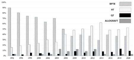
Over the years the choice of graft had changed across the world. The ACL study group ahead of their annual meeting in 2020 at Kitzbühel, Austria published the trend of graft usage over the last 3 decades. (Figure 1).

From the early 1990s till 2006 the BPTB graft showed a downward trend and after 2006 the number of ACLRs using hamstring autograft began to rise. Though during 2008-2010 there was interest in shifting to allografts, currently, its usage as a primary graft has come down except in the setting of multi-ligament or revision settings 4,14,18.

Quadriceps tendon (QT) autograft has increased in frequency since 2014 and peaked at over 10% in 2018 3. However, the majority (60%) still prefer hamstring autograft as their first choice.

Figure 1: Showing trend of ACL graft choice over the past 3 decades (Courtesy: Redrawn from Arnold MP et al. KSSTA Jan 2021. DOI: 10.1007/s00167-021-06443-9 PMID: 33486558)
ojmpc_149_01.jpg

On-going research

Pharmaceuticals – Alendronate, given locally or systemically was noted to improve bone tunnel mineralization, reduce peri tunnel bone loss and enhance graft-tunnel integration after six weeks. Subcutaneous parathyroid hormone showed enhanced thickness and microarchitecture of trabecular bone on CT scans. Oral Simvastatin was shown to promote bone formation 31.

Growth factors:

Growth factors can stimulate proliferation, migration and differentiation of cells. Platelet-derived growth factor, insulin-like growth factors and basic fibroblast growth factor can stimulate proliferation of fibroblasts while transforming growth factor-beta can increase matrix synthesis of tendon cells. They can increase the strength and stiffness of the healed ligaments. However, as their biological half-life is short, very high doses and repeated injections are often required 33,40.


Gene therapy:

Gene therapy is considered as the best method for local administration of growth factors. It can play a significant role in tissue-engineered ACL grafts. There are two ways in which the genetic material may be transferred to the tissue. First is via in-vivo transfer of the gene within a vector which is then directly applied to the target tissues.

The second method involves harvesting the target tissues from the body, transfecting the vector into it and allowing them to grow in an in-vitro culture media. They are transferred back to the target area once tissues mature.

The cells transduced by these vectors can act as a source of molecules capable of healing tissues. Viral and non-viral vectors can be used to deliver the genetic materials into the cells. Non-viral transfers are easier and have lower toxicity and low immunogenicity. However, viral gene vectors are more efficient.

It is important to remove pathogenic genes before transfer. Insertional mutagenesis, abnormal regulation of cell growth, development of malignancy and chronic overexpression of growth factor proteins are potential complications of these transfers 10,40,41.

Stem cell therapy:

Most of the reported results of mesenchymal stem cells (MSC) are based on animal studies. MSCs can secrete soluble factors which alter the tissue microenvironment and help to repair tissue. Bone marrow is a rich source to acquire mesenchymal stem cells.

These stem cells have greater transdifferentiation capability compared to stem cells from other sources. Micro-computed tomography and biomechanical analysis showed that BMP gene delivery led to enhanced bone formation at the graft-bone interface, osteointegration and superior biomechanical properties of the graft 32,35,41.

Platelet-rich plasma (PRP) may promote ligamentization, but there is less evidence to suggest that it enhances osseointegration 31. PRP increases the expression of collagen proteins, reduce apoptosis and stimulate fibroblast metabolic activity. However, most published studies on PRP combined with a collagen scaffold and mesenchymal stems cells have not been able to show any significant role of PRP in the acceleration of healing of soft tissue graft in a bone tunnel in ACLR 12,28,31,42.

Conclusion

We need to understand that there is “No one-size-fits-all” graft. It is very important to have an appropriate discussion with the patients as each patient has individualized goals and desires to do after reconstruction and this needs to be discussed in detail before selecting the best graft option for the patient.

Hamstring grafts appear to be a good all-around graft choice with fewer donor site complications and good results. BPTB is the best graft choice for professional sportspersons who are participating in sports at a very competitive level and wishing to have an early return to sports. Outcomes with quadriceps seem to lie between BPTB and Hamstrings and may be considered as an alternative graft option in indicated cases.

Fresh frozen, non-irradiated allografts and augmentation with synthetic grafts may be considered in multiligament / revision setting in less active patients after explaining the pros and cons with the patient. Biological augmentation of graft is still in the experimental phase and we need to wait for long term results.

References

1. Dallo I, Chahla J, Mitchell JJ, Pascual-Garrido C, Feagin JA, LaPrade RF. Biologic Approaches for the Treatment of Partial Tears of the Anterior Cruciate Ligament: A Current Concepts Review. Orthopaedic Journal of Sports Medicine. 2017 Jan 1;5(1):1–26. [Crossref][PubMed][Google Scholar]

2. Ahmed MA, Xie ZG, Ahsan SM. Review of Graft Choices for Anterior Cruciate Ligament. jmscr. 2016 Dec 6;04(12):14402–14114. [Crossref][PubMed][Google Scholar]

3. Arnold MP, Calcei JG, Vogel N, Magnussen RA, Clatworthy M, Spalding T, et al. ACL Study Group survey reveals the evolution of anterior cruciate ligament reconstruction graft choice over the past three decades. Knee Surg Sports Traumatol Arthrosc [Internet]. 2021 Jan 24 [cited 2021 May 6]; Available from: http://link. springer.com/10.1007/s00167-021-06443-9 [Crossref][PubMed][Google Scholar]

4. Dhammi I, Kumar S, Rehan-Ul-Haq. Graft choices for anterior cruciate ligament reconstruction. Indian J Orthop. 2015;49(2):127–8. [Crossref][PubMed][Google Scholar]

5. Tapasvi S, Jain S, Shyam A. BTB Vs Hamsrtings – Is There a Winner Yet ? Asian Journal of Arthroscopy. 2016 Jun;1(1):11–5. . [Crossref][PubMed][Google Scholar]


6. Ranjan R, Asif N. Choices of graft for anterior cruciate ligament reconstruction. Saudi J Sports Med. 2016;16(1):7–14. [Crossref][PubMed][Google Scholar]

7. Feller JA, Webster KE. A Randomized Comparison of Patellar Tendon and Hamstring Tendon Anterior Cruciate Ligament Reconstruction. Am J Sports Med. 2003 Jul 1;31(4):564–73. [Crossref][PubMed][Google Scholar]

8. Webster KE, Feller JA, Hartnett N, Leigh WB, Richmond AK. Comparison of Patellar Tendon and Hamstring Tendon Anterior Cruciate Ligament Reconstruction: A 15-Year Follow-up of a Randomized Controlled Trial. Am J Sports Med. 2016 Jan 1;44(1):83–90. [Crossref][PubMed][Google Scholar]

9. Shaerf DA. Anterior cruciate ligament reconstruction best practice: A review of graft choice. WJO. 2014;5(1):23–9. [Crossref][PubMed][Google Scholar]

10. Sundararajan SR, Sambandam B, Rajasekaran S. Future Trends In Grafts Used In ACL Reconstruction. Asian Journal of Arthroscopy. 2016 Jun;1(1):29–34. [Crossref][PubMed][Google Scholar]

11. Cerulli G, Placella G, Sebastiani E, Tei MM, Speziali A, Manfreda F. ACL Reconstruction: Choosing the Graft. joints. 2013;1(1):18–24. [Crossref][PubMed][Google Scholar]

12. Vaishya R, Agarwal AK, Ingole S, Vijay V. Current Trends in Anterior Cruciate Ligament Reconstruction: A Review. Cureus [Internet]. 2015 Nov 13 [cited 2021 May 6]; Available from: http://www. cureus.com/articles/3376-current-trends-in-anterior-cruciate-ligament-reconstruction-a-review [Crossref][PubMed][Google Scholar]

13. Kyung H-S. Graft considerations for successful anterior cruciate ligament reconstruction. Knee Surg & Relat Res. 2019 Dec;31(1):1–2. [Crossref][PubMed][Google Scholar]

14. Lanoue P. Does an ACL autograft, allograft or synthetic graft lead to improved long term knee stability? [Internet] [Physician Assistant Studies]. [Minneapolis]: Augsburg University; 2020. Available from: https://idun. augsburg.edu/etd/1060 [Crossref][PubMed][Google Scholar]

15. Campos GC de, Nunes LFB, Arruda LRP, Teixeira PEP, Amaral GHA, Alves Junior W de M. Current panorama of anterior cruciate ligament reconstruction surgery in Brazil. Acta ortop bras. 2019 Jun;27(3):146–51. [Crossref][PubMed][Google Scholar]

16. Rahardja R, Zhu M, Love H, Clatworthy MG, Monk AP, Young SW. Effect of Graft Choice on Revision and Contralateral Anterior Cruciate Ligament Reconstruction: Results From the New Zealand ACL Registry. Am J Sports Med. 2020 Jan 1;48(1):63–9. [Crossref][PubMed][Google Scholar]

17. Grassi A, Carulli C, Innocenti M, Mosca M, Zaffagnini S, Bait C, et al. New Trends in Anterior Cruciate Ligament Reconstruction: A Systematic Review of National Surveys of the Last 5 Years. Joints. 2018 Sep;06(03):177–87. [Crossref][PubMed][Google Scholar]

18. Mistry H, Metcalfe A, Colquitt J, Loveman E, Smith NA, Royle P, et al. Autograft or allograft for reconstruction of anterior cruciate ligament: a health economics perspective. Knee Surg Sports Traumatol Arthrosc. 2019 Jun;27(6):1782–90. [Crossref][PubMed][Google Scholar]

19. Perkins CA, Busch MT, Christino M, Herzog MM, Willimon SC. Allograft Augmentation of Hamstring Anterior Cruciate Ligament Autografts Is Associated With Increased Graft Failure in Children and Adolescents. Am J Sports Med. 2019 Jun;47(7):1576–82. [Crossref][PubMed][Google Scholar]

20. Stergios PG, Georgios KA, Konstantinos N, Efthymia P, Nikolaos K, Alexandros PG. Adequacy of Semitendinosus Tendon Alone for Anterior Cruciate Ligament Reconstruction Graft and Prediction of Hamstring Graft Size by Evaluating Simple Anthropometric Parameters. Anatomy Research International. 2012 Jul 29;2012:1–8. [Crossref][PubMed][Google Scholar]

21. Hurley ET, Calvo-Gurry M, Withers D, Farrington SK, Moran R, Moran CJ. Quadriceps Tendon Autograft in Anterior Cruciate Ligament Reconstruction: A Systematic Review. Arthroscopy: The Journal of Arthroscopic & Related Surgery. 2018 May;34(5):1690–8. [Crossref][PubMed][Google Scholar]

22. Kumar V K, Narayan S K, Vishal R B. A study on peroneus longus autograft for anterior cruciate ligament reconstruction. Int J Res Med Sci. 2019 Dec 25;8(1):183–8. [Crossref][PubMed][Google Scholar]

23. Macaulay AA, Perfetti DC, Levine WN. Anterior Cruciate Ligament Graft Choices. Sports Health. 2012 Jan;4(1):63–8. [Crossref][PubMed][Google Scholar]

24. Sun J, Wei X, Li L, Cao X, Li K, Guo L, et al. Autografts vs Synthetics for Cruciate Ligament Reconstruction: A Systematic Review and Meta‐Analysis. Orthop Surg. 2020 Apr;12(2):378–87. [Crossref][PubMed][Google Scholar]


25. Yang J, Dong Y, Wang J, Chen C, Zhu Y, Wu Y, et al. Hydroxypropylcellulose Coating to Improve Graft-to-Bone Healing for Anterior Cruciate Ligament Reconstruction. ACS Biomater Sci Eng. 2019 Apr 8;5(4):1793–803. [Crossref][PubMed][Google Scholar]

26. Herald J, Kakatkar S. Synthetic Grafts in Anterior Cruciate Ligament Reconstruction. Asian Journal of Arthroscopy. 2016 Jun;1(1):16–9. [Crossref][PubMed][Google Scholar]

27. Ebert JR, Annear PT. ACL Reconstruction Using Autologous Hamstrings Augmented With the Ligament Augmentation and Reconstruction System Provides Good Clinical Scores, High Levels of Satisfaction and Return to Sport, and a Low Retear Rate at 2 Years. Orthopaedic Journal of Sports Medicine. 2019 Oct 1;7(10):1–10. [Crossref][PubMed][Google Scholar]

28. Riediger MD, Stride D, Coke SE, Kurz AZ, Duong A, Ayeni OR. ACL Reconstruction with Augmentation: a Scoping Review. Curr Rev Musculoskelet Med. 2019 Jun;12(2):166–72. [Crossref][PubMed][Google Scholar]

29. Rothrauff BB, Kondo E, Siebold R, Wang JH, Yoon KH, Fu FH. Anterior cruciate ligament reconstruction with remnant preservation: current concepts. J ISAKOS. 2020 May;5(3):128–33. [Crossref][PubMed][Google Scholar]

30. Bashaireh K, Audat Z, Radaideh AM, Aleshawi AJ. The Effectiveness of Autograft Used in Anterior Cruciate Ligament Reconstruction of the Knee: Surgical Records for the New Generations of Orthopedic Surgeons and Synthetic Graft Revisit. ORR. 2020 Jun;Volume 12:61–7. [Crossref][PubMed][Google Scholar]

31. Hexter AT, Thangarajah T, Blunn G, Haddad FS. Biological augmentation of graft healing in anterior cruciate ligament reconstruction: a systematic review. The Bone & Joint Journal. 2018 Mar;100-B(3):271–84. [Crossref][PubMed][Google Scholar]

32. Yates EW, Rupani A, Foley GT, Khan WS, Cartmell S, Anand SJ. Ligament Tissue Engineering and Its Potential Role in Anterior Cruciate Ligament Reconstruction. Stem Cells International. 2012;2012:1–6. [Crossref][PubMed][Google Scholar]

33. Mahapatra P, Horriat S, Anand BS. Anterior cruciate ligament repair – past, present and future. J EXP ORTOP. 2018 Dec;5(1):1–10. [Crossref][PubMed][Google Scholar]

34. Lavender C, Johnson B, Kopiec A. Augmentation of Anterior Cruciate Ligament Reconstruction With Bone Marrow Concentrate and a Suture Tape. Arthroscopy Techniques. 2018 Dec;7(12):e1289–93. [Crossref][PubMed][Google Scholar]

35. Sciarretta FV. History of anterior cruciate ligament surgery. JASSM. 2020 Jul 15;1:90–7. [Crossref][PubMed][Google Scholar]

36. Benson DM, Hopper GP, Wilson WT, Mackay GM. Anterior Cruciate Ligament Reconstruction Using Bone–Patellar Tendon–Bone Autograft With Suture Tape Augmentation. Arthroscopy Techniques. 2021 Feb;10(2):e249–55. [Crossref][PubMed][Google Scholar]

37. Lai VJ, Reynolds AW, Kindya M, Konicek J, Akhavan S. The Use of Suture Augmentation for Graft Protection in ACL Reconstruction: A Biomechanical Study in Porcine Knees. Arthroscopy, Sports Medicine, and Rehabilitation. 2021 Feb;3(1):e57–63. [Crossref][PubMed][Google Scholar]

38. Lau BC, Rames J, Belay E, Riboh JC, Amendola A, Lassiter T. Anterolateral Complex Reconstruction Augmentation of Anterior Cruciate Ligament Reconstruction: Biomechanics, Indications, Techniques, and Clinical Outcomes. JBJS Rev. 2019 Nov;7(11):e5–e5. [Crossref][PubMed][Google Scholar]

39. Cerciello S, Batailler C, Darwich N, Neyret P. Extra-Articular Tenodesis in Combination with Anterior Cruciate Ligament Reconstruction. Clinics in Sports Medicine. 2018 Jan;37(1):87–100. [Crossref][PubMed][Google Scholar]

40. Martinek V, Huard J, Fu FH. Gene Therapy in Tendon Ailments. In: Maffulli N, Renström P, Leadbetter WB, editors. Tendon Injuries [Internet]. London: Springer-Verlag; 2005 [cited 2021 May 6]. p. 307–12. Available from: [Article][Crossref][PubMed][Google Scholar]

41. Bez M, Kremen TJ, Tawackoli W, Avalos P, Sheyn D, Shapiro G, et al. Ultrasound-Mediated Gene Delivery Enhances Tendon Allograft Integration in Mini-Pig Ligament Reconstruction. Molecular Therapy. 2018 Jul;26(7):1746–55. [Crossref][PubMed][Google Scholar]

42. Andriolo L, Di Matteo B, Kon E, Filardo G, Venieri G, Marcacci M. PRP Augmentation for ACL Reconstruction. BioMed Research International. 2015;2015:1–15. [Crossref][PubMed][Google Scholar]


Disclaimer / Publisher's Note

The statements, opinions and data contained in all publications are solely those of the individual author(s) and contributor(s) and not of Journals and/or the editor(s). Journals and/or the editor(s) disclaim responsibility for any injury to people or property resulting from any ideas, methods, instructions or products referred to in the content.