Orthopaedic Journal of MP Chapter

Publisher: Madhya Pradesh Orthopaedic Association www.mpioa.com
E-ISSN:2582-7243, P-ISSN:2320-6993
2021 Volume 27 Number 1 Jan-Jun

Surgical Management of Acromioclavicular Joint Injuries by Ligament Reconstruction Using Mersilene Tape and Ethibond

Patidar A1*, Chauhan A2, Aggarwal A3, Singh V4, Sharma S5

1* Ashish Patidar, Associate Professor, Department of Orthopaedics, RD Gardi Medical College and Associated Hospital, Ujjain, MP, India.

2 A Chauhan, Department of Orthopaedics, RD Gardi Medical College and Associated Hospital, Ujjain, MP, India.

3 A Aggarwal, Department of Orthopaedics, RD Gardi Medical College and Associated Hospital, Ujjain, MP, India.

4 V Singh, Department of Orthopaedics, RD Gardi Medical College and Associated Hospital, Ujjain, MP, India.

5 SK Sharma, Department of Orthopaedics, RD Gardi Medical College and Associated Hospital, Ujjain, MP, India.

Background: Acromioclavicular (AC) injuries account for 9% to 12% of all shoulder injuries. Rockwood grade IV to VI AC injuries require surgical fixation, which can be done by Mersilene tape reconstruction, K-wire transfixation, hook plates, reconstruction using autografts, or suture anchors. But no gold standard procedure has been established till date.

Material and Methods: 12 patients of AC joint disruption treated by surgical reconstruction using mersilene tape and ethibond suture were evaluated functionally using Visual analog scale (VAS) and Constant and Murley scores and radiological for re-displacement and fixation.

Results: The mean age in the group was 46.6 years (range 26 to 61), with male to female ratio of 3:1. Mean delay in surgery was 11 days (range 4 to 14 days), mean blood loss was 100 ml and mean duration of surgery was 54 min. The mean pre-operative VAS score improved from 6.41 to post-operative score of 2.68 and 1.25 at 6 and 12 months respectively. Constant Murley score improved from a mean pre-operative score of 51 to a post-operative score of 88.33 and 92.08 at 6 and 12 months respectively. At the final follow up all the patients had satisfactory results in terms of pain, cosmetic correction and movements and strength of the shoulder. The AC joint was clinically as well as radiologically stable in all the cases.

Conclusion: Anatomic reconstruction of AC joint disruption requires reconstruction of both coracoclavicular ligament as well as acromioclavicular ligament to achieve stability in both superior-inferior as well as antero-posterior plane, which can be achieved by mersilene tape fixation augmented by 5-0 ethibond suture leading to excellent results.

Keywords: Acromioclavicular injuries, Mersilene tape, Ethibond, Ligament reconstruction

Corresponding Author How to Cite this Article To Browse
Ashish Patidar, Associate Professor, Department of Orthopaedics, RD Gardi Medical College and Associated Hospital, Ujjain, MP, India.
Email:
Patidar A, Chauhan A, Aggarwal A, Singh V, Sharma S, Surgical Management of Acromioclavicular Joint Injuries by Ligament Reconstruction Using Mersilene Tape and Ethibond. ojmpc. 2021;27(1):28-32.
Available From
https://ojmpc.com/index.php/ojmpc/article/view/131
Manuscript Received Review Round 1 Review Round 2 Review Round 3 Accepted
2021-06-06 2021-06-12 2021-06-18 2021-06-24 2021-06-30
Conflict of Interest Funding Ethical Approval Plagiarism X-checker Note
Authors state no conflict of interest. Non Funded. The conducted research is not related to either human or animals use. 13.23 All authors have accepted responsibility for the entire content of this manuscript and approved its submission.

© 2021by Patidar A, Chauhan A, Aggarwal A, Singh V, Sharma Sand Published by Madhya Pradesh Orthopaedic Association. This is an Open Access article licensed under a Creative Commons Attribution 4.0 International License https://creativecommons.org/licenses/by-nc/4.0/ unported [CC BY NC 4.0].

Introduction

Injuries in and around the shoulder joint, including acromioclavicular joint injuries, are common occurrence in active young adults. Acromioclavicular (AC) joint injuries account for 9% to 12% of all shoulder injuries. Sports related incidents account for 25–50% of these acromioclavicular (AC) separations [1-4].

These injuries are grouped according to the Rockwood classification system into six groups [5]. Grades I and II injuries represent strain and partial tearing of supporting ligaments and are treated conservatively with excellent results. Surgical management is typically indicated for patients with grades IV to VI AC joint injuries.

For patients with grade III injuries, there is a debate regarding the optimal treatment strategy. Various operative techniques have been proposed. Current treatment focuses on anatomical reconstruction of coraco-clavicluar (CC) ligaments which show better outcome in biomechanical comparisons [3,4].

These techniques involve reconstruction of the conoid and trapezoid ligaments through anatomically-based tunnels in the clavicle. A variety of stabilization methods have been used for the AC joint, including mersilene tape reconstruction, K-wire trans fixation, hook plates, reconstruction using autografts, and suture anchors. But no gold standard procedure has been established. We evaluated the outcomes of surgical reconstruction of AC joint disruption by mersilene tape and ethibond suture, which provides enhanced tensile strength to the fixation.

Material and Method

This study was conducted at our centre from March 2017 to March 2019, in patients of AC joint disruption treated by surgical reconstruction by mersilene tape and ethibond suture, after obtaining Institutional Ethical Committee clearance.

All patients of age more than 18 years, with isolated, closed, grade III or more as per Rockwood classification system of AC joint disruption, presenting within 2 weeks of injury were included in the study. Patients having associated injuries, more than two weeks old injury, chronic AC joint injury or with co morbidities were excluded from our study.

All patients were evaluated by thorough history followed by complete physical exam and range of motion estimation. AP, lateral and Zanca views of the shoulder joint were done and injury was classified as per Rockwood classification system.

After stabilizing the patients haemo-dynamically, fitness for surgery was obtained and following this all the patients were treated surgically with reconstruction of coraco-clavicular ligaments using mersilene tape augmented with ethibond no 5 in general anaesthesia in supine position with a sand bag beneath the scapula of the operating side.

A 5 cm vertical incision was given 3 cm medial to the AC joint centred over the coracoid process.

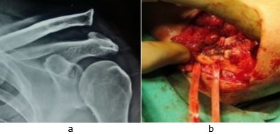
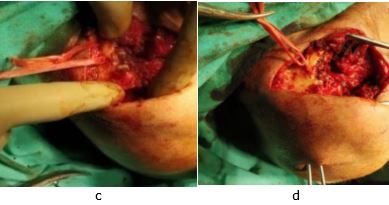
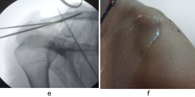
Blunt dissection was done to expose the clavicle, coracoid and the acromion, taking care of the haemostasis. With the help of 4.5 mm drill bit, a hole was made in the clavicle for the conoid ligament at around 3cm proximal to the acromio-clavicular joint slightly posterior to the midline of the clavicle from above downwards. Similarly, another hole was made in clavicle for the trapezoid ligament around 1.5cm proximal to the acromio-clavicular joint slightly anterior to the midline on the clavicle from above downwards to correctly reproduce the anatomic location of the respective ligaments. Mersilene tape augmented with 5-0 ethibond suture was passed under the coracoid process in a figure of eight manner and then passed through these holes and tied over clavicle to correct superior displacement and to replicate the anatomy (fig. 1). Another drill hole was then made in the acromion with the help of 4.5 mm drill bit from antero-medial to posterolateral direction for reconstruction of the acromioclavicular ligament by another Mersilene tape augmented with 5-0 ethibond suture, which was passed across joint to tie knot anteriorly to hold antero-posterior displacement. A 2mm smooth Kirschner wire was used to transfix the acromioclavicular joint temporarily, to provide additional support to the mersilene tape.

Figure 1: X ray Zanca view (a) of 50 years’ male with AC joint disruption. Intraoperative photograph (b to d) and fluoroscopic view (e) showing ligament reconstruction by mersilene tape, ethibond and transfixing k wires. 4 weeks follow up clinical photograph (f) showing healed scar after k wire removal.
ojmpc_131_01a.jpg
ojmpc_131_01c.jpg
ojmpc_131_01e.jpg


Post-operatively, patients were given cuff and collar pouch for 2 weeks, following which suture removal was done. The shoulder was immobilized in a sling for 4 weeks, after which the K-wire was removed and range of motion exercises were started. Strengthening exercises were done at 3 months postoperatively. Patients were followed regularly till minimum follow up of one year.

All the patients were evaluated functionally using Visual analog scale (VAS) and Constant and Murley scores and radiological for re-displacement and fixation.

Results

12 cases of Rockwood type III to V AC joint injuries with mean age of 46.6 years (range 26 to 61) were included in the study. Right shoulder was involved in 8 out of 12 cases. Males were predominantly affected with male to female ratio being 3:1. Mechanism of injury was fall in 10 cases, whereas 2 had a road traffic accident.

The mean delay in surgery was 11 days (range 4 to 14 days). The mean blood loss in surgery was around 100 ml and mean duration of surgery was 54 min. The mean pre-operative VAS score improved from 6.41 to post-operative score of 2.68 and 1.25 at 6 and 12 months respectively.

Constant Murley score improved from a mean pre-operative score of 51 to a post-operative score of 88.33 and 92.08 at 6 and 12 months respectively. At the final follow up all the patients had satisfactory results in terms of pain, cosmetic correction and movements and strength of the shoulder.

The AC joint was clinically as well as radiologically stable in all the cases with normal alignment and anatomical reduction of the AC joint, which was maintained even at final follow up. No case had postoperative wound complications, loss of fixation or osteolysis. None of the case required any revision surgery.

Discussion

Grades I and II injuries are treated conservatively with excellent results. Surgical treatment of the disrupted AC joint is well established in types IV, V, and VI. But the management of grade III injuries remains controversial and continues to evolve ranging from non-operative treatment to older surgical techniques.

Prospective studies comparing non-operative and operative treatment of these injuries have shown similar results with no great advantage of either treatment. Though many surgical procedures have been reported in the literature to manage these injuries, it has been difficult to achieve good long term functional outcome in AC joint disruptions. Surgical techniques used are such as excision of distal end of clavicle, K-wires or Bosworth screw fixation [6]. Cooper's first described surgical fixation of an AC dislocation in 1861 [7]. Weaver and Dunn first described the treatment of these injuries through excision of the lateral end of the clavicle and transfer of the coraco-acromial ligament to the rest of clavicle [8].

Since the transferred ligament was weaker than the native coraco-clavicular ligaments, recurrence of the dislocation was a common complication. Numerous modifications of this technique have been reported to reduce the risk of secondary dislocations with varied results like Mumford et al and Cadenat et al [9-10]. Hardware prominence and loss of fixation has been a common complication of other procedures.

We evaluated the outcomes of surgical reconstruction of AC joint disruption in 12 cases by Mersilene tape and ethibond suture and found excellent results. A similar study was done by Deshpande et al, in which they showed outcome of reconstruction of acromio-clavicular ligament and coraco-clavicular ligament using mersilene tape as to correct the antero-posterior displacements and the superior-inferior displacements respectively [11].

Mandice et al confirmed improved surgical outcomes, when Mersilene tape fixation techniques were augmented with fiber wires with restoration of shoulder joint to near normal anatomic and functional shoulder joint without donor site morbidity [12].

Hence we augmented our Mersilene tape reconstruction for both acromio-clavicular ligament and coraco-clavicular ligament, with 5-0 ethibond suture for more strength and so our results were better functionally as compared to their study in terms of function, disability, pain, and satisfaction.

Mc Connell et al in cadaveric specimens tested the stiffness of three different methods of fixation i.e. coraco-clavicular Bosworth screw (CC Screw), a coraco-clavicular sling of Mersilene tape (CC Sling), and a Hook Plate used in acute disruption of the acromio-clavicular (AC) joint and compared it with baseline to see which fixation most closely replicate the stiffness of healthy cadaveric AC specimens (Intact). They showed that the coracoclavicular sling using mersilene tape was significantly less stiff than the intact joint or the other methods of fixation [13].

In their study, they have used only mersilene tape for fixation of coraco-clavicular sling, whereas we in our study have used mersilene tape along with ethibond to increase its mechanical and tensile strength and used them as a sling for both coraco-clavicular and acromio-clavicular fixation, so as to maintain the anatomy of the joint in both the planes.

Haung et al in a retrospective comparative analysis of single coracoclavicular suture fixation with mersilene tape versus hook plate concluded that both hook plate and mersilene tape fixations provides comparable clinical outcomes, but hook plate may need removal of implant [14], but mersilene tape did not require removal.

Studies by Fakuda et al [15], Urist et al [16] and Lee et al [17] confirmed that acromioclavicular ligaments provide support in the antero-posterior plane and coraco-clavicular ligaments provide stability in superior plane. Debski et al biomechanically confirmed that the conoid and trapezoid ligaments act separately but synergistically in restraining antero-posterior and superior loading of the AC joint [18].


Beitzel et al in their study concluded that both the CC ligaments and the AC ligaments should be repaired anatomically to control the optimal physiologic function (translation and rotation) [19].

Hence we reconstructed both the ligaments for providing both antero-posterior and superior stability at the earliest i.e. in fresh cases. Further, the importance of early fixation was stressed by study of Rolf et al who compared with the results of delayed surgical reconstruction after conservative treatment versus re-surgery after primary failure and revealed a statistically significant better outcome in the early reconstruction group, regarding the Constant score, the degree of AC joint reduction, numbers of complications and patient’s satisfaction [20].

Our study is limited by lack of control, fewer numbers of patients and lesser follow-up. Further studies with larger group and longer follow-ups are suggested.

Conclusion

Anatomic reconstruction of AC joint disruption requires reconstruction of both coracoclavicular ligament as well as acromioclavicular ligament to achieve stability in both superior-inferior as well as antero-posterior plane, which can be achieved by mersilene tape fixation augmented by 5-0 ethibond suture leading to excellent results.

References

1. Costic RS, Labriola JE, Rodosky MW, Debski RE. Biomechanical rationale for development of anatomical reconstructions of coracoclavicular ligaments after complete acromioclavicular joint dislocations. Am J Sports Med 2004;32(8):1929-36. . [Crossref][PubMed][Google Scholar]

2. Dias JJ, Gregg PJ. Acromioclavicular joint injuries in sport. Recommendations for treatment. Sports Med 1991;11(2):125-32. [Crossref][PubMed][Google Scholar]

3. Lemos MJ. The evaluation and treatment of the injured acromioclavicular joint in athletes. Am J Sports Med 1998;26(1):137-44. . [Crossref][PubMed][Google Scholar]

4. Meister K. Injuries to the shoulder in the throwing athlete. Part one: Biomechanics / pathophysiology / classification of injury. Am J Sports Med 2000;28(2):265-75. [Crossref][PubMed][Google Scholar]

5. Rockwood CA, Young CD. Disorders of the acromioclavicular joint. In: Rockwood CA, Matsen FA editors. The Shoulder. 2nd edi. Philadelphia, PA. WB Saunders Co;1990:pp 413-76 [Crossref][PubMed][Google Scholar]

6. Larsen E, Bjerg-Nielsen A, Christensen P. Conservative or surgical treatment of acromioclavicular dislocation: a prospective, controlled, randomized study. J Bone Joint Surg Am. 1986;68:552-5. [Crossref][PubMed][Google Scholar]

7. Cooper ES. New method for treating longstanding dislocations of the scapuloclavicular articulation. Am J Med Sci. 1861;41:389–92. doi: 10.1097/00000441-186104000-00006 [Crossref][PubMed][Google Scholar]

8. Weaver JK, Dunn HK. Treatment of acromioclavicular injuries, especially complete acromio clavicular separation. J Bone Joint Surg Am. 1972;54:6:1187–94. [Crossref][PubMed][Google Scholar]

9. Cadenat FM. The treatment of dislocations and fractures of the outer end of the clavicle. Int Clin. 1917;27:145–69. [Crossref][PubMed][Google Scholar]

10. Mumford EB. Acromioclavicular dislocation. a new operative treatment. J Bone Joint Surg. 1941;23:799–802 [Crossref][PubMed][Google Scholar]

11. Deshpande SB, Munde AS, Naik NP. A novel method of surgical management of Rockwood type III-V acromioclavicular joint injuries. Al Ameen J Med Sci 2015;8(3):206-11. . [Crossref][PubMed][Google Scholar]

12. Mandice CJ, Kumar SM, Anandan H. Functional and radiological outcomes of acromioclavicular joint reconstruction in type iii disruption without allograft with synthetic materials. Int J Orthop Sci. 2017;3:4:836-9. [Crossref][PubMed][Google Scholar]

13. McConnell AJ, Yoo DJ, Zdero R, et al. Methods of operative fixation of the acromioclavicular joint: a biomechanical comparison. J Orthop Trauma. 2007;21(4):248-53. [Crossref][PubMed][Google Scholar]

14. Huang YC, Yang SW, Chen CY, Lin KC, Renn JH. Single coracoclavicular suture fixation with Mersilene tape versus hook plate in the treatment of acute type V acromioclavicular dislocation: a retrospective analysis. J Orthop Surg Res. 2018;13(1):110. [Crossref][PubMed][Google Scholar]

15. Fukuda K, Craig EV, An KN, Cofield RH, Chao EYS. Biomechanical study of the ligamentous system of the acromioclavicular joint. J Bone Joint Surg Am. 1986;68(3):434-40. [Crossref][PubMed][Google Scholar]


16. Urist MR. Complete dislocations of the acromioclavicular joint: the nature of the traumatic lesion and effective methods of treatment with an analysis of forty-one cases. J Bone Joint Surg. 1946;28(4):813-37. [Crossref][PubMed][Google Scholar]

17. Lee KW, Debski RE, Chen CH. Functional evaluation of the ligaments at the acromioclavicular joint during anteroposterior and superoinferior translation. Am J Sports Surg 1997;25:858–62. . [Crossref][PubMed][Google Scholar]

18. Debski RE, Parsons IM, Woo SLY. Effect of capsular injury on acromioclavicular joint mechanics. J Bone Joint Surg 2001;83-A:1344–51. . [Crossref][PubMed][Google Scholar]

19. Beitzel K, Obopilwe E, Apostolakos J, Cote MP, Russell RP, Charette R, Singh H, Arciero RA, Imhoff AB, Mazzocca AD. Rotational and translational stability of different methods for direct acromioclavicular ligament repair in anatomic acromioclavicular joint reconstruction. Am J Sports Med. 2014;42(9):2141-8. [Crossref][PubMed][Google Scholar]

20. Rolf O, Weyhern AHV, Ewers A, Boehm D, Gohike F. Acromioclavicular dislocation Rockwood III–V: results of early versus delayed surgical treatment. Arch Orthop Trauma Surg 2008;28(10):1153-7. . [Crossref][PubMed][Google Scholar]

Disclaimer / Publisher's Note

The statements, opinions and data contained in all publications are solely those of the individual author(s) and contributor(s) and not of Journals and/or the editor(s). Journals and/or the editor(s) disclaim responsibility for any injury to people or property resulting from any ideas, methods, instructions or products referred to in the content.