Orthopaedic Journal of MP Chapter

Publisher: Madhya Pradesh Orthopaedic Association www.mpioa.com
E-ISSN:2582-7243, P-ISSN:2320-6993
2021 Volume 27 Number 1 Jan-Jun

Efficacy of Microscopic Posterior Cervical Laminectomy for Multilevel Compressive Cervical Myelopathy: A Long Term Analysis

Asati S1, Raut S2, Ruparel S3, Patel A4, Chaddha R5, Kundnani V6*

1 S Asati, Center of Excellence in Spine Surgery, Mumbai Institute of Spine Surgery and Bombay Hospital and Medical Research Centre and Mumbai and Lilavati Hospital and Medical research Centre, Mumbai, Maharashtra, India.

2 S Raut, Center of Excellence in Spine Surgery, Mumbai Institute of Spine Surgery and Bombay Hospital and Medical Research Centre and Mumbai and Lilavati Hospital and Medical research Centre, Mumbai, Maharashtra, India.

3 S Ruparel, Center of Excellence in Spine Surgery, Mumbai Institute of Spine Surgery and Bombay Hospital and Medical Research Centre and Mumbai and Lilavati Hospital and Medical research Centre, Mumbai, Maharashtra, India.

4 A Patel, Center of Excellence in Spine Surgery, Mumbai Institute of Spine Surgery and Bombay Hospital and Medical Research Centre and Mumbai and Lilavati Hospital and Medical research Centre, Mumbai, Maharashtra, India.

5 R Chaddha, Center of Excellence in Spine Surgery, Mumbai Institute of Spine Surgery and Bombay Hospital and Medical Research Centre and Mumbai and Lilavati Hospital and Medical research Centre, Mumbai, Maharashtra, India.

6* Vishal Kundnani, Center of Excellence in Spine Surgery, Mumbai Institute of Spine Surgery and Bombay Hospital and Medical Research Centre and Mumbai and Lilavati Hospital and Medical research Centre, Mumbai, Maharashtra, India.

Background: Cervical spondylotic myelopathy and ossified posterior longitudinal ligament (OPLL) are the two most common causes of compressive multilevel cervical myelopathy. These may cause progressive neurological deterioration and require surgical treatment. There is no gold standard treatment available. Anterior surgery is associated with morbidity and complications in multilevel cases because of which posterior surgeries are preferred, which have shown good clinical outcomes. We determined the long-term efficacy of microscopic posterior cervical laminectomy for multilevel compressive cervical myelopathy.

Material and Methods: We reviewed 110 patients with multilevel compressive cervical myelopathy who underwent posterior cervical laminectomy from January 2007 to December 2014. Patients with age ≥45 years, C2-C7 cobb’s angle ≥ 10º, compression at ≥3 levels and a minimum of 5 years follow-up were included in the study. Demographic data, pre and post-operative clinical parameters (visual analog scale [VAS], Nurick’s grading and modified Japanese orthopaedic association [mJOA] score), radiological parameters (C2-C7 Cobb’s Angle), peri-operative parameters, complications and recovery rate were evaluated.

Results: The mean age of the patients was 55.6 years (44-74) with M: F 68:42. The mean blood loss and mean operative time was 93.9 ml and 96.6 minutes. There was significant improvement (p<0.05) in VAS (3.7±1.5 to 1.9±0.8), Nurick’s grading (3.3±0.9 to 1.8±0.6) and mJOA score (8.3±1.4 to 13.9±1.8). At final follow-up 61.8% patients’ maintained cervical lordosis, 21.8% changes to a straight spine and 16.3% became kyphotic. Intraoperatively 7 patients had a dural tear. 3 patients showed neurological deterioration postoperatively and 3 had unilateral C5 palsy which improved within 6 months period. 19% had an excellent outcome, 39% had good, 33.6% had fair and 8.1% patients had a poor outcome.

Conclusion: Microscopic posterior cervical laminectomy is the gold standard surgical procedure in patients with multilevel compressive cervical myelopathy with good recovery and clinical outcomes in properly selected patients. In long term it may causes progression of kyphosis, without any significant clinical affection.

Keywords: Cervical spondylotic myelopathy, OPLL, Cervical laminectomy, CSF leak, C5 palsy, Neurological deterioration

Corresponding Author How to Cite this Article To Browse
Vishal Kundnani, , Center of Excellence in Spine Surgery, Mumbai Institute of Spine Surgery and Bombay Hospital and Medical Research Centre and Mumbai and Lilavati Hospital and Medical research Centre, Mumbai, Maharashtra, India.
Email:
Asati S, Raut S, Ruparel S, Patel A, Chaddha R, Kundnani V, Efficacy of Microscopic Posterior Cervical Laminectomy for Multilevel Compressive Cervical Myelopathy: A Long Term Analysis. ojmpc. 2021;27(1):15-21.
Available From
https://ojmpc.com/index.php/ojmpc/article/view/130
Manuscript Received Review Round 1 Review Round 2 Review Round 3 Accepted
2021-06-06 2021-06-12 2021-06-18 2021-06-24 2021-06-30
Conflict of Interest Funding Ethical Approval Plagiarism X-checker Note
Authors state no conflict of interest. Non Funded. The conducted research is not related to either human or animals use. 12.65 All authors have accepted responsibility for the entire content of this manuscript and approved its submission.

© 2021by Asati S, Raut S, Ruparel S, Patel A, Chaddha R, Kundnani Vand Published by Madhya Pradesh Orthopaedic Association. This is an Open Access article licensed under a Creative Commons Attribution 4.0 International License https://creativecommons.org/licenses/by-nc/4.0/ unported [CC BY NC 4.0].

Introduction

Multilevel compressive cervical myelopathy is the commonest cause of spinal cord impairment in the elderly population [1]. It comprises cervical spondylotic myelopathy (CSM), ossification of the posterior longitudinal ligament (OPLL), ossification of the ligamentum flavum (OLF), and degenerative disc disease (DDD). These structural changes, narrows the spinal canal and reduces the space available for the spinal cord, leading to neurological deficit. Surgical decompression is the gold standard procedure for preventing the progression of neurological deficits in these patients [2,3].

Good surgical results can be obtained by various surgical procedures done either anterior or posterior, such as anterior corpectomy and fusion, laminectomy alone or laminoplasty and laminectomy with fusion (LF). Patients with less than 3 segments involved can be managed by anterior surgeries, whereas when the involvement is more than 3 segments, posterior surgery is the gold standard [4].

The long-term efficacy of cervical laminectomy is already described in literature in past, but long-term follow-up data in Indian subcontinent is scanty. Here we are presenting long term outcome of microscopic posterior cervical laminectomy for multilevel compressive cervical myelopathy, with respect to functional recovery, complications and radiological outcomes.

Materials and methods

After ethical committee approval, we retrospective analyzed 118 patients (8 lost to follow-up) with multilevel compressive cervical myelopathy who underwent microscopic posterior cervical laminectomy in a single hospital by a single surgeon from January 2007 to December 2014. Preoperatively all the patients had signs and symptoms of long tract involvement, such as hand clumsiness, gait disturbance, and hyperreflexia in lower limbs. Patients with age ≥45 years, compression at 3 or more levels, C2-C7 cobb’s angle ≥ 10º (lordotic) and a minimum of 5 years’ follow-up after microscopic posterior cervical laminectomy were included in the study.

Patients with only axial neck pain without myelopathy, instability on dynamic x-ray’s, fracture/infection/metabolic disorders, revision surgery or having developmentally narrow spinal canal were excluded from the study. Demographic data (age, sex, duration of illness to presentation, and co-morbidities), pre‑ and post‑operative clinical parameters (neck pain score ‑ visual analogue scale [VAS], Nurick’s grading and modified Japanese orthopaedic association [mJOA]), radiological parameters (Sagittal cervical Cobb’s Angle),

perioperative parameters (operative time, blood loss, and hospital stay), postoperative complications (infection, root palsy or neurological worsening) were evaluated. The recovery rate was calculated as per hirabayashi [5], recovery rate (%) = [postoperative JOA score - preoperative JOA score]/ [18 - preoperative JOA score] * 100. A recovery rate of > 75% was considered an excellent outcome, 50%–75% as good outcome, 25%–49% as fair outcome, and < 25% was considered a poor outcome.

All patients were operated under general anaesthesia, in prone position on padded bolsters. Padding was done under all the bony prominences and ocular pressure was checked after positioning. The neck was placed in neutral or in mild flexion. The arms were strapped by the side (figure 1).

After confirmation in C-Arm a standard midline posterior exposure from C3 to C6 was carried out up to the lamina‑facet junction taking care to preserve the attachments to C2 and C7. The dissection was restricted just lateral to the lamina‑facet junction and the soft tissues attachments over the facet joints were preserved.

The furrow at the junction of the lamina and the facet joints was marked at all levels requiring laminectomy. Under microscopic guidance the gutters were created on both sides using a high‑speed cutting burr (Midas burr) till the inner cortex were reached.

1‑mm Kerrison rongeur was used to remove the flavum up to the lateral gutters created. The rongeur was used to complete the furrows on either side all the way up to the C2–C3 interlaminar space. The laminectomy was completed by lifting the laminae en bloc from the caudal end, and gentle dissection was performed for any adhesion between the ligamentum flavum and dura. Undercutting of C2 and C7 laminae with C4-C5 foraminotomy was done in all the cases to provide adequate decompression.

Postoperatively patients were encouraged to sit up in bed 24 h after the surgery. Patients were mobilized out of bed on the 2nd postoperative day using a soft cervical collar which was discontinued after suture removal. Patients were sequentially followed up at 4 weeks, 3 months, 6 months, 12 months and then annually. At each follow-up clinical and radiological evaluation was done.

The statistical analysis was carried out using a paired student t‑test. Differences were considered statistically significant at P < 0.05. Statistical analysis was done using SPSS software 20.0 (SPSS Inc., Chicago, IL, USA).


Figure 1:Positioning of the patient in well-padded bolsters, arms by the side strapped and head secured.
ojmpc_130_01.jpg

Results

118 patients fulfilled the inclusion criteria, out of whom 8 patients were lost to follow up and hence 110 patients were included in the study. Out of 110 patients 63 had degenerative cervical spondylotic myelopathy (CSM) and 47 had cervical OPLL. The mean age of the patients was 55.6 years (range 44 to 74 years) with Male to female ration of 68:42.

The mean duration of presentation of illness was 3.2 months (range 1 to 8 months) and the mean duration of follow-up period was 7.6 years (range 5 to 11 years). 48 patients had ≥ 2 comorbidities, 44 had < 2 and 18 had no associated comorbidities. 21 patients were non-ambulatory at the time of presentation (table 1).

The mean blood loss was 95.9 ml (range 70 to 180 ml) and the mean operative time was 95.6 mins (range 82 to 140 min). The mean duration of hospital stay was 4.8 days (range 3 to 8 days) (table 2). VAS demonstrated significant improvement (p<0.05) from pre-op 3.7±1.5 to post-op 1.9±0.8.

There was significant difference (p<0.05) in Nurick’s grading preop 3.3±0.9 to post op 1.8±0.6. mJOA score improved from pre-op 8.3±1.4 to post-op 13.9±1.8 (p<0.05) (Table 3) the mean recovery rate at final follow-up was 57.7%. Out of 110 patients 21 (19%) had an excellent outcome, 43 (39%) had good, 37 (33.6%) had fair and 9 (8.1%) patients had a poor outcome (table 3).

Out of 110, 68 (61.8%) patients maintained cervical lordosis, 24 (21.8%) changes to a straight spine and 18 (16.3%) became kyphotic (kyphosis greater than +5°, straight from -5º to +5°, lordosis less than -5º) (table 3).

Intraoperatively 7 (6.3%) patients had a dural tear, although no repair was done intraoperatively, as the tear was small. There was no case of post-operative CSF leak.

Superficial infection developed in 6 (5.4%) individuals for which intravenous antibiotics were given till the healing and there was no case of deep infection or wound dehiscence.

3(3.6%) patients showed neurological deterioration just after the surgery, out of which 2 showed decrease in their mJOA score by 5 points and 1 showed decrease in mJOA by 3 points.
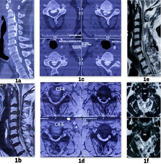
Figure 2:Cervical CT scan [(1a) sagittal, (1c) Axial] showing mixed type of OPLL from C2-C6 with significant canal compression (>50%) and myelomalacia [MRI sagittal (1b), axial (1d)] changes in the spinal cord. Post-operative MRI [(1e) sagittal, (1f) axial] after multilevel laminectomy showing well decompressed canal.
ojmpc_130_02.jpg

Figure 3:53 year old male patient with multilevel cervical spondylotic myelopathy operated by microscopic posterior multilevel laminectomy. Immediate AP(a) & lateral (b) & postoperative lateral flexion (c) and extension (d) x ray images at 5 year follow-up showing maintained cervical lordosis.
ojmpc_130_03.jpg

Over the time mJOA improved to their preoperative level. All these 3 cases were patients operated for cervical OPLL. 3 (3.6%) patients had an isolated unilateral C5 palsy which was transient in nature and improved within 6 months period with regular physiotherapy (table 4).


Table 1: Demographics

VariablesN=110
Age55.6
(44-74)
Gender (M: F)68:42
BMI31.9
Comorbidities
(DM, HTN, BA, HIV)
≥248
<244
No comorbidity18
Surgical indicationCSM63
Cervical OPLL47
Duration symptoms (months)3.2
Duration of follow-up (years)7.6 (5-11)

Discussion

Surgical decompression is the gold standard treatment for multilevel cervical compressive myelopathy. Various techniques include anterior, posterior or combined approach [6].

The disadvantages associated with anterior approach are increased blood loss and surgical time, post-operative dysphagia, hematoma formation and fusion‑related complications [4,7-9]. Studies have shown that in general elderly patients have higher mortality, postoperative complications, and bony nonunion rates than younger patients [7,8].

Puvanesarajah et al demonstrated significantly increased rates of surgical complications and mortality after anterior cervical fusion [9]. Posterior decompression (laminectomy) with or without fusion are reliable and effective in treating multilevel cases [10].

Table 2: Perioperative parameters

ParametersMean
Operative time (minutes)95.6 (82-140)
Blood loss (ml)95.9 (70-180)
Hospital stay (days)4.8 (3-8)

Table 3: Clinical and radiological outcomes:

ParametersPre-opPost-opp-value
VAS3.7±1.51.9±0.8<0.05
Nurick’s score3.3±0.91.8±0.6<0.05
mJOA score8.3±1.413.9±1.8<0.05
mJOA Recovery rate (%)Excellent (>75%)21 (19%)
Good (50%-75%)43 (39%)
Fair (25%-49%)37 (33.6%)
Poor (<25%)9 (8.1%)

The incidence of kyphotic change after multi-level laminectomy is approximately 20% [11].

Table 4: Complications:

ComplicationsN (%)
IntraoperativeDural tear7 (6.3%)
Early post-operativeSuperficial infection6 (5.4%)
Deep infection0
CSF leak0
C5 palsy3 (3.6%)
Neurological deterioration3 (3.6%)
Late post-operativeAxial neck pain24 (21.8%)
Progressive kyphosis18 (16.3%)
Delayed Neurological deterioration0
Reoperation0

The different causes include young age, preoperative kyphosis, and aggressiveness of posterior soft tissue resection, extent of facetectomy or capsule resection and multiplicity of laminectomy level. Facet injury is the most significant cause of postoperative kyphosis [12].

To overcome the disadvantages associated with laminectomy procedure laminoplasty came into light which preserves the posterior elements that protect the spinal cord against external forces and might decrease the incidence of neurological deterioration caused by falls by providing spinal stability [5,13,14]. Despite a variety of laminoplasty techniques, its advantages over laminectomy remains unclear. A review of literature revealed that the general result in the long-term follow-up of laminoplasty patients is similar to that in laminectomy patients [14].

Lee et al in his study suggested that there was no significant difference between cervical lordosis overtime in patients operated by laminectomy alone, laminoplasty and laminectomy with fusion [6]. Kaptain et al reported on 46 patients undergoing laminectomy and concluded that the development of a postoperative deformity (kyphosis) was more than twice as likely in patients with a “straight” preoperative spine (loss of lordosis) than in those with a normal lordosis [10].

In our study in 21.8% curvature changed to a straight spine and 16.3% became kyphotic. This change in the cervical alignment had no significant effect on the clinical outcomes of the patients and none of our patient required surgical intervention for that. In a study by Lee et al, 70.6% maintained their original curvature or improved from straight spine to lordosis [14]. The reasons for the maintenance of cervical lordosis in our study can be because of meticulous dissection and preservation of facet capsule during laminectomy under microscopic guidance and the selection of only those patients who has a C2-C7 cobbs ≥ 10º.


The mean recovery rate in present study at final follow-up was 57.7% which is more than the study done Kato et al [12] where at last evaluation it was 32.8% and comparable to Lee et al [14] study with a recovery rate of 56.3%. We observed that history of trauma, high BMI, a low preoperative mJOA score and late presentation were associated with a delayed recovery while higher preoperative JOA score and younger age at surgery were associated with early recovery.

Progression of OPLL was seen in 65.4% of the cases without any delayed neurological deficit, which is lesser than suggested by Hori et al [15] (in 71% at 10 years) and more than suggested by Chiba et al [16] where it was 56.5% of patients after 2 years.

The risk of durotomy during laminectomy is 0.3%–13% and can be up to 18% with revision surgery [17]. Singhatanadgige et al found the incidence of C5 palsy was higher 9.6%–25% following laminoplasty with fusion compared to 0%–8% in laminoplasty group [18]. Lee et al observed 5.9% C5 palsy in his study [14]. In our study the incidence of C5 palsy was 3.6% which recovered spontaneously within 6 months’ period. Axial neck pain may be the most frequently reported complication of laminectomy, with reports of its incidence ranging anywhere from 16%–48%.

We observed 21.9% incidence of axial neck pain which was manageable with medications. Bartels et al in his clinical randomized trial did not find a difference in the neurologic outcome or quality of life between laminectomy alone and laminectomy fusion groups at an average follow‑up of 18.3 months which suggest that laminectomy alone may be safe and effective in patients with preserved cervical lordosis and a stable cervical spine, without preoperative spinal instability [19].

As per our results and understanding we suggest microscopic posterior cervical laminectomy for patients with compressive cervical myelopathy ≥3 levels, without instability and C2-C7 cobb’s angle >10°.

Complications associated with fixation like higher incidences of C5 palsy, pseudoarthrosis, more surgical time in old and debilitated patients can be avoided. There are chances of post-laminectomy kyphosis which can be prevented by proper patient selection and OPLL progression which we think is only radiological without any clinical effect. This is a single surgeon, single hospital study and is the only study done on this pathology in Indian subcontinent with a long-term follow-up.

There are several limitations in this study. first, the study is retrospective and represents the experience of a single surgeon in a single institute.

Though it is retrospective in nature all the data was collected prospectively. Second, our inclusion criteria were narrow because of which it cannot we used in large number of patients. Third, there is no comparative group in our study. To determine the actual efficacy multicentric comparative studies are needed.

Conclusion

Microscopic posterior cervical laminectomy is the gold standard surgical procedure in patients with multilevel compressive cervical myelopathy with good recovery and clinical outcomes in properly selected patients. In long term it may causes progression of kyphosis, without any significant clinical affection.

References

1. Kalsi‑Ryan S, Karadimas SK, Fehlings MG. Cervical spondylotic myelopathy: The clinical phenomenon and the current pathobiology of an increasingly prevalent and devastating disorder. Neuroscientist 2013;19:409‑21. . [Crossref][PubMed][Google Scholar]

2. Isogai N, Nagoshi N, Iwanami A, Kono H, Kobayashi Y, Tsuji T, et al. Surgical treatment of cervical spondylotic myelopathy in the elderly: Outcomes in patients aged 80 years or older. Spine (Phila Pa 1976) 2018;43:E1430‑6. . [Crossref][PubMed][Google Scholar]

3. Fehlings MG, Wilson JR, Kopjar B, Yoon ST, Arnold PM, Massicotte EM, et al. Efficacy and safety of surgical decompression in patients with cervical spondylotic myelopathy: Results of the AO Spine North America prospective multi‑center study. J Bone Joint Surg Am 2013;95:1651‑8. . [Crossref][PubMed][Google Scholar]

4. Liu W, Hu L, Chou PH, et al. Comparison of anterior decompression and fusion versus laminoplasty in the treatment of multilevel cervical ossification of the posterior longitudinal ligament: a systematic review and meta-analysis. Ther Clin Risk Manag 2016;12:675–85. . [Crossref][PubMed][Google Scholar]

5. Hirabayashi K, Satomi K. Operative procedure and results of expansive open-door laminoplasty. Spine (Phila Pa 1976) 1988;13:870–6. . [Crossref][PubMed][Google Scholar]

6. Lee CH, Jahng TA, Hyun SJ, et al. Expansive laminoplasty versus laminectomy alone versus laminectomy and fusion for cervical ossification of the posterior longitudinal ligament. is there a difference in the clinical outcome and sagittal alignment. Clin Spine Surg 2016. 29:9–15 [Crossref][PubMed][Google Scholar]


7. Hayashi K, Matsumura A, Konishi S, Kato M, Namikawa T, Nakamura H, et al. Clinical outcomes of posterior lumbar interbody fusion for patients 80 years of age and older with lumbar degenerative disease: Minimum 2 years’ follow‑up. Global Spine J 2016;6:665‑72. . [Crossref][PubMed][Google Scholar]

8. Puvanesarajah V, Jain A, Shimer AL, Singla A, Shen F, Hassanzadeh H, et al. Complications and mortality following one to two‑level anterior cervical fusion for cervical spondylosis in patients above 80 years of age. Spine (Phila Pa 1976) 2017;42:509‑14. . [Crossref][PubMed][Google Scholar]

9. Zhong CJ, Xu WH. Laminectomy Followed by Fusion in Treating Severe Ossification of the Posterior Longitudinal Ligament. Fujian Province: Fujian Medical University, 2009;301:1–49. . [Crossref][PubMed][Google Scholar]

10. Kaptain GJ, Simmons NE, Replogle RE, Pobereskin L. Incidence and outcome of kyphotic deformity following laminectomy for cervical spondylotic myelopathy. J Neurosurg (2 Suppl) 2000;93:199-204. . [Crossref][PubMed][Google Scholar]

11. Zdeblick TA, Abitbol J-J, Kunz DN, McCabe RP, Garfin S. Cervical stability after sequential capsule resection. Spine 1993;18:2005-8. . [Crossref][PubMed][Google Scholar]

12. Kato Y, Iwasaki M, Fuji T, Yonenobu K, Ochi T. Long-term follow-up results of laminectomy for cervical myelopathy caused by ossification of the posterior longitudinal ligament. J Neurosurg 1998;89:217–23. . [Crossref][PubMed][Google Scholar]

13. Ratliff JK, Cooper PR. Cervical laminoplasty: a critical review. J Neurosurg 2003;98(3):230–8. . [Crossref][PubMed][Google Scholar]

14. Lee SE, Chung CK, Jahng T, Kim H. Long-term outcome of laminectomy for ossification of the posterior longitudinal ligament. J Neurosurg Spine. 2013;18(5):465-71. [Crossref][PubMed][Google Scholar]

15. Hori T, Kawaguchi Y, Kimura T. How does the ossification area of the posterior longitudinal ligament progress after cervical laminoplasty. Spine (Phila Pa 1976) 2006. 31:2807–12. [Crossref][PubMed][Google Scholar]

16. Chiba K, Yamamoto I, Hirabayashi H, Iwasaki M, Goto H, Yonenobu K, et al. Multicenter study investigating the postoperative progression of ossification of the posterior longitudinal ligament in the cervical spine: a new computer-assisted measurement. J Neurosurg Spine 2005;3:17–23. . [Crossref][PubMed][Google Scholar]

17. Jason Pui Yin Cheung. Keith Dip-Kei Luk. Complications of Anterior and Posterior Cervical Spine Surgery Asian Spine J 2016. 10(2):385-400. [Crossref][PubMed][Google Scholar]

18. Singhatanadgige W, Limthongkul W, Valone F, et al. Outcomes following laminoplasty or laminectomy and fusion in patients with myelopathy caused by ossification of the posterior longitudinal ligament: a systematic review. Global Spine J 2016;6:702–9. . [Crossref][PubMed][Google Scholar]

19. Bartels RH, Groenewoud H, Peul WC, Arts MP. Lamifuse: Results of a randomized controlled trial comparing laminectomy with and without fusion for cervical spondylotic myelopathy. J Neurosurg Sci 2017;61:134‑9. . [Crossref][PubMed][Google Scholar]

Disclaimer / Publisher's Note

The statements, opinions and data contained in all publications are solely those of the individual author(s) and contributor(s) and not of Journals and/or the editor(s). Journals and/or the editor(s) disclaim responsibility for any injury to people or property resulting from any ideas, methods, instructions or products referred to in the content.