Orthopaedic Journal of MP Chapter

Publisher: Madhya Pradesh Orthopaedic Association www.mpioa.com
E-ISSN:2582-7243, P-ISSN:2320-6993
2021 Volume 27 Number 1 Jan-Jun

Micro-Endoscopic Tubular Minimal Invasive Spine Surgery - Overview

Raut S1, Asati S2, Patel A3, Ruparel S4, Chaddha R5, Kundnani V6*

1 S Raut, Center of Excellence in Spine Surgery, Bombay Hospital and Medical Research Centre and Mumbai Lilavati Hospital and Medical Research Center Mumbai, Mumbai, Maharashtra.

2 S Asati, Center of Excellence in Spine Surgery, Bombay Hospital and Medical Research Centre and Mumbai Lilavati Hospital and Medical Research Center Mumbai, Mumbai, Maharashtra.

3 A Patel, Center of Excellence in Spine Surgery, Bombay Hospital and Medical Research Centre and Mumbai Lilavati Hospital and Medical Research Center Mumbai, Mumbai, Maharashtra.

4 S Ruparel, Center of Excellence in Spine Surgery, Bombay Hospital and Medical Research Centre and Mumbai Lilavati Hospital and Medical Research Center Mumbai, Mumbai, Maharashtra.

5 R Chaddha, Center of Excellence in Spine Surgery, Bombay Hospital and Medical Research Centre and Mumbai Lilavati Hospital and Medical Research Center Mumbai, Mumbai, Maharashtra.

6* Vishal Kundnani, Center of Excellence in Spine Surgery, Bombay Hospital and Medical Research Centre and Mumbai Lilavati Hospital and Medical Research Center Mumbai, Mumbai, Maharashtra, India.

Damage to paraspinal muscles as by caused by conventional open posterior lumbar spinal surgery can lead to inferior clinical and functional results. Minimally invasive approach to lumbar spine by microscopic, endoscopic or micro-endoscopic techniques using specialised instruments via neuro-vascular planes using muscle splitting approach to accesses the pathological site can reduce or minimise these complications. MIS techniques have demonstrated less blood loss, less postoperative pain, decreased need of analgesics post operatively, faster rehabilitation, shorter hospital stays and lower infection rates as compared to open techniques. while achieving equally efficacious results.

A thorough knowledge of anatomy of posterior spinal structures and understanding of the instruments used in minimal invasive spine surgery is of paramount importance. This article focuses on the anatomy, history, basics, instrumentation and indications used in minimally invasive lumbar spine surgeries.

Keywords: Spine, Micro-Endoscopic, Minimal Invasive

Corresponding Author How to Cite this Article To Browse
Vishal Kundnani, , Center of Excellence in Spine Surgery, Bombay Hospital and Medical Research Centre and Mumbai Lilavati Hospital and Medical Research Center Mumbai, Mumbai, Maharashtra, India.
Email:
Raut S, Asati S, Patel A, Ruparel S, Chaddha R, Kundnani V, Micro-Endoscopic Tubular Minimal Invasive Spine Surgery - Overview. ojmpc. 2021;27(1):4-9.
Available From
https://ojmpc.com/index.php/ojmpc/article/view/129
Manuscript Received Review Round 1 Review Round 2 Review Round 3 Accepted
2021-06-06 2021-06-12 2021-06-18 2021-06-24 2021-06-30
Conflict of Interest Funding Ethical Approval Plagiarism X-checker Note
Authors state no conflict of interest. Non Funded. The conducted research is not related to either human or animals use. 13.34 All authors have accepted responsibility for the entire content of this manuscript and approved its submission.

© 2021by Raut S, Asati S, Patel A, Ruparel S, Chaddha R, Kundnani Vand Published by Madhya Pradesh Orthopaedic Association. This is an Open Access article licensed under a Creative Commons Attribution 4.0 International License https://creativecommons.org/licenses/by-nc/4.0/ unported [CC BY NC 4.0].

Introduction

Posterior lumbar spinal surgery, is among the most commonly performed spinal surgery. It inherently causes damage to surrounding posterior paraspinal muscles. This morbidity due to posterior lumbar spinal surgery, is mostly attributed to damage of paraspinal muscles, excision or injury of midline posterior interspinous and supraspinous ligaments or due to associated blood loss during surgery. Among the different surgical approaches to the spine, it appears that injury to the muscles and ligaments is greatest when using conventional posterior midline approach.

Injury to paraspinal muscles can be caused by direct injury caused by dissection, thermal injury as by electrocautery, compression injury as by forceful retractors of by denervation. It can lead to atrophy of muscles with subsequent loss of function, thus giving rise to inferior clinical and functional results [1]. These can be reduced or minimised by minimally invasive approaches to lumbar spine by microscopic, endoscopic or micro-endoscopic techniques using specialised instruments.

The goal of any lumbar spine surgery is to achieve adequate decompression of spinal cord and nerve roots, attainment of fusion and maintenance / restoration of sagittal alignment. Minimally invasive spine (MIS) surgery also aims towards attainment of these goals, but via minimal invasive approach by minimal incision and soft tissue damage. A thorough knowledge of anatomy of posterior spinal structures and understanding of the instruments used in minimal invasive spine surgery is of paramount importance and shall benefit to optimise the learning curve of MIS. This article focuses on the anatomy, history, basics, instrumentation and indications used in minimally invasive lumbar spine surgeries.

Anatomy of posterior paraspinal muscles

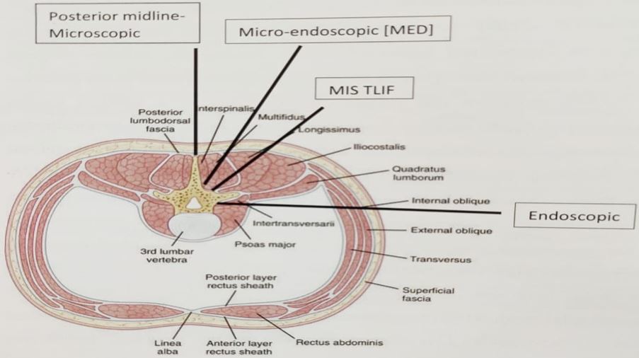
The posterior paraspinal muscles are responsible for controlled movements of lumbar spine while maintaining its stability. These are composed of two muscle groups figure 1.

1. The deep paramedian transverse spinalis group which includes the multifidus, interspinalis and intertransversarii.

2. The superficial and lateral erector spinae muscles which include the longissimus and iliocostalis.

All receive their innervation from the dorsal rami (table 1) [2].

Principles of MISS surgery:

To preserve the spinal anatomy as much as possible while addressing the pathology optimally to reduce morbidity and achieve targeted goal of surgery.

Rationale of using MISS technique:

Minimal invasive spinal surgery uses muscle splitting approach and accesses the pathological site through known neuro-vascular planes. Surgeon can move from one compartment to other, only after incising the fascia over the other compartment so as to prevent disruption of the neurovascular supply of muscles.

The safe surgical corridor for MIS-TLIF is the neurovascular plane between the multifidus and longissimus muscle. When approaching the spinal canal, laminae or facet joint, as in micro-endoscopic decompression and discectomies, a trans-multifidus compartment approach is used. When placing percutaneous pedicle screws or posterolateral onlay fusion, a transerector spinae approach is used (fig 1).

Thus minimal invasive spinal surgery strives to minimize muscle injury and preserve bone ligament complex, providing early recovery. Kim et al compared trunk muscle strength between patients treated with open posterior instrumentation and percutaneous instrumentation and found that, latter group displayed 50% improvement in extension strength [3].

Lee et al studied markers of skeletal injury and found that markers return to baseline in 3 days in MIS group whereas open group required 7 days [4]. Similarly, Stevens et al assessed post-surgical [6 months] MRI sequences of patients undergoing open and MIS TLIF and found marked intermuscular and intramuscular oedema in the open group as compared to normal appearance of multifidus in MIS group [5].

Figure 1:Anatomy of paraspinal muscles
ojmpc_129_01.jpg


Table 1: Showing origin, insertion, nerve supply & action of posterior paraspinal muscles

MuscleOriginInsertionNerve supplyPrime action
MultifidusSpinous process and lateral surface of laminaMammillary
processes of
caudal vertebra two to five levels below
Medial branch
of dorsal rami
Prime
stabilizer of spinal column
Erector spinaeLongissimus Transverse process
Iliocostalis
Tips of transverse process & adjacent fascia
Longissimus
Posterior superior iliac spine
Iliocostalis
Ventral edge of iliac crest
Longissimus
Intermediate
branch dorsal rami
Iliocostalis
Lateral branch
of dorsal rami
Move the trunk to Extension, lateral bending and rotation
Interpsinalis, Intertransversarii and short rotatorsIntertransverse and interspinous ligamentsIntertransverse and interspinous ligamentDorsal ramiProprioceptive sensors

History

Spine surgeons around the world are in a constant attempt to achieve optimum surgical results with minimum collateral damage and to overcome drawbacks of traditional conventional surgeries. This is how Minimally Invasive Spine Surgery (MISS) came into use. MISS is the most advanced and least invasive form of spine surgery as it reduces the morbidity of a conventional technique and achieves the surgical goals. Reports of MISS procedures date back to the early 20th century [6].

The development of microscopic, fluoroscopic and endoscopic systems came into existence only in 1990s and from then MISS gained momentum. Tubular access to the lumbar disc was first reported by Faubert and Caspart in 1991. Tubular access minimised muscle damages and decreased blood loss considerably. Tubes became popular due to the easy access to contralateral side. Problems like degenerative disc, lumbar canal stenosis, listhesis etc can be dealt with the tubular technique. Micro-endoscopic discectomy was then described by Foley and Smith in 1997 [7].

The evolution of endoscopes was very well accepted by orthopaedic surgeons. Later microscope was introduced and added in this tubular technique by around 2003. Microscope gave better magnification and illumination giving better surgical outcomes. Fusion for treating instability patterns was also possible with minimal invasive spinal surgery, as the percutaneous pedicle screws came into existence.

Instruments for MISS

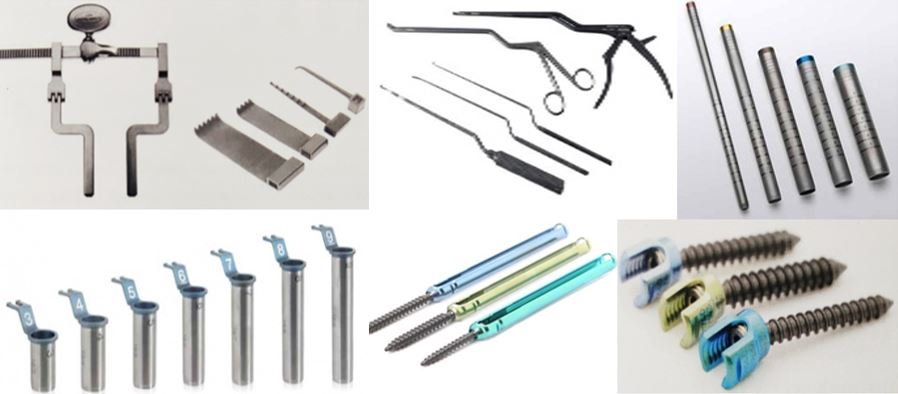
Instruments for standard microscopic, micro-endoscopic and endoscopic surgery may vary depending on the type of surgery to access the bony spine. Standard microlumbar decompression requires unilateral approach to access bony spine for decompression. Instruments required for MIS are as follows (fig 2).

Figure 2: Instruments used for MIS (a) Mc Cullohs Retractors (b)MIS instruments (c) Serial dilators (d) tubular retractors) (e) Percutaneous pedicle screw instrumentation (f) cannulated pedicles screw
ojmpc_129_02.jpg

1. C-arm and Microscope: A good quality C-arm and microscope are essential for successful MIS surgery. Surgery should not be contemplated unless anatomical landmarks are clearly seen through the C-arm. Microscope with assistant eye piece as well, with good focussing depth and light adjustment provides adequate magnification and illumination for decompression through narrow working channels.

2. Mc Cullohs Retractors: in which one blade fits into interspinous process and the wide blade sits on paraspinal muscles over the corresponding facet joint, allowing for unilateral exposure are used. Varying sizes and depth blades are available, corresponding to the depth of exposure required.

3. Serial dilators: These are concentric tubes used sequentially for serial dilation decreasing the need for muscle stripping during the exposure.

4. Tubular retractors: These cylindrical retractors allow the surgical corridor to be opened after serial dilation. Tubular retractors are preferred to blades as these are thin walled (0.9mm). The retractor allows for appropriately sized working channel ranging from 14mm to 25mm choosing appropriate depth size is important as it prevents the muscle from intruding into the field of view. The retractors can be fixed or expandable. Expandable retractors provide a larger working channel after docking.


5. Table mounted retractor holder: In MIS, retractor holder, which is table mounted is used to hold the tubular retractor in place than self-retaining. In self-retaining mechanism, constant pressure is exerted on tissues thus causing damage, whereas the pressure exerted by MIS table mounted retractors is undetectable.

6. MIS instruments: The MIS instruments for spinal decompression and fusion procedures are same as that of open techniques except that these are long and bayonetted which helps the surgeon to work through narrow working channel and under the microscope.

7. Burr: A high speed burr with long and thin shaft is usually required for decorticating and thinning bony elements

8. Percutaneous pedicle screw instrumentation: Instruments and pedicle screws required for percutaneous techniques are different. A Jamshedi type needle [Cook's needle] with trocar and cannula, which are gently tapped with mallet to reach isthmus of pedicle is required for pedicle marking. After trocar removal, cannulated tap over a K-wire is inserted through the cannula. Once tapping is done, cannulated pedicle screws are inserted over the guide wires inserted previously. These are connected with a sleeve during insertion which later help in passage of rods.

9. Percutaneous Rods: These rods have a bullet tip which ensure easy percutaneous passage. They are usually pre bent to accommodate for lumbar lordosis. Also they have an attachment for secure connection to rod insertor.

10. Rod insertion systems. There are two types of rod insertor systems. First, when the rod is inserted through screw heads and other is Pivot mechanism when rod is inserted after creation of passage above and across screw heads. Inner screws inserted help in gradual approximation of the rod to the screw heads through the sleeves.

Indications:

The indications of minimally invasive spine surgery are similar to open traditional surgical indications. MIS is equally efficacious with Micro-endoscopic discectomies and decompressions. Similarly, it is effectively used in cases where instrumentation and fusion is required like spondylolisthesis, degenerative scoliosis and trauma. MIS is advantageous in revision spine surgeries as it provides a native surgical approach free of scar tissue. MISS now is widely used for dealing lumbar disc herniations, lumbar canal stenosis,

cervical disc herniations, lumbar spondylolisthesis, spine infections, tumours, spinal deformities and spinal trauma and the spectrum is increasing day by day (Table 2).

Table 2: Indications for MIS

Spinal Degenerative ConditionsMicro-endoscopic discectomyMicro-endoscopic decompressionDegenerative instabilityCervical Lamino-foraminotmyMISS C1-2 Trans-articular Screw Fixation
Spinal InfectionsTranspedicular biopsy
Endoscopic decompression and debridement
Endoscopic drainage of epidural abscess
Anterior/Transforaminal debridement and reconstruction
Spinal TraumaPercutaneous Vertebroplasty/ Kyphoplasty
Percutaneous pedicle screw fixations
Anterior minimal access decompression and stabilization supplement with percutaneous screws
Spinal deformitiesAdult deformities- Anterior/ Lateral minimal access- XLIF/ALIF/OLIF with percutaneous screws
Congenital and Adolescent deformities
Spinal TumorsIntra and extra medullary tumors

Contraindications:

Obesity [BMI> 40), advanced spondylolisthesis (Grade 3 or 4) and previous instrumentation that requires open approach for extension or removal are all relative contraindications. In these patients, MIS is technically demanding and has high rate of complications as the working length through the tube increases [8].

Advantages: MIS techniques are advantageous in [9-12] as it:

1. Minimizes muscular trauma & denervation.

2. No trauma to paravertebral muscles on contralateral side.

3. Bilateral decompression can be done through unilateral approach.

4. Preservation of posterior ligamentous tension band.

5. Significant reduction of risk associated with dead space after conventional laminectomies.

6. Decrease chances of infection.

7. Small incision, better cosmesis.

8. Early mobilization, negligible postoperative wound pain, decreased need of analgesics post-operatively and an early start to rehabilitation.

9. Minimal blood loss

10. Shorter hospitalization


11. Early mobilization ensures decreased postoperative complications such as DVT, UTI, or pneumonia.

12. Very less chances of increasing instabilities even in grade I spondylolisthesis.

13. Lesser chances of wrong level surgeries.

Drawbacks: The MIS is has following drawbacks, which can be overcome by experience -

1. Radiation exposure: Fluoroscopically guided pedicle screw placement exposes surgeon to increases dose of radiation. Although, with gain in experience and advent of navigation exposure to radiation is markedly decreased [13].

2. Operative time: Studies have shown that the operative time for screw insertion is longer than conventional method, but this time reduced as surgeon gains experience.

3. Learning Curve: MIS has a steep learning curve. Technical difficulty of the process and lack of training opportunities adds to this drawback.

Future Trends:

MED is gaining popularity because of its advantage over conventional methods as it increases precision and accuracy of the surgeon thus making the job a lot easier and will not be surprising with each passing day to see MED soon being accepted as a gold standard technique worldwide. Widespread applications to tackle numerous spinal pathologies with safety and achieving excellent clinical and functional outcomes have prompted most surgeons to want to perform MIS procedures.

Author's viewpoint:

The author feels that the first surgery to venture with minimally invasive surgery would be micro endoscopic decompression (MED). After successfully operating around 5-6 cases one can plan for micro endoscopic discectomy and then gradually MIS TLIF, each after 5-6 cases. One must not feel shy to convert an MIS surgery into an open procedure if at any point the goals of surgery are compromised. One must adequately counsel the patient pre operatively for such a consequence.

Conclusion

MIS surgery aims to achieve better clinical and functional results through minimal soft tissue injury and bone resection while attaining the goals of spinal decompression and fusion.

The instruments required for MIS technique are different and one must have a thorough knowledge of these before contemplating minimally invasive surgery. This technique has a steep learning curve and has its own contra-indications and limitations. It is an important tool in the armamentarium of a spine surgeon and should consider this technique taking into account its advantages and clinical results.

References

1. Gille O, Jolivet E, Dousset V, Degrise C, Obeid I, Vital JM, Skalli W. Erector spinae muscle changes on magnetic resonance imaging following lumbar surgery through a posterior approach. Spine (Phila Pa 1976). 2007;15;32(11):1236-4. [Crossref][PubMed][Google Scholar]

2. Bogduk N. Functional anatomy of the spine. Handb Clin Neurol. 2016;136:675-88. [Crossref][PubMed][Google Scholar]

3. Kim KT, Lee SH, Suk KS, Bae SC. The Quantitative Analysis of Tissue Injury Markers After Mini-Open Lumbar Fusion. Spine (Phila Pa 1976). 2006;15;31(6):712-6. [Crossref][PubMed][Google Scholar]

4. Lee JC, Jang HD, Shin BJ. Learning Curve and Clinical Outcomes of Minimally Invasive Transforaminal Lumbar Interbody Fusion: Our Experience in 86 Consecutive Cases. Spine (Phila Pa 1976). 2012;15;37(18):1548-57. [Crossref][PubMed][Google Scholar]

5. Stevens KJ, Spenciner DB, Griffiths KL, et al. Comparison of Minimally Invasive and Conventional Open Posterolateral Lumbar Fusion Using Magnetic Resonance Imaging and Retraction Pressure Studies. J Spinal Disord Tech. 2006;19(2):77-86. [Crossref][PubMed][Google Scholar]

6. Pool J. Direct visualization of dorsal nerve roots of the cauda equina by means of myeloscope. Archives of Neurology and Psychiatry. 1938;39:1308-12. [Crossref][PubMed][Google Scholar]

7. MM FKTS. Microendoscopic discectomy techniques in Neurosurgery. 1997;3:301-7. . [Crossref][PubMed][Google Scholar]

8. Rosen DS, Ferguson SD, Ogden AT, Huo D, Fessler RG. Obesity and self-reported outcome after minimally invasive lumbar spinal fusion surgery. Neurosurgery. 2008;63(5):956-60. [Crossref][PubMed][Google Scholar]


9. Dhall SS, Wang MY, Mummaneni PV. Clinical and radiographic comparison of mini-open transforaminal lumbar interbody fusion with open transforaminal lumbar interbody fusion in 42 patients with long-term follow-up. J Neurosurg Spine. 2008;9(6):560-5. [Crossref][PubMed][Google Scholar]

10. Park Y, Ha JW. Comparison of one-level posterior lumbar interbody fusion performed with a minimally invasive approach or a traditional open approach. Spine (Phila Pa 1976). 2007;1;32(5):537-43. [Crossref][PubMed][Google Scholar]

11. Peng CW, Yue WM, Poh SY, Yeo W, Tan SB. Clinical and radiological outcomes of minimally invasive versus open transforaminal lumbar interbody fusion. Spine (Phila Pa 1976). 2009;1;34(13):1385-9. [Crossref][PubMed][Google Scholar]

12. Vazan M, Gempt J, Meyer B, Buchmann N, Ryang YM. Minimally invasive transforaminal lumbar interbody fusion versus open transforaminal lumbar interbody fusion: a technical description and review of the literature. Acta Neurochir (Wien). 2017;159(6):1137-1146. [Crossref][PubMed][Google Scholar]

13. Kim CW, Lee YP, Taylor W, Oygar A, Kim WK. Use of navigation-assisted fluoroscopy to decrease radiation exposure during minimally invasive spine surgery. Spine J. 2008;8(4):584-90. [Crossref][PubMed][Google Scholar]

Disclaimer / Publisher's Note

The statements, opinions and data contained in all publications are solely those of the individual author(s) and contributor(s) and not of Journals and/or the editor(s). Journals and/or the editor(s) disclaim responsibility for any injury to people or property resulting from any ideas, methods, instructions or products referred to in the content.