Orthopaedic Journal of MP Chapter

Publisher: Madhya Pradesh Orthopaedic Association www.mpioa.com
E-ISSN:2582-7243, P-ISSN:2320-6993
2020 Volume 26 Number 2 Jul-Dec

Philos Plate For Fixation Of Supracondylar Dome Osteotomy

Mohindra M1, Gautam V2, Meena A3*, Gupta N4, Desai J5, Saikia S6

1 M Mohindra, Central Institute of Orthopaedics Vmmc and Safdarjung Hospital, New Delhi, India.

2 VK Gautam, Central Institute of Orthopaedics Vmmc and Safdarjung Hospital, New Delhi, India.

3* Amit Meena, Senior Resident, Central Institute of Orthopaedics Vmmc and Safdarjung Hospital, New Delhi, India.

4 N Gupta, Central Institute of Orthopaedics Vmmc and Safdarjung Hospital, New Delhi, India.

5 J Desai, Central Institute of Orthopaedics Vmmc and Safdarjung Hospital, New Delhi, India.

6 S Saikia, Central Institute of Orthopaedics Vmmc and Safdarjung Hospital, New Delhi, India.

Background: Various corrective osteotomies and different modes of fixation have been described to correct genu valgum deformity. We evaluated the results of dome osteotomy for genu valgum which was stabilized with proximal humerus internal locking system (PHILOS) plate.

Material and Methods: 39 cases of Genu Valgum deformity in 24 patients treated by dome osteotomy and fixed with PHILOS plate (15 patients had bilateral deformities and 9 patients had unilateral deformity) were evaluated clinically (intermalleolar distance and tibio-femoral angle) and radiologically (tibio-femoral angle).

Results: Pre-operative mean intermalleolar distance, clinical tibio-femoral angle and radiological tibio-femoral angle pre-operatively improved from 17.5 cm (range 11 to 24), 19.250 (range 140 to 240) and 20.90 (range 150 to 260) to postoperative 2.25 cm (range 0 to 4 cm), 7.750 (range 40 to 100) and 8.950 (range 50 to 110) respectively. The mean pre-operative LDFA was 74.850 (range 670 to 830) whereas post-operative mean value was 86.90 (range 830 to 900). The mean Bostman knee score improved significantly from 20.8 (range 18 to 22) to 29.1 (range 27 to 30). 2 patients (2 limbs) had good score i.e. between 20 to 27, while rest all the patients had excellent score between 28 to 30. Improvement in intermalleolar distance, tibio-femoral angle and LDFA was statistically significant (P< 0.001)

Conclusion: Dome osteotomy with PHILOS plate fixation is reasonable cost effective, easy, available and viable option for treatment of genu valgum with excellent short to midterm results and without complication as seen by wedge osteotomy.

Keywords: Genu Valgum, Dome Osteotomy, PHILOS plate

Corresponding Author How to Cite this Article To Browse
Amit Meena, Senior Resident, , Central Institute of Orthopaedics Vmmc and Safdarjung Hospital, New Delhi, , India.
Email:
Mohindra M, Gautam V, Meena A, Gupta N, Desai J, Saikia S, Philos Plate For Fixation Of Supracondylar Dome Osteotomy. ojmpc. 2020;26(2):75-80.
Available From
https://ojmpc.com/index.php/ojmpc/article/view/122
Manuscript Received Review Round 1 Review Round 2 Review Round 3 Accepted
2020-12-07 2020-12-13 2020-12-19 2020-12-25 2020-12-31
Conflict of Interest Funding Ethical Approval Plagiarism X-checker Note
Authors state no conflict of interest. Non Funded. The conducted research is not related to either human or animals use. 12.22 All authors have accepted responsibility for the entire content of this manuscript and approved its submission.

© 2020by Mohindra M, Gautam V, Meena A, Gupta N, Desai J, Saikia Sand Published by Madhya Pradesh Orthopaedic Association. This is an Open Access article licensed under a Creative Commons Attribution 4.0 International License https://creativecommons.org/licenses/by-nc/4.0/ unported [CC BY NC 4.0].

Introduction

Genu valgum is a common deformity in childhood which may originate from the distal femur, proximal tibia or the knee joint, but it usually originates from the distal femur, which may be confirmed by clinical examination as well as by various angle measurements on radiographs [1,2]. Although deformities up to the age of 12 years can be corrected by epiphysiodesis with stapling, mini-plate or external fixator, but when the patient presents after physeal closure, corrective osteotomy is the treatment of choice [3].

Various types of corrective osteotomies of the distal femur have been described in the literature which includes lateral opening wedge, medial closing wedge, dome osteotomy, wedge-less spike osteotomy, and wedge-less ‘V’ osteotomy [4-14].

Dome osteotomy, in which, circular bone cuts are made, allows deformity correction and overcomes complications associated with wedge osteotomy like length discrepancy, fragment mismatch, delayed union or segment translation. These advantages have made dome osteotomy the standard treatment, but the choice of implant to stabilize the osteotomy is debatable. Wide ranges of fixation methods have been tried, ranging from cast, k wires and external fixator to plates [15-20].

We evaluated the results of dome osteotomy for genu valgum stabilized with proximal humerus internal locking system (PHILOS) plate.

Material and Methods

This prospective study was performed at our center from May 2015 to May 2017 in 39 cases of Genu Valgum deformity in 24 patients treated by dome osteotomy and fixed with PHILOS plate (15 patients had bilateral deformities and 9 patients had unilateral deformity).

All the patients were selected from the outpatient department of our institute considering the inclusion criteria and were scrutinized through clinical assessment, radiological and biochemical investigations. The patients with genu valgum deformity with age more than 15 years having a tibio-femoral angle of more than 150 and an intermalleolar distance (IMD) of more than 10 cm were included in the study. Patients with 2-year minimum follow up were only included in the study. Patients who had unstable knee with evidence of subluxation, severe collateral ligament instability, and sagittal plane deformity (fixed flexion deformity) or genu recurvatum were excluded from the study.

All patients were investigated for metabolic and developmental disorders. Preoperative deformity assessment was done clinically (intermalleolar distance and tibio-femoral angle) and radiologically (tibio-femoral angle). Clinically the intermalleolar distance was measured in a standing position with the patella facing forward, the knees extended and the medial surface of the knees touching each other.

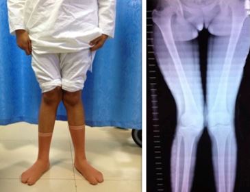
Clinical tibio-femoral angle was assessed by measuring the angle between the line drawn from the anterior superior iliac spine to the center of the patella and the line joining the center of the patella to the center of the ankle joint. Radiological assessment was done on full length standing anteroposterior radiographs of the affected limb including the hip, knee and ankle joint (Figure 1).

The radiological tibio-femoral angle was measured as angle formed between the anatomical axes of tibia and femur. The lateral distal-femoral angle (LDFA) was defined as the angle between the mechanical axis of the femur (line from center of femoral head to center of knee joint) and the articular surface of the distal femur.

Patients who were considered suitable for surgery underwent a deformity correction by a supracondylar femoral dome osteotomy, with fixation of osteotomy using the PHILOS plate. All patients were operated under spinal anesthesia in supine position with tourniquet on a standard operating table. The knee was kept flexed, with the help of a sand bag. An ECG electrode was fixed on the skin overlying the center of the femoral head that was confirmed by image intensifier. A 10 cm long incision was made over the anterolateral aspect of the distal one third of thigh extending to superolateral border of the patella and whole of anterior supracondylar region of femur was exposed by developing plane between rectus femoris and vastus lateralis and splitting vastus intermedius.

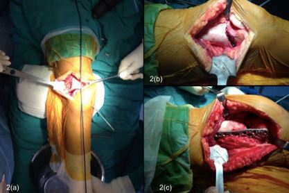
The dome was marked using a divider, which could be fixed at a particular radius. One limb of the divider was kept over the CORA (center of rotation of angulation), which was at the center of knee joint or femoral condyle level in all cases. This point corresponds approximately to the lower border of patella with the knee in full extension, and after correlating this point with the joint line, one limb of the divider was sunk into this part of patella and the radius of the dome was adjusted about 5 mm proximal to the adductor tubercle. An arc was now drawn over the lower femur. Multiple drill holes were made antero-posteriorly over the marked dome, perforating both femoral cortices. The dome osteotomy was completed by joining the drilled hole using a thin osteotome (Figure 2a).

Care was taken to keep the knee flexed so that neurovascular structures fall away. Now by holding the upper leg and keeping the knee straight, the distal fragment was rotated at the osteotomy site thereby correcting the deformity. In some cases, the medial most portion of the proximal fragment hampered the rotation of distal fragment. A triangular bony projection was occasionally removed from this area to facilitate rotation, in these cases. The alignment was checked using a telescopic rod (Fig 2b) about 1-meter-long so that center of hip, knee and ankle become co-linear. The osteotomy was temporary fixed with a K wire or steinman pin percutaneously for provisional stability anteromedial to posterolaterally. Following this, osteotomy was fixed using appropriate length PHILOS plate applied laterally (fig 2c). Stability as well as the correction was confirmed intra-operatively under image intensifier. After applying a suction drain, the surgical wound was closed in layers.


Postoperatively, a long knee brace was applied intermittently for 4 weeks, and was be removed during range of movement exercises which were started from third day. The drain was removed at 24-48 hours after surgery and the amount of blood collected in the drain was noted. Sutures were removed at 2 weeks. Knee range of motion and quadriceps exercises started from third postoperative day. By using two axillary crutches patient was allowed ambulation, but no weight-bearing was permitted for the first 3 weeks.

This was followed by toe-touch walking for 3 weeks, and further one axillary crutch was used for the next month with gradual transition to full weight-bearing as per tolerance. Patients were followed up regularly at 4 weeks, 12 weeks, 6 months, 12 months and 24 months’ post-surgery.

Postoperatively, the patients were re-evaluated clinically and radiologically for the alignment, correction achieved, range of motion at knee and union of the osteotomy. Intermalleolar distance, tibio-femoral angle and lateral distal-femoral angle (LDFA) were calculated. Functional outcome was evaluated by Bostman knee score and patients with score between 28 to 30 were classified as having excellent outcome, a score between 20 to 27 was good and a score below 20 was classified as unsatisfactory. Statistical analysis was done by student'st-test between values obtained preoperatively and postoperatively and two-tailedP< 0.05 was considered statistically significant.

Results

39 osteotomies were carried out on 24 patients of genu valgum. 15 patients had bilateral deformities whereas 9 patients had unilateral deformity. The average age at operation was 16.01 years (range 14 to 20). There were 16 females and 8 males.

The mean intermalleolar distance pre-operatively was 17.5 cm (range 11 to 24), which postoperatively improved to 2.25 cm (range 0 to 4 cm). The mean pre-operative clinical tibio-femoral angle was 19.250 (range 140 to 240) and radiological tibio-femoral angle was 20.90 (range 150 to 260) which postoperatively improved to mean value was 7.750 (range 40 to 100) and 8.950 (range 50 to 110) respectively. The Mean pre-operative LDFA was 74.850 (range 670 to 830) whereas post-operative mean value was 86.90 (range 830 to 900). The mean Bostman knee score improved significantly from 20.8 (range 18 to 22) to 29.1 (range 27 to 30) (table 1).

2 patients (2 limbs) had good score i.e. between 20 to 27, while rest all the patients had excellent score between 28 to 30. Improvement in intermalleolar distance, tibio-femoral angle and LDFA was statistically significant (P< 0.001). All the patients were happy with the cosmetic correction except one and none of the patients complained of any hardware prominence. All patients regained 900 knee flexion within 2 weeks of surgery and almost full ROM at 3 months after the surgery except one patient, who had deep infection at the incision site, which responded to antibiotics and healed in 3 months (Figure 3).

One patient with epiphyseal dysplasia complained of pain in the knees and in spite of a good cosmetic correction was not happy. Four knees had slightly hypertrophic scars.

Discussion

Genu valgum in adolescents and young adults is a frequent cause of orthopaedic referral [1-3]. The abnormal biomechanical loads on the

Table 1:Pre-operative and post-operative clinical and radiological parameters

Parameters (n=39)PreoperativePostoperativeP
Intermalleolar distance in cm (range)17.5±2.2 (11-24)2.25±0.7 (0-4)<0.001
Clinical tibiofemoral angle (range)19.25±2.1 (14-24)7.75±0.8 (4-10)<0.001
Radiological tibiofemoral angle (range)20.9±2.1 (15-26)8.95±0.8 (5-11)<0.001
Lateral distal-femoral angle (range)74.85±2.1 (67-83)86.9±0.6 (83-90)<0.001
Boatsman knee score (range)20.8±1.3 (18-22)29.1±0.6 (27-30)<0.001

Figure 1:Preoperative clinical photograph (a) and full length antero-posterior X rays (b) of the patient which shows genu valgum deformity.
ojmpc_122_01.jpg

Figure 2:Intraoperative photo (a to c) of a patient of genu valgum with dome osteotomy & provisional fixation with K wire, alignment rod to check the correction & osteotomy stabilized by PHILOS plate.
ojmpc_122_02.jpg


Figure 3:12 weeks postoperative antero-posterior (a) and lateral (b) radiograph of knee showing union at osteotomy site and correction achieved.
ojmpc_122_03.jpg

knee due to lateralization of the mechanical axis in genu valgum may lead to anterior knee pain, patello-femoral instability, circumduction gait, and difficulty in running [4-11]. Hence surgical intervention is needed to improve the biomechanics, which results in improved appearance, gait and function in significant valgus deformity [3].

Deformity correction in the coronal plane may be achieved using realignment osteotomies like lateral opening wedge, medial closing wedge, dome osteotomy, wedge-less spike osteotomy, and wedge-less ‘V’ osteotomy [4-14], which can be fixed with k wire, external fixators or plate [15-20]. This realignment osteotomy corrects the limb alignment and decreases the risk of arthritis development and progression [21-23].

We evaluated the results of 39 cases of dome osteotomy for genu valgum in 24 patients (15 bilateral and 9 unilateral) with mean age of 16 years, which were stabilized with proximal humerus internal locking system (PHILOS) plate and found statistically significant improvement in intermalleolar distance, tibio-femoral angle and LDFA, post-operatively. Dome Osteotomy (DO) is a cylindrical osteotomy with corresponding bone cuts, which rotate around the central axis of a circle with no bone resection, which ensures no limb length discrepancy. When this central axis corresponds with the Centre of rotation of axis (CORA), correction of the deformity can be attained without translation of the bone axes and it is then called as focal dome osteotomy [1-3].

We preferred dome osteotomy instead of a wedge osteotomy as closing wedge results in limb shortening whereas opening wedge results in delayed union with more restrictive weight-bearing (absent bone contact). The DO provides a large surface and maximizes bone contact, which results in optimal healing. Small bone spikes are produced by multiple drill holes after low-energy dome osteotomy.

These small bony spikes interdigitate at the osteotomy site after acute deformity correction and provide additional stability. Not only it reduced segment motion during osteotomy fixation but it also reduced the stress on the plate and screws, allowing early rehabilitation, range of motion and weight-bearing.

The dome osteotomy also allows high degrees of correction in the coronal plane. The DO is technically demanding surgery because performing the osteotomy as an arc needs care, precision and expertise to maintain the circular contour to ensure perfect segment rotation and bone contact and to avoid inadvertent propagation.

Further, for osteotomy to be effective it is important that osteotomy should be done at CORA. In our study, the preoperative radiographic planning showed the CORA to be at the knee joint or femoral condyle level in all cases.

This CORA was the center of the circle upon which the DO was created and hence it ensured a focal dome osteotomy and so we could achieve full correction with minimal translation of the distal bone fragment in all our cases.

Finding appropriate fixation device for supracondylar osteotomies in adolescent is relatively a tedious job and one needs to arrange for specialized pediatric angled blade-plates, angle-stable plates or intramedullary nails, which are costlier [2,11].

Seah et al found no significant differences in their comparative study of internal versus external fixation for distal femoral osteotomies, and concluded that the fixation method should be left to the discretion of the surgeon [24].

We used the adult Proximal humerus Interlocking System (PHILOS), routinely used in adult proximal humeral fractures to avoid more expensive and remotely available specialized pediatric implants while retaining its advantage of ability and contour which fits to match the desired correction.

PHILOS provides advantages like no need for post-operative plaster immobilization, early rehabilitation hence decreases chances of knee joint stiffness and quadriceps atrophy and decreased chances of fixation loss hence decrease incidence of non-union at osteotomy site. In adolescents, the antero-posterior dimensions of the distal femur are not large enough to place a condylar buttress plate, but the PHILOS plate is reasonable wide and stable suitable for fixation until osteotomy union.

Further, PHILOS plate is widely available and cost effective compared to customized paediatric plates which are expensive as well as availability is an issue in the developing countries. Although, the PHILOS plate is a relatively weak implant in comparison with the condylar buttress plate, but we found it to be sufficient for fixation in adolescents and we did not encounter any implant failure.

The most common, described complications following corrective angular osteotomies are non-union and failure of fixation [25]. Use of Dome osteotomy and PHILOS plate, in our study, provided good bone apposition and stable fixation which prevented these complications. Our study is limited by small number of cases, and relatively shorter follow up. Future studies involving a larger number of cases, longer follow ups and those comparing the use of plates with an external fixator may be useful.


Conclusion

Correction of Genu valgum deformity by dome osteotomy via lateral approach, can be accomplished very well using PHILOS for fixation, without complication of wedge osteotomy. Being a cost effective and easily available implant, the PHILOS has encouraging results in short to mid-term follow up.

References

1. Paley D, Tetsworth K. Mechanical axis deviation of the lower limbs. Preoperative planning of uniapical angular deformities of the tibia or femur. Clin Orthop Relat Res 1992;280:48-64. [Crossref][PubMed][Google Scholar]

2. Espandar R, Mortazavi SM, Baghdadi T. Angular deformities of the lower limbs in children. Asian J Sports Med 2010;1(1):46-53. . [Crossref][PubMed][Google Scholar]

3. Gautam VK, Maini L, Gupta R, Sabharwal A, Arora S. Flexion test in the coronal plane deformities of the knee. J Clin Orthop Trauma 2013;4:115-8. . [Crossref][PubMed][Google Scholar]

4. Aglietti P, Stringa G, Buzzi R. Correction of valgus knee deformity with a supracondylar V osteotomy. Clin Orthop Relat Res 1987;217:214-20. . [Crossref][PubMed][Google Scholar]

5. Brinkman JM, hurscheler C, Agneskirchner JD, Freiling D, Van Heerwaarden RJ. Axial and torsional stability of supracondylar femur osteotomies: biomechanical comparison of the stability of five different plate and osteotomy configurations. J Knee Surg Sports Traumatol Arthrosc 2010;19(4):579-87. . [Crossref][PubMed][Google Scholar]

6. Dewilde TR, Dauw J, Vandenneucker H, Bellemans J. Opening wedge distal femoral varus osteotomy using the Puddu plate and calcium phosphate bone cement. Knee Surg Sports Traumatol Arthrosc 2013;21(1):249-54. . [Crossref][PubMed][Google Scholar]

7. Dietz FR, Weinstein SL. Spike osteotomy for angular deformities of the long bones in children. J Bone Joint Surg Am 1988;70:848-52. . [Crossref][PubMed][Google Scholar]

8. Gorji J. Genu valgum : treatment by osteotomy. Clin Orthop Relat Res 1971;81:178-80. . [Crossref][PubMed][Google Scholar]

9. Healy WL, Anglen JO, Wasilewski SA, Krackow KA. Distal femoral varus osteotomy. J Bone Joint Surg Am 1988;70(1):102-9. . [Crossref][PubMed][Google Scholar]

10. Lin HP. Dome shaped osteotomy with external compression fixation in treatment of genu varum and valgum deformities (in Chinese). Zhonghua WaiKe Za Zhi 1982;20(5):296-7. . [Crossref][PubMed][Google Scholar]

11. McDermott AG, Finklestein JA, Farine I, Boynton EL, MacIntosh DL, Gross A. Distal femoral varus osteotomy for valgus deformity of the knee. J Bone Joint Surg Am 1988;70:110-6. . [Crossref][PubMed][Google Scholar]

12. Omidi-Kashani F, Hasankhani IG, Mazlumi M, Ebrahimzadeh MH. Varus distal femoral osteotomy in young adults with valgus knee. J Orthop Surg Res 2009;4:15. . [Crossref][PubMed][Google Scholar]

13. Puddu G, Cipolla M, Cerullo G, Franco V, Gianni E. Which osteotomy for a valgus knee. Int Orthop 2010. 34:239-47. [Crossref][PubMed][Google Scholar]

14. Scheffer MM, Peterson HA. Opening-wedge osteotomy for angular deformities of long bones in children. J Bone Joint Surg Am 1994:76:325-34. . [Crossref][PubMed][Google Scholar]

15. Hankemeier S, Paley D, Pape HC, Zeichen J, Gosling T, Krettek C. Knee para-articular focal dome osteotomy. Orthopade 2004;33(2):170–7. . [Crossref][PubMed][Google Scholar]

16. Paley D. Osteotomy concepts and frontal plane realignment. In: Herzenberg JE (ed) Principles of deformity correction, 1st edn. Springer, Berlin, 2002. p 112 [Crossref][PubMed][Google Scholar]

17. Paley D. Hardware and osteotomy considerations. In: Herzenberg JE (ed) Principles of deformity correction, 1st edn. Springer, Berlin, 2002. pp 300 [Crossref][PubMed][Google Scholar]

18. Sundaram NA, Hallett JP, Sullivan MF. Dome osteotomy of the tibia for osteoarthritis of the knee. J Bone Joint Surg (Br). 1986;68(5):782–6. [Crossref][PubMed][Google Scholar]

19. Watanabe K, Tsuchiya H, Sakurakichi K, Matsubara H, Tomita K. Acute correction using focal dome osteotomy for deformity about knee joint. Arch Orthop Traum Surg. 2008;128(12):1373–8. [Crossref][PubMed][Google Scholar]

20. Böstman O, Kiviluoto O, Nirhamo J. Comminuted displaced fractures of the patella. Injury. 1981;13(3):196–202. [Crossref][PubMed][Google Scholar]

21. Sharma L, Song J, Felson DT, Cahue S, Shamiyeh E, Dunlop DD. The role of knee alignment in disease progression and functional decline in knee osteoarthritis. JAMA 2001;286:188-95. . [Crossref][PubMed][Google Scholar]

22. Gugenheim JJ, Brinker MR. Bone realignment with use of temporary external fixation for distal femoral valgus and varus deformities. J Bone Joint Surg. 2003;85-A(7):1229–37. [Crossref][PubMed][Google Scholar]


23. Heervarden RJV, Wymenga AB, Freiling D, Staubli AE. Indications-planning-surgical techniques using plate fixators. In: Lobenhoffer P, Heervarden RJV, Staubli AE, Jakob RP (eds) Osteotomies around the knee, 1st edn. Thieme Medical Publishers, New York, 2008. pp 147 [Crossref][PubMed][Google Scholar]

24. Seah MKT, Shafi R, Fragomen AT, Rozbruch SR. Distal femoral osteotomy. is internal fixation better than external. Clin Orthop Relat Res 2011. 469(7):2003–11 [Crossref][PubMed][Google Scholar]

25. Wang JW, Hsu CC. Distal femoral varus osteotomy for osteoarthritis of the knee. Surgical technique. J Bone Joint Surg Am 2008;88(Suppl 1):100–8. [Crossref][PubMed][Google Scholar]

Disclaimer / Publisher's Note

The statements, opinions and data contained in all publications are solely those of the individual author(s) and contributor(s) and not of Journals and/or the editor(s). Journals and/or the editor(s) disclaim responsibility for any injury to people or property resulting from any ideas, methods, instructions or products referred to in the content.