Orthopaedic Journal of MP Chapter

Publisher: Madhya Pradesh Orthopaedic Association www.mpioa.com
E-ISSN:2582-7243, P-ISSN:2320-6993
2020 Volume 26 Number 1 Jan-Jun

External Fixator As A Definitive Treatment For Tibial Diaphyseal Fractures

Jain S1, Patel P2*, Gupta S3

1 S Jain, Department of Orthopaedics, Gajra Raja Medical College, Gwalior, Madhya Pradesh, India.

2* Prasant Patel, Senior Resident, Department of Orthopaedics, Gajra Raja Medical College, Gwalior, Madhya Pradesh, India.

3 S Gupta, Department of Orthopaedics, Gajra Raja Medical College, Gwalior, Madhya Pradesh, India.

Background: Precarious blood supply, subcutaneous nature and lack of soft-tissue cover of the shaft of the tibia make these fractures vulnerable to open fractures with high rate of nonunion and infection. External fixators have been used to treat these open tibial fractures as temporary mode of fixation. We evaluated the role of external fixator as a definitive treatment for tibial diaphyseal fractures.

Materials and methods: 57 patients with open tibial diaphyseal fracture with various degree of soft tissues injuries, treated with external fixator as definitive fixation were included in the study. The outcome, rate of union and complications were assessed.

Results: 57 patients with mean age 34.4 (range 18 to 59 years) were included. 45 were male while 12 were females. Mean duration of trauma to surgery interval 26.5 hrs. Mean time for dynamization was 7.44 weeks. 50 patients had union with mean time of union 22.4 weeks, while 7 patients had nonunion. 13 patients had pin tract infection, out of which 7 infections healed by oral antibiotics while 6 patients eventually had pin loosening requiring change of pin under local anaesthesia. One patient had malunion.

Conclusion: External fixator is a very useful method for treatment of open tibial diaphyseal fractures which eliminates the need of second surgery and allows bone and soft tissue healing without increasing morbidity when applied properly.

Keywords: Open tibal fracture, External fixation, Dynamization

Corresponding Author How to Cite this Article To Browse
Prasant Patel, Senior Resident, Department of Orthopaedics, Gajra Raja Medical College, Gwalior, Madhya Pradesh, India.
Email:
Jain S, Patel P, Gupta S, External Fixator As A Definitive Treatment For Tibial Diaphyseal Fractures. ojmpc. 2020;26(1):34-39.
Available From
https://ojmpc.com/index.php/ojmpc/article/view/102
Manuscript Received Review Round 1 Review Round 2 Review Round 3 Accepted
2020-06-06 2020-06-12 2020-06-18 2020-06-24 2020-06-30
Conflict of Interest Funding Ethical Approval Plagiarism X-checker Note
Authors state no conflict of interest. Non Funded. The conducted research is not related to either human or animals use. 13.39 All authors have accepted responsibility for the entire content of this manuscript and approved its submission.

© 2020by Jain S, Patel P, Gupta Sand Published by Madhya Pradesh Orthopaedic Association. This is an Open Access article licensed under a Creative Commons Attribution 4.0 International License https://creativecommons.org/licenses/by-nc/4.0/ unported [CC BY NC 4.0].

Introduction

Tibial diaphysis fractures are among commonly occurring long-bone fractures. These fractures are frequently present as open fractures due subcutaneous nature of tibia and increasing high velocity trauma due to two wheeler and pedestrian accidents. The precarious blood supply, lack of soft-tissue cover of the shaft of the tibia and increased incident of open fracture make these fractures vulnerable to nonunion and infection. The rate of infection may be as high as 52% in grade-IIIB open fractures [1,2].

To reduce these complications aggressive treatment is required which include proper intravenous antibiotics treatment, repeated soft-tissue debridement and stable fixation of the fracture [3].

Various methods for fracture stabilization include plating, intramedullary nailing or external fixator application. Use of plating or intramedullary nailing in patients with open tibial-shaft fracture is controversial with increased risk of infection [4-6].

External fixator is especially useful, as damage controlled orthopaedics, as temporary fixation of fracture, but later needs to be converted to internal fixation with reamed or unreamed intramedullary nail [7].

Hence we conducted this study to evaluated external fixator as a definitive mode of treatment modality for tibial diaphyseal fractures and assessed its outcome, rate of union and complications.

Material and Methods

This prospective study is conducted on 60 patients of open tibial diaphsis fracture treated with external fixator as definitive method of treatment at our centre. The study was approved by institutional ethical review committee and written informed consent was obtained from all the patients before inclusion into study.

Patients with open tibial diaphyseal fracture with any grade or degree of soft tissues injuries, between 16 to 65 years were included in this study. Patients with concomitant fracture in same limb, pathological fracture, and fracture associated with bone loss, neurovascular injury or with fracture extended to joint were excluded from study.

After stabilizing the patients heamo-dynamically and after advanced trauma life support resuscitation, thorough irrigation of the wound and third generation intravenous cephalosporin were given. Routine blood & radiological investigations were carried out. Radiological examination included Antero-Posterior and lateral view of leg including knee and ankle joint.

The fractures were classified according to the AO/OTA classification and Gustilo-Anderson classification [8,9]. All patients were planned for debridement and fixation of fracture by external fixation under spinal anaesthesia in supine position.

Initially the surgical debridement with removal of all dead necrotic tissues, removing the free loose bone pieces was done this was followed by stabilization of the fracture with application of uniplanar AO type external fixator, under image intensifier holding the reduction manually.

For application of external fixator, at least two 5.0 mm cortical Schanz screw, with radial preload, were inserted with T handle manually in each proximal and distal end of tibia after drilling. Attempts were made to avoid cancellous area of upper and lower end of tibia if fracture pattern permitted; else 6mm cancellous Schanz screws were used. After manual reduction maintaining length, axial and rotational alignment, and two connecting rods were connected to schanz screw with the help of AO clamps.

More schanz screws were fixed and connected to rod with clamps on either end of fracture if needed for stability. Axial loading was done for simple transverse and short oblique fractures by unlocking pin to rod nuts of the clamp pins and re fixing it in bending stress towards the fracture for pins nearer to fracture and bending stress in the direction away from the fracture for the far pins. Pins were subjected to bending stress in reverse manner for comminuted fractures.

Postoperatively systemic antibiotic were continued for 5 days and dressing done regularly and the wound was left as such to heal, with secondary intention and dressings.

Patients were encouraged to attain knee and ankle range of motion (ROM) depending on the patient pain tolerance. Axial dynamization and loading were individualized and was started once patient became painless on walking or could walk with minimal pain (Figure 1).

Figure 1:Illustration showing how dynamization done by crosswise loosening of tube nuts (lower tube nuts of one rod and upper tube nuts of other rod) as marked by arrow.
ojmpc_102_01.jpg


After dynamization weight bearing was encouraged. Patients were followed regularly. Once clinical or radiological union was achieved, i.e. no pain or mobility at fracture site and union in 3 cortices in anteroposterior and lateral view respectively, the external fixator frame was removed and patient was put on patellar tendon bearing (PTB) cast for further 6 week to consolidate the union.

All patients were assessed for union, time to union, alignment and associated complications like infection, nonunion, malunion, reoperations etc. Normal healing was defined as union within 6 months, delayed union as healing between 6 and 9 months, and nonunion as the absence of healing even after 9 months, whereas malunion was defined when there was more than 50 of varus or valgus alignment, more than 100 of anteroposterior alignment, or more than 1 inch of shortening was considered as malunion.

Results

Total of 60 patients of open tibial fractures were operated with external fixator as definitive mode of treatment. Among these 3 patients were lost to follow up, so total 57 patients with mean age 34.4 (range 18 to 59 years) were included in our study, out of which, 45 were male while 12 were females.

50(88%) patients had RTA injuries, 3(5%) had injuries due to assault and 4(7%) had injuries due to fall of heavy object over limb. 9 (15.79%) patients had associated injuries in upper or contralateral limb. As per Gustilo-Anderson Classification, type IIIB was most common type seen 31 (54.39%) patients, whereas 2(3.51%) patients had type I injury, 8(14.04%) patients had type II injury, and 16(28.07%) patients had type IIIA injury.

As per AO type fracture, 21(36.84%) had A, 23(40.35%) patients had B and 13(22.80%) patients had C type injuries. In subtypes B2 was the most common injury pattern seen amongst all. The mean duration of injury to presentation of patient to hospital was 15.8 hours (1hour – 240 hours), and mean delay in surgery was 26.5 hrs (range 9 to 248 hrs).

40 and 15 patients were operated within 24 hrs and within 3 days respectively. Mean time for dynamization was 7.44 weeks (range 5 to 10 weeks). Most of the patients i.e. 30 (52.63%) patients were dynamized by 6th or 7th week after surgery, whereas dynamization was done in 2 (3.51%), in 21(36.84%) and in 4(7.02%) patients after 4th to 5th week, 8th to 9th week and after 9th week respectively.

Except of 7 patients who had non-union, all 50 (87.7%) patients had union, in mean time of 22.4 weeks (range 15 to 29 weeks) (fig 2). Of the 7 non-union 6 were of Gustilo Anderson type IIIB (RR:1.576) and one was of IIIA (RR:0.509). None of nonunion was encountered in type I and II type injuries. 14 patients had complications, one had malunion (anteroposterior angulation >100) while 13 patients had pin tract infection, 7 of which healed by oral antibiotics while 6 eventually had pin loosening requiring change of pin under local anaesthesia.

Figure 2: Clinical photo (a) and preoperative X rays AP (b) and lateral (c) view of 38 year male patient with open tibial fracture and immediate post-operative X rays AP (d) and lateral (e), 8 weeks follow AP (f) and lateral (g) and at final follow AP (h) and lateral (i), who was treated with external fixator showing good union.
ojmpc_102_02a.jpg
ojmpc_102_02d.jpg
ojmpc_102_02h.jpg


Discussion

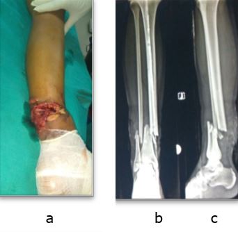
Open tibial fractures are among most common fractures in young adults encountered at various trauma centre [9-11]. As most of these patients are young adults who sustained these fractures belong to physically highly active and productive age group, they need optimal treatment to get back to their previous work capacity as early as possible and avoid long term complications.

Open tibial fractures with inherent less soft tissue coverage and added soft tissue trauma by injury poses higher risk of postoperative complications like wound dehiscence and infection [4-6]. Hence treatment of open tibial fractures demands tissue friendly surgical procedures as well as adequate fracture fixation.

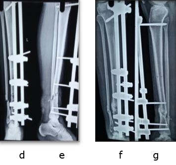
External Fixator application is a commonly used technique for compound tibia fractures, its main benefit being its less invasive nature. Disadvantage of external fixator is lower stability as compared to other methods of fixation. Other limitations of the external fixator are pin tract infections, pin loosening, re-displacement, less useful in osteoporotic fracture, delayed union, non-union and malunion.

In developing country like ours, where patient load is very high and resources are limited, it is difficult getting patient into operation theatre twice especially when it is not an emergency. Irregular follow-ups, low socioeconomic group, high cost of reoperation, poor hygiene and associated pin tract infection makes conversion of temporary external fixation to definitive internal fixation, difficult [12].

Giannoudis in 96 open tibial fractures showed over 17% infection rate after conversion of external fixation to IM nailing [13]. Hence, external fixator itself is preferred as definitive treatment modality of treatment because it eliminates the second surgery, indirectly reduces the patient loads waiting for surgery and avoids the risk associated with second surgery and anaesthesia [12].

We conducted this study to confirm the role of external fixator as definitive fixation method of treatment for open tibial fractures in 57 patients with mean age 34 years and found it to be very effective with rapid healing, and few complications.

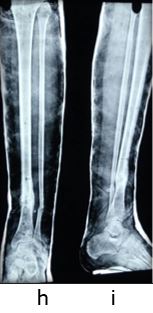
In our series, 50 (87.7%) had union, in mean time of 22.4 weeks (range 15 to 29 weeks). Similar union rate was seen by studies of Kumar (97% in 37 patients), Beltsios (91 % in 241 patients) and of Emani (95% in 62 patients) [14-16].

The mean time to union by Beltsios was 25 weeks in open fractures whereas in Emami series union time was 22 week, but 22% had delayed union [15,16].

In metaanalysis done by Bhandari et al directly comparing external fixators and unreamed IM nails, it is shown that there is no statistically significant difference between the two with respect to union, delayed union, deep infection and chronic osteomyelitis,

but external fixation was associated with statistically significant increased rate of malunion and reoperations, whereas unreamed nailing showed a statistically significant increase in the rate of failure of the implant [17].

In our study, there were 15 (18.99%) delayed union and 7 (10.94%) nonunions compared to 8 to 15% non-union and 9 to 39% delayed union in reported series by Giannoudis et al, Beltsios et al, Emani et al, Kimmel et al and Velazco A [13,15-19].

The reported incidence of malunion in Beltsios series was 1.8%, Kimmel series of 26% and Giannoudis series was 20%, but in our series only one patient (1.7%) had malunion [13,15,18].

The probable reason for this is because we tried to achieve anatomic reduction before applying the external fixator, which could have led the fracture to unite without malunion. Further, the reported incidence of pin tract infection is 32 to 80% while the incidence of deep infection is 16.2%, with average 4% developing chronic osteomyelitis [3,18,19],

but in our series we encounter 13 (22.8%) patients of pin tract infection, out of which 7 infection healed by oral antibiotics and only 6 (10.52%) requiring pin revision due to loosening. Low incidence of pin tract infection in our series is attributed to proper technique of pin insertion, preloading and adequate pin site dressing and care [20].

Thus external fixator, as a definitive treatment modality to achieve union in patients of open tibial fractures provides early and strong bone union due to inherent benefits of less tissue damage and minimal disturbance of fracture site biology.

But care must be taken to achieve proper reduction and to avoid pin related associated complications of infection / loosening by following proper pin insertion technique, pre-tensioning the pins and doing regular pin site care. For better functional outcomes range of motion, dynamization and weight bearing should be started early to promote healing by converting fixator frame to less rigid allowing axial micromovements at fracture site.

Conclusion

External fixator for compound tibial diaphyseal fractures is a very useful modality of treatment which eliminates the need of re-operation and allows bone and soft tissue healing without increasing morbidity when applied properly. Pin tract infection and loosening are common complications, but it can be reduced by proper technique.

References

1. Gustilo RB, Mendoza RM, Williams DN. Problems in the management of type III (severe) open fractures: a new classification of type III open fractures. J Trauma. 1984;24(8):742–6. [Crossref][PubMed][Google Scholar]


2. Ajmera A, Verma A, Agrawal M, Jain S, Mukherjee A. Outcome of limb reconstruction system in open tibial diaphyseal fractures. Indian J Orthop. 2015;49:429-35. [Crossref][PubMed][Google Scholar]

3. Patzakis MJ, Wilkins J, Moore TM. Considerations in reducing the infection rate in open tibial fractures. Clin Orthop. 1983;178:36–41. [Crossref][PubMed][Google Scholar]

4. Chapman MW. The role of intramedullary fixation in open fractures. Clin Orthop. 1986;212:26–34. [Crossref][PubMed][Google Scholar]

5. Chiron HS. Open fractures of the shaft of the tibia treated by intramedullary nailing. Bull Hosp Joint Dis. 1982;42:92-102. [Crossref][PubMed][Google Scholar]

6. Fairbank AC, Thomas D, Cunningham B, Curtis M, Jinnah RH. Stability of reamed and unreamed intramedullary tibial nails: a biomechanical study. Injury. 1995;26(7):483–5. [Crossref][PubMed][Google Scholar]

7. Giannoudis PV, Papakostidis C, Roberts C. A review of the management of open fractures of the tibia and femur. J Bone Joint Surg Br. 2006;88(3):281–9. [Crossref][PubMed][Google Scholar]

8. Kellam JF, Meinberg EG. Tibia. J Orthop Trauma. 2018;32:1:53-55. (AO/OTA.Classification of Fractures—Long Bones aotrauma.aofoundation.org) [Crossref][PubMed][Google Scholar]

9. Gustilo RB, Anderson JT. Prevention of infection in the treatment of one thousand and twenty-five open fractures of long bones: retrospective and prospective analyses. J Bone Joint Surg Am. 1976;58:453–458. [Crossref][PubMed][Google Scholar]

10. Zettas JP, Zettas P, Thanasophon B. Injury patterns in motorcycle accidents. J trauma. 1979;19(11):833–6. [Crossref][PubMed][Google Scholar]

11. Li X, Yun Z, Li X, Wang Y, Yang T, Zheng L, et al. Orthopedic injury in electric bicycle-related collisions. Traffic Inj Prev. 2017;19;18(4):437–40. [Crossref][PubMed][Google Scholar]

12. Banskota B, Shrestha S, Chaudhary RK, Rajbhandari T, Rijal S, Shrestha BK, et al. Patterns of Orthopaedic Injuries among Motorbike Accident Admissions Presenting to a Tertiary Care Hospital in Kathmandu. J Nepal Health Res Counc. 2016;14(32):51–7. [Crossref][PubMed][Google Scholar]

13. Giannoudis PV, Papakostidis C, Roberts C. A review of the management of open fractures of the tibia and femur. J Bone Joint Surg Br. 2006;88:281–289. [Crossref][PubMed][Google Scholar]

14. Kumar DR, Sujai DS, Chethan D, Nanjundappa DH, Swamy DS. Results of open fractures of tibia treated by external fixator as primary and definitive procedure. Int J Orthop Sci. 2017;1;3(1):179–81. [Crossref][PubMed][Google Scholar]

15. Beltsios M, Savvidou O, Kovanis J, Alexandropoulos P, Papagelopoulos P. External fixation as a primary and definitive treatment for tibial diaphyseal fractures. Strateg Trauma Limb Reconstr. 2009;4(2):81–7. [Crossref][PubMed][Google Scholar]

16. Emami A, Mjöberg B, Karlström G, Larsson S. Treatment of closed tibial shaft fractures with unilateral external fixation. Injury. 1995 Jun;26(5). 299-303 [Crossref][PubMed][Google Scholar]

17. Bhandari M, Guyatt GH, Swiontkowski MF, Schemitsch EH. Treatment of open fractures of the shaft of the tibia. Bone Jt J. 2001;1;83–B(1):62–8. [Crossref][PubMed][Google Scholar]

18. Kimmel RB. Results of treatment using the Hoffmann external fixator for fractures of the tibial diaphysis. J Trauma. 1982;22(11):960–5. [Crossref][PubMed][Google Scholar]

19. Velazco A, Fleming LL. Open fractures of the tibia treated by the Hoffmann external fixator. Clin Orthop. 1983;180:125–32. [Crossref][PubMed][Google Scholar]

20. Moroni A, Vannini F, Mosca M, Giannini S. State of the art review: techniques to avoid pin loosening and infection in external fixation. J Orthop Trauma. 2002;16(3):189–95. [Crossref][PubMed][Google Scholar]

Disclaimer / Publisher's Note

The statements, opinions and data contained in all publications are solely those of the individual author(s) and contributor(s) and not of Journals and/or the editor(s). Journals and/or the editor(s) disclaim responsibility for any injury to people or property resulting from any ideas, methods, instructions or products referred to in the content.